
Leprosy
Reading File
Searching sources...
Finding Sources
Searching PubMed
leprosy Hansen disease
Reading File
Finding Sources
Reading File
Reading File
Reading File
Finding Sources
Reading File
Reading File
Reading File
Loading Image
Reading File
Finding Sources
I now have comprehensive information to write a thorough response on leprosy. Let me compile everything.
Leprosy (Hansen Disease)
Overview
Leprosy is a slowly progressive infection caused by Mycobacterium leprae (and occasionally M. lepromatosis) that mainly affects the skin and peripheral nerves. Despite its low communicability, approximately 200,000 new infections occur per year, predominantly in low-resource tropical nations - especially Southeast Asia, East Africa, and Brazil. - Robbins, Cotran & Kumar Pathologic Basis of Disease
Causative Organism
- Obligate intracellular pathogen that cannot be cultured in vitro
- Grows optimally at 32-34°C (the temperature of human skin), explaining its predilection for cool peripheral tissues
- Proliferates in dermal macrophages and disseminates hematogenously
- Secretes no toxins; virulence depends on its cell wall, particularly the lipid PGL-1, which is critical for host cell invasion
- BCG vaccination confers partial cross-protection (M. leprae is genetically similar to M. tuberculosis)
- The organism inhibits mitochondrial energy metabolism to evade the immune response
Transmission
- Primarily via human respiratory secretions
- Rare zoonotic transmission from armadillos has been documented
- Overall communicability is low; most exposed individuals never develop disease
Classification
Ridley-Jopling Spectrum (Immunological Basis)
| Type | Abbreviation | Immune Response | Bacteria |
|---|---|---|---|
| Tuberculoid | TT | Strong Th1, IFN-γ, IL-2; granulomas | Few (paucibacillary) |
| Borderline tuberculoid | BT | Moderate Th1 | Few |
| Borderline | BB | Mixed | Moderate |
| Borderline lepromatous | BL | Weak Th1 | Many |
| Lepromatous | LL | Weak Th1, relative Th2/Treg dominance | Many (multibacillary) |
WHO Classification (Operational - for treatment)
- Paucibacillary (PB): up to 5 skin lesions, slit-skin smear negative
- Multibacillary (MB): 6 or more skin lesions, or smear-positive
Pathogenesis
The host T-cell response determines the disease pattern:
- Tuberculoid leprosy: Strong Th1 response with classically activated (M1) macrophages, IL-2, IFN-γ, and a Th17 component. Microbial burden is low; granulomas form. Analogous to M. tuberculosis - IFN-γ mobilizes effective macrophage killing.
- Lepromatous leprosy: Weak Th1 response, relative Th2 and regulatory T-cell predominance. Bacteria multiply unchecked. Occasional antibody production against M. leprae antigens forms immune complexes that can cause erythema nodosum, vasculitis, and glomerulonephritis.
Clinical Features
Tuberculoid Leprosy
- Localized, flat, red lesions that enlarge with indurated, elevated, hyperpigmented margins and depressed pale centers (central healing)
- Skin lesions are anesthetic - definite loss of sensation (light touch, pain, temperature)
- Asymmetric involvement of large peripheral nerves; nerve damage can be severe
- Nerves enclosed within granulomatous reactions; small twigs are destroyed
- Complications: chronic skin ulcers, contractures, paralyses, autoamputation of digits, keratitis from facial nerve involvement
Lepromatous Leprosy
- Affects skin, peripheral nerves, anterior eye chamber, upper airways (to larynx), testes, hands, and feet
- CNS and vital organs are spared (too warm for bacterial growth)
- Macular, papular, or nodular lesions on face, ears, wrists, elbows, and knees
- Progressive coalescing nodules produce the characteristic leonine facies
- Symmetric nerve involvement (unlike tuberculoid); ulnar and peroneal nerves particularly affected
- Skin lesions are hyposthetic or anesthetic
- Nasal: persistent inflammation, bacilli-laden discharge
- Testes: destruction of seminiferous tubules → sterility
- Lymph nodes and spleen: foamy macrophage aggregates
Histopathology
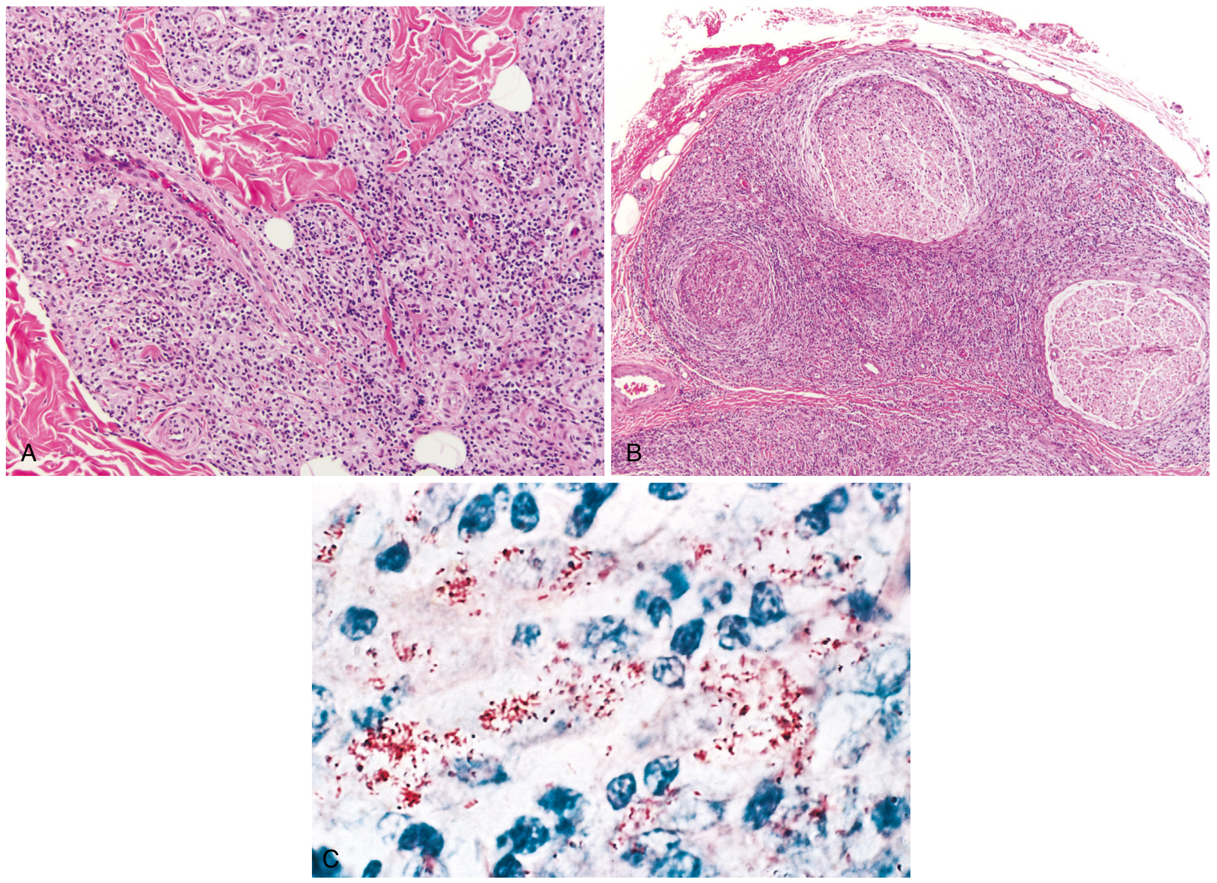
Fig. 8.33 from Robbins Pathology - (A) Tuberculoid: granulomatous dermal inflammation around adnexa, vessels, and nerves; (B) Lepromatous: large nerve bundle infiltration; (C) Acid-fast bacilli (red rods) within macrophages (blue-stained nuclei), lepromatous form.
- Tuberculoid: Granulomas resembling tuberculosis; bacilli almost never found (paucibacillary)
- Lepromatous: Large aggregates of lipid-laden macrophages called lepra cells, filled with masses ("globi") of acid-fast bacilli; abundant bacteria (multibacillary)
Cardinal Signs (Diagnosis)
A clinical diagnosis requires at least one of three cardinal signs:
- Hypopigmented or erythematous skin lesion(s) with definite loss or impairment of sensation
- Involvement of peripheral nerves demonstrated by definite thickening with sensory impairment (greater auricular, ulnar, radial, median, lateral popliteal, posterior tibial, sural, superficial peroneal nerves are the most commonly palpated)
- Positive AFB in slit-skin smears, biopsy, or positive PCR
Diagnostic Tools
Slit-Skin Smear
- Taken from 4 sites: right earlobe, forehead, chin, left buttock (men) or left upper thigh (women)
- Stained with Ziehl-Neelsen reagent; examined by light microscope
- Bacteriological index graded on a logarithmic scale from 0 to 6
Skin Biopsy
- Confirms diagnosis, classifies disease, supports diagnosis of reactions, and determines cure post-MDT
- Macular lesions: biopsy from the middle; plaques: from the active indurated edge
PGL-1 Antibody Test (Serology)
- PGL-1 is a specific lipid on the M. leprae cell wall
- Positive in 90-95% of multibacillary cases; only 25-60% of paucibacillary cases
- ML flow test (immunochromatographic lateral-flow assay): 92-97% sensitivity in MB, 32-40% in PB
- A newer quantitative UPC-LFA test has higher specificity in paucibacillary disease
Lepromin (Mitsuda) Test
- Intradermal injection of inactivated M. leprae; induration read at 3-4 weeks
- Measures cellular immunity to M. leprae
- Negative in lepromatous (BL/LL) leprosy → indicates lack of protective cellular response
PCR
- Detects as few as 1-10 bacilli; positive in 60-75% of smear-negative cases
- Multiplex PCR (RLEP, SodA, 16S rRNA genes) useful for early diagnosis and screening household contacts
Nerve Function Assessment
- Semmes-Weinstein monofilament test is the gold standard; ballpoint pen used in the field
- Voluntary muscle testing: eye closure (facial), finger abduction (ulnar), thumb opposition (median), wrist extension (radial), ankle extension (peroneal)
- Ultrasound imaging can detect early nerve enlargement
- Nerve conduction studies: sensory parameters affected months before clinical tests become positive
Differential Diagnosis
| Lesion Type | Differentials |
|---|---|
| Hypopigmented macules | Pityriasis alba, vitiligo, pityriasis versicolor, pityriasis rosea, sarcoidosis, post-kala-azar dermal leishmaniasis |
| Plaques/nodules | Granuloma annulare, cutaneous sarcoidosis, cutaneous leishmaniasis, lupus erythematosus, mycosis fungoides, secondary syphilis |
| ENL lesions | Erythema nodosum (other causes), nodular vasculitis, cutaneous polyarteritis nodosa |
| Mononeuropathy | Diabetes, amyloidosis, myxedema |
| Polyneuropathy (acute) | Guillain-Barré syndrome, toxic polyneuropathy |
Leprosy Reactions
Immune-mediated inflammatory episodes that can cause rapid, irreversible nerve damage and represent medical emergencies.
Type 1 Reaction (Reversal Reaction - T1R)
- Can occur in tuberculoid, borderline tuberculoid, or borderline lepromatous patients
- Cell-mediated immune activation attacking M. leprae antigen
- Affected nerves enlarge and become tender; sudden loss of nerve function can be permanent
- May also be subacute/chronic with limited symptoms but severe underlying damage
- Histology: perivascular and perineural edema, large numbers of lymphocytes; bacilli reduced
Type 2 Reaction (Erythema Nodosum Leprosum - ENL)
- Occurs in 50% of BL or lepromatous patients, usually within a few years of starting MDT or during pregnancy
- Mechanism: immune complex-mediated vasculitis with local complex formation, elevated TNF-α, complement membrane attack complex
- Systemic symptoms: fever, myalgias, arthralgias, anorexia
- Characteristic skin lesions: erythematous, subcutaneous/dermal nodules widely distributed, NOT at existing lesion sites; favor extensor arms and medial thighs (unlike classic erythema nodosum)
- Multisystem involvement: conjunctivitis, neuritis, keratitis, iritis, synovitis, nephritis, orchitis, hepatosplenomegaly, lymphadenopathy
- Labs: elevated ESR and CRP, neutrophilia
- Histology: leukocytoclastic vasculitis
Lucio Phenomenon
- Uncommon; occurs in diffuse lepromatous leprosy ("la bonita" type), mainly in western Mexico
- Purpuric macules → bullous lesions → rapid ulceration (especially below the knees)
- Lacks neutrophilia and systemic symptoms (differs from ENL)
- Not triggered by MDT initiation; often the initial presenting event
Treatment
WHO-Recommended Multidrug Therapy (MDT)
| Drug | Paucibacillary | Multibacillary |
|---|---|---|
| Dapsone | 100 mg/day (adult) | 100 mg/day (adult) |
| Rifampin | 600 mg/month (adult) | 600 mg/month (adult) |
| Clofazimine | Not included* | 50 mg/day + 300 mg/month (adult) |
| Duration | 6 months | 12 months |
Note: In 2018, WHO suggested adding clofazimine to paucibacillary MDT, but implementation varies due to concerns about skin discoloration and compliance.
Children age 10-14: rifampin 450 mg monthly, dapsone 50 mg/day; younger children receive weight-adjusted doses. - Harrison's Principles of Internal Medicine 22E
Drug Mechanisms and Side Effects
- Rifampin: Inhibits DNA-dependent RNA polymerase; given monthly in leprosy (different from TB dosing). Risk of hepatotoxicity is low at monthly intervals; harmless urine discoloration.
- Dapsone: Blocks folic acid synthesis; weakly bactericidal; half-life ~28 hours. Risks: mild hemolysis, anemia, rarely psychosis. "DDS syndrome" (dapsone hypersensitivity syndrome) - a severe adverse event, typically 6 weeks after starting treatment.
- Clofazimine: Causes skin discoloration (the main compliance concern in paucibacillary disease).
Relapse Monitoring
- Paucibacillary: 2 years of monitoring post-treatment
- Multibacillary: at least 5 years
- Relapse can be difficult to distinguish from T1R in paucibacillary disease; a therapeutic trial of glucocorticoids (improvement within 4 weeks = T1R; no response = relapse)
Drug-Resistant Leprosy
- Rifampin resistance: treat with at least two second-line drugs (clarithromycin, minocycline, or a quinolone - ofloxacin, levofloxacin, or moxifloxacin) plus clofazimine for 6 months, then clofazimine plus one second-line drug for 18 months
- Dual rifampin + ofloxacin resistance: clarithromycin + minocycline + clofazimine for 6 months, then clarithromycin or minocycline + clofazimine for 18 months
Treatment of Reactions
Type 1 (Reversal Reaction):
- Prednisolone 1 mg/kg/day (max 60-80 mg; WHO field protocol: 40 mg/day)
- Taper by 5 mg every 2 weeks over 20 weeks (better outcomes than a 12-week taper)
- Assess every 2 weeks; include nerve function assessment
- Reaction flares often return when dose falls below 10-20 mg/day - balance prolonged use risks with benefit
Type 2 (ENL):
- Mild first-time ENL: aspirin + pentoxifylline
- Most effective rapid-acting drug: thalidomide 100-200 mg once or twice daily (blacklisted in many countries due to teratogenicity; strict contraception required for women of childbearing age)
- Multisystem ENL (eyes, testes, kidneys, joints): longer treatment course needed
- Prednisolone is the alternative where thalidomide is unavailable
Complications and Disability
- Chronic skin ulcers from loss of protective sensation
- Contractures and fixed-joint deformities
- Bone absorption and autoamputation of digits
- Keratitis and corneal ulceration from facial nerve paralysis (lagophthalmos)
- Sterility (testicular destruction in lepromatous leprosy)
- "Silent neuritis" or "quiet nerve paralysis" - progressive nerve damage with minimal symptoms
Key Points Summary
| Feature | Tuberculoid | Lepromatous |
|---|---|---|
| Immune response | Strong Th1, granulomas | Weak Th1, Th2/Treg dominance |
| Skin lesions | Few, well-defined, anesthetic, dry | Many, symmetric, hyposthetic, nodular |
| Nerve involvement | Asymmetric, severe, few nerves | Symmetric, widespread |
| Bacteria | Few (paucibacillary) | Many (multibacillary) |
| Lepromin test | Positive | Negative |
| Antibodies | Low | High (non-protective) |
| Smear | Negative | Positive |
| Systemic spread | Absent | Present (not CNS/vital organs) |
Recent Evidence: A 2025 meta-analysis (PMID 41082563) examined leprosy relapse rates after MDT; a 2024 systematic review (PMID 39308868) updated leprosy immunopathogenesis - both aligned with current textbook content without indicating major changes to WHO treatment protocols.
Sources: Robbins, Cotran & Kumar Pathologic Basis of Disease; Harrison's Principles of Internal Medicine 22E (2025); Andrews' Diseases of the Skin - Clinical Dermatology
This is a shared conversation. Sign in to Orris to start your own chat.