
Is this app free?
Yes, I'm Orris, and I'm completely free to use! There are no subscription fees, paywalls, or hidden charges. Just ask me anything and I'll do my best to help — whether it's research, writing, medical questions, data analysis, document creation, or general knowledge.
Okay detail me about heart conduction pathway
Reading File
The cardiac conduction system carries electrical impulses from the SA node through the AV node, His bundle, bundle branches, and Purkinje fibers to coordinate the heartbeat. Let me pull the full details from the medical library.
Searching sources...
Finding Sources
Finding Sources
Reading File
Reading File
Reading File
Reading File
Reading File
Loading Image
Here is a detailed account of the cardiac conduction system, drawn from Gray's Anatomy for Students and Fuster and Hurst's The Heart, 15th Edition.
The Cardiac Conduction System
The cardiac conduction system is a network of specialized myocytes that initiates and coordinates the contraction of the atria and ventricles in a precise, unidirectional sequence. It consists of four main components:
- Sino-atrial (SA) node
- Atrioventricular (AV) node
- AV bundle (Bundle of His) with right and left bundle branches
- Subendocardial plexus of Purkinje fibers
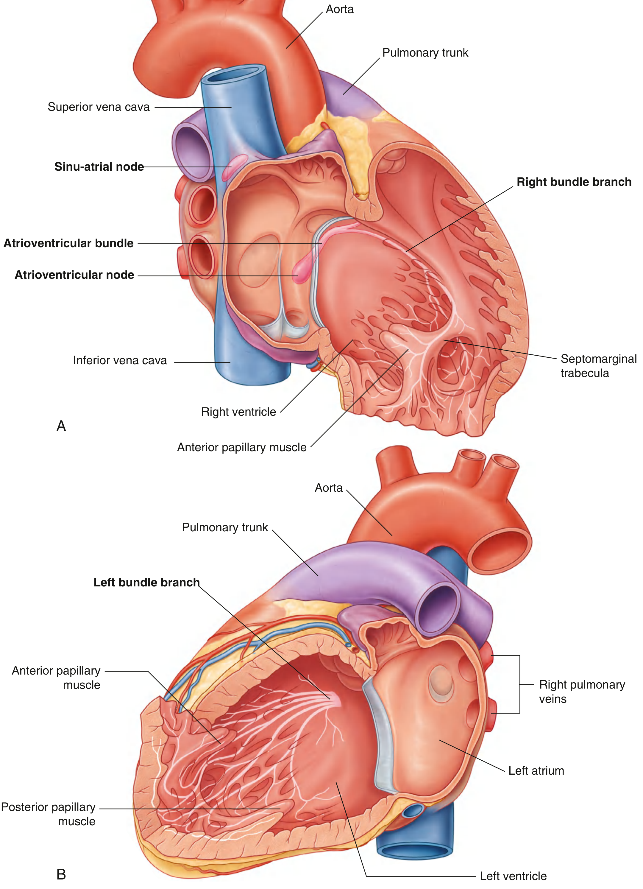
1. Sino-Atrial (SA) Node - The Pacemaker
- Location: At the superior end of the crista terminalis, at the junction of the superior vena cava and right atrium. This is the embryological junction between the sinus venosus and the atrium proper.
- Shape: Tadpole-like; ~13.5 mm long in the adult. The "head" lies close to the epicardium, the "tail" extends toward the endocardium, and it can extend more than 50% of the crest length toward the inferior vena cava.
- Function: Spontaneously generates electrical impulses (action potentials) at a rate of 60-100 beats/min, making it the dominant cardiac pacemaker.
- Innervation: Richly supplied by both sympathetic chains and vagus nerve, which modulate its firing rate.
- Signal spread: Excitation spreads across both atria, triggering atrial contraction (the P wave on ECG).
2. Internodal Conduction and Bachmann's Bundle
- While early anatomists described three discrete internodal tracts (anterior, middle, posterior), their existence as discrete pathways has never been definitively demonstrated.
- Instead, the arrangement of muscle bundles - the terminal crest, the rim of the oval fossa, and particularly Bachmann's bundle - facilitates conduction between the nodes.
- Bachmann's bundle is a prominent interatrial muscle bridge running across the roof of the atria, anterosuperior to the fossa ovalis. It facilitates interatrial synchrony by causing early activation of the anterior left atrium.
3. Atrioventricular (AV) Node
- Location: Near the apex of the triangle of Koch, close to the opening of the coronary sinus, adjacent to the attachment of the septal cusp of the tricuspid valve, within the atrioventricular septum.
- Size: Approximately 5 mm long and wide in the adult.
- Key feature: In the normal heart (without accessory pathways), the AV node provides the only pathway of electrical continuity between the atria and ventricles.
- Function: Receives excitation from the atria and introduces a physiological delay (~0.1 sec), allowing the atria to finish contracting before the ventricles are activated (corresponds to the PR interval on ECG).
- The compact AV node has inferior extensions - a right extension that courses adjacent to the septal leaflet hinge line, and a left extension toward the mitral vestibule. These are the targets for catheter ablation in AV nodal reentrant tachycardia (AVNRT).
- Blood supply: The AV nodal artery mostly arises from the dominant coronary artery (right in ~85% of people).
4. Bundle of His (AV Bundle)
- The penetrating bundle of His is a direct continuation of the compact AV node. It passes through the central fibrous body coursing anterocephalad, then runs along the lower border of the membranous interventricular septum.
- The bundle is sandwiched between the membranous septum and the crest of the muscular ventricular septum.
- It is surrounded by a thin fibrous sheath throughout its course, insulating it from surrounding myocardium and preventing premature activation.
- After a short distance, it bifurcates into the right and left bundle branches.
5. Bundle Branches
Right Bundle Branch (RBB)
- Cord-like in structure; descends through the musculature of the ventricular septum.
- Continues on the right side of the interventricular septum toward the apex of the right ventricle.
- Enters the septomarginal trabecula (moderator band), which carries it to the base of the anterior papillary muscle.
- Then fans out into the Purkinje network.
Left Bundle Branch (LBB)
- Fans out into interconnecting fascicles (not cord-like) as it descends in the subendocardium of the septal surface of the left ventricle.
- Classically described as dividing into:
- Left anterior fascicle - toward the anterolateral LV wall
- Left posterior fascicle - toward the inferoposterior LV wall
- Continues into the Purkinje fiber network.
6. Purkinje Fiber Network
- Both bundle branches divide into progressively finer branches, eventually forming the subendocardial plexus of Purkinje fibers.
- These run in the subendocardium and through the thickness of the ventricular walls and septum, making direct contact with working myocardium.
- Key property: Purkinje fibers conduct impulses very rapidly (~4 m/s), far faster than ordinary myocardium, enabling near-simultaneous activation of the entire ventricular mass.
- Activation begins at the papillary muscles and apex, then moves toward the arterial outflow tracts - this ensures the ventricles squeeze blood upward and outward efficiently.
Summary of the Conduction Sequence
| Step | Structure | Event |
|---|---|---|
| 1 | SA Node | Impulse generated (~70/min); atria depolarize (P wave) |
| 2 | Internodal pathways / Bachmann's bundle | Excitation spreads across atria; interatrial synchrony |
| 3 | AV Node | Impulse delayed ~0.1 sec (PR interval); atria finish contracting |
| 4 | Bundle of His | Impulse penetrates fibrous skeleton into ventricles |
| 5 | Right & Left Bundle Branches | Rapid conduction down both sides of interventricular septum |
| 6 | Purkinje Fibers | Simultaneous activation of ventricular myocardium from apex upward (QRS complex) |
Autonomic Regulation
The autonomic nervous system modulates the conduction system:
- Sympathetic (norepinephrine): Increases SA node firing rate, speeds AV conduction, increases force of contraction.
- Parasympathetic/Vagus (acetylcholine): Slows SA node (bradycardia), slows AV conduction (increases PR interval), can cause AV block in excess.
Both systems contribute fibers to the cardiac plexus, which branches to supply nodal tissue, the conduction system, coronary vessels, and myocardium.
Clinical Relevance
- Coronary artery disease disrupting blood supply to the conduction system can produce dysrhythmias, AV blocks, or bundle branch blocks.
- AVNRT (most common SVT) involves the dual pathway physiology of the AV node - ablation of the slow pathway (right inferior extension) is curative.
- Bundle branch blocks delay ventricular activation on the affected side, widening the QRS complex.
- His bundle pacing is an emerging technique that relies on precise anatomical knowledge of the His bundle location relative to the membranous septum.
Sources: Gray's Anatomy for Students, p. 247-249 | Fuster and Hurst's The Heart, 15th Edition, p. 1045-1047
I was just tesr8ng you you are the perfect partner from now on
Glad the test went well! I'm here whenever you need me — whether it's medicine, research, document creation, data analysis, or anything else. Just ask away.
This is a shared conversation. Sign in to Orris to start your own chat.