
Sinus and fistula in full detail including examination
sinus fistula diagram surgical tract
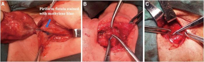
This composite of three intraoperative clinical photographs illustrates the surgical management of a pyriform sinus fistula (PSF) in the cervical region. Panel A demonstrates the identification of the fistula tract, which is visually enhanced using methylene blue dye. The tract appears as a distinct blue-stained tubular structure located medial to the inferior angle of the thyroid cartilage within the surgical field. Panels B and C show the progressive surgical steps of ligation and excision. In Panel B, surgical instruments (forceps and clamps) are used to isolate and stabilize the blue-stained fistula for suture ligation. Panel C depicts the final stages where the fistula is excised from the surrounding soft tissues, with surgical retractors providing exposure of the hypopharyngeal surgical site. The series highlights the importance of dye injection for intraoperative visualization of the fistula's anatomical course to ensure complete resection and reduce the risk of recurrence, which is the gold standard treatment for congenital branchial pouch anomalies.
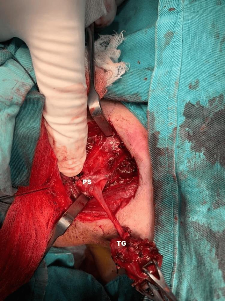
This clinical photograph provides an intraoperative view of the surgical excision of a branchial fistula tract in the right neck. The operative field is exposed through a surgical incision, with drapes and retractors maintaining visibility. A gloved finger and surgical forceps are seen manipulating the tissues. Two key structures are labeled: 'TG' (Thyroid Gland) and 'PS' (Pyriform Sinus). A distinct, elongated, fibrous fistula tract is clearly visible, extending from the superior pole of the right thyroid gland toward the right pyriform sinus. The thyroid tissue appears hemorrhagic and partially dissected, reflecting the surgical manipulation required to isolate the tract. The image illustrates the complex anatomical relationship between the branchial fistula and adjacent endocrine and pharyngeal structures, specifically highlighting the pathway where a fourth branchial pouch sinus typically traverses. This material is educationally relevant for otorhinolaryngology and endocrine surgery trainees to understand the surgical anatomy of congenital neck fistulas.
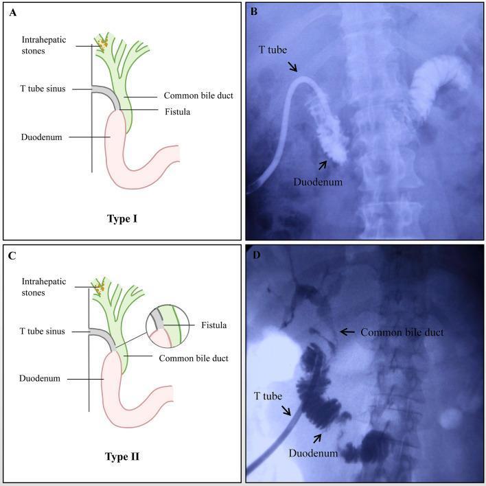
A multi-panel figure illustrating two types of T-tube sinus tract duodenal fistulas. Panels A and C are anatomical diagrams showing the relationship between intrahepatic stones, the common bile duct, the T-tube sinus tract, and the duodenum. Panels B and D are fluoroscopic images (T-tube cholangiography) demonstrating these complications. In Type I (A and B), the diagram shows the T-tube unintentionally inserted directly into the duodenum through a fistula; the corresponding cholangiogram in Panel B shows contrast material filling the duodenum with an absence of opacification in the biliary tree, indicating complete diversion or distal blockage of the sinus tract. In Type II (C and D), the diagram depicts a fistula allowing communication between the T-tube sinus, duodenum, and common bile duct; the cholangiogram in Panel D shows initial contrast filling of the duodenal loop followed by retrograde opacification of the common bile duct and intrahepatic ducts. The image serves as an educational reference for hepatobiliary complications following procedures for intrahepatic stone extraction, specifically the diagnostic presentation of iatrogenic fistulous tracts on imaging.
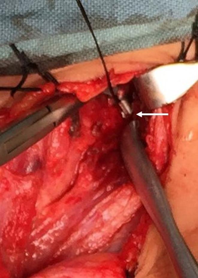
This clinical photograph captures an intraoperative view of a surgical neck dissection for the management of a recurrent pyriform sinus fistula (PSF). The surgical field shows exposed deep cervical fascia and soft tissues. A white arrow points to the remaining fistula tract, which appears as a small, cylindrical, pale-colored tubular structure a few millimeters in diameter. Key surgical instruments in the field include a Senn retractor being used on the right to provide lateral traction for better visualization of the deeper structures, and hemostatic forceps or a clamp on the left manipulating adjacent tissue. Black silk sutures are visible at the superior margin of the incision. The image illustrates the complex anatomy and the surgical challenge of identifying and excising the complete tract in recurrent cases to prevent future infection or abscess formation.
Can you make it
Ok now complete
anal fistula Parks classification intersphincteric suprasphincteric
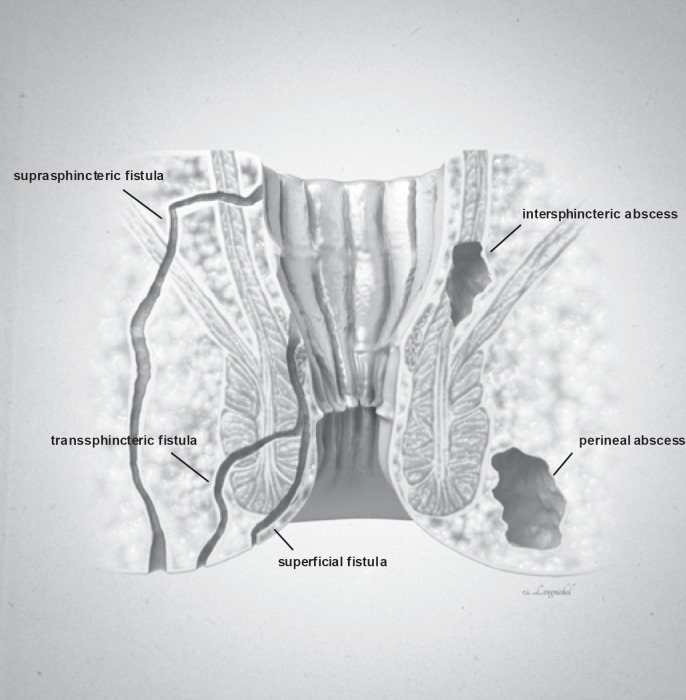
**Imaging Modality:** Medical illustration/Anatomical diagram. **Anatomical Region:** Coronal cross-section of the anorectal region, including the anal canal, rectum, internal and external anal sphincters, and surrounding perianal soft tissues. **Observed Pathology:** The illustration classifies various types of anorectal fistulas and abscesses based on their anatomical trajectory and location. **Characteristic Visual Features:** * **Fistula Tracts:** Three distinct epithelial-lined tracks are labeled. A **superficial fistula** is shown confined to the perianal skin and distal anal canal. A **transsphincteric fistula** is depicted crossing both the internal and external sphincter muscles into the ischioanal fossa. A **suprasphincteric fistula** is illustrated looping above the puborectalis muscle before descending to the skin surface. * **Abscess Formations:** Two localized collections of fluid/pus are identified. An **intersphincteric abscess** is positioned within the space between the internal and external sphincter muscles. A **perineal (perianal) abscess** is shown as a larger pocket located in the subcutaneous tissue adjacent to the anal verge. **Key Diagnostic Features:** The diagram emphasizes the relationship between inflammatory tracts and the sphincter complex, following the Parks classification system for fistula-in-ano. Suitable for clinical education on perianal sepsis and surgical planning.
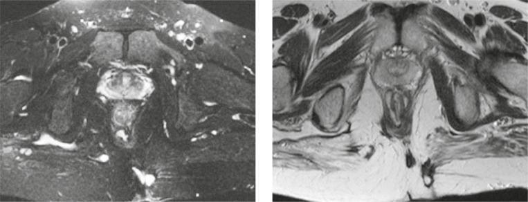
This diagnostic imaging set consists of axial MRI scans of the pelvic region focusing on the perianal area, utilizing T2-weighted Fat Suppressed (left) and T2-weighted turbo spin-echo (right) sequences. The images demonstrate an intersphincteric perianal fistula with a characteristic horseshoe-shaped configuration. The fistulous tract exhibits high signal intensity (hyperintense) on the T2-weighted sequences, effectively contrasting against the low signal intensity of the surrounding anal sphincter muscles. This tract is located within the intersphincteric space, bounded by the internal and external anal sphincters. The primary opening is visualized on the left posterior aspect of the anal canal. The high-signal area suggests active inflammation or fluid collection within the tract. Key anatomical structures visible include the rectum, levator ani muscle, and the ischioanal fossa. These images are clinically significant for the classification of perianal fistulae according to the Parks or St James's University Hospital classification systems, assisting in surgical planning by defining the relationship between the inflammatory tract and the sphincteric complex.
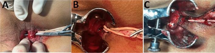
This composite of three clinical photographs (A, B, C) illustrates a surgical procedure for high trans-sphincteric or suprasphincteric anal fistulas, specifically the 'Decompression and Drainage Seton' (DADS) technique. Panel A shows the perianal surgical field where a seton has been rerouted through the intersphincteric plane and tied loosely around the external anal sphincter (EAS); a curved artery forceps is seen inserted through the internal opening to identify the intersphincteric tract. Panel B demonstrates the surgical exposure of the anal canal using a bivalve speculum, where the internal sphincter is being incised via electrocautery to open the fistula tract. Panel C displays the final stages of the procedure, showing the marsupialization of the operative wound. Interrupted absorbable sutures (polyglactin 3-0) are visible along the edges of the opened fistula tract, securing the mucosa to the underlying tissue from distal to proximal to facilitate continuous drainage and secondary intention healing. This visual series highlights key steps in sphincter-preserving proctological surgery for complex fistula-in-ano.
SINUS AND FISTULA - Complete Surgical Overview
PART 1: SINUS
Definition
Key: One opening = Sinus. Two openings = Fistula.

Structure of a Sinus
- External opening (usually on skin)
- A tract lined with granulation tissue
- A deep blind end (often an abscess cavity, sequestrum, or foreign body)
- The tract may become epithelialised over time
Classification of Sinus
| Type | Example |
|---|---|
| Congenital | Remnants of embryonic ducts (branchial sinus, pre-auricular sinus, thyroglossal sinus, pilonidal sinus of natal cleft) |
| Acquired | Post-infective, post-surgical, foreign body, malignancy |
- Granulation tissue lined - most acute/subacute sinuses
- Epithelium lined - chronic sinuses (e.g. pilonidal sinus, pre-auricular sinus)
Causes of Sinus (Acquired)
| Letter | Cause | Example |
|---|---|---|
| A | Abscess inadequately drained | Perianal abscess → sinus |
| T | Tuberculosis (chronic infection) | Cervical TB, osteomyelitis |
| O | Osteomyelitis | Chronic bone infection discharging |
| M | Malignancy | Malignant ulcer forming sinus |
| I | Inadequate surgical drainage | Post-op collection |
| C | Crohn's disease / retained foreign body | Suture material, ingrown hair |
- Actinomycosis - multiple discharging sinuses ("sulphur granules")
- Retained foreign body - non-absorbable sutures, mesh
- Radiation - post-radiotherapy tissue breakdown
Why Does a Sinus Persist? (Factors preventing healing)
- S - Sequestrum / foreign body present
- I - Inadequate drainage (dependent or gravity drainage not achieved)
- N - Neoplasm (malignant change in wall)
- U - Undrained cavity distal to the opening
- S - Specific infection (TB, actinomycosis, Crohn's)
- Distal obstruction
- Epithelialisation of the tract (prevents contraction)
- Radiation damage
- Immunosuppression / poor host
Examination of a Sinus
History
- Duration, intermittent vs. continuous discharge
- Nature of discharge: pus, blood, serous, faecal, bile, urine, air
- Previous surgery or trauma
- Previous TB, Crohn's, actinomycosis, malignancy
- Constitutional symptoms (fever, weight loss)
Inspection
- Site and number of openings
- Skin around opening: induration, redness, excoriation
- Nature of discharge
- Surrounding skin changes: hyperpigmentation, scarring, skin tethering
Palpation
- Tenderness
- Induration along tract
- Palpate for deep underlying mass (abscess, lymph node, bone)
- Feel for sequestrum (bony fragment)
- Regional lymph nodes
Probing (Gentle)
- Use a malleable silver/lacrimal probe
- Determine:
- Depth and direction of the tract
- Whether the probe meets a hard structure (bone/sequestrum/foreign body)
- Whether the probe enters a cavity
- Never force the probe - may create false passages
- Note the direction: toward a joint, bowel, bone
Special Tests
- Sinogram/Fistulogram - contrast injected into sinus, X-ray taken; shows extent, communication with deep structures
- Methylene blue injection - to trace the tract and find internal opening
- Biopsy of tract wall - mandatory to exclude malignancy or specific infection (TB)
- Bacteriology swab of discharge
- USS / MRI - to define deep tracks (especially in perianal, pilonidal sinuses)
Management of a Sinus
- Biopsy tract wall to exclude malignancy or specific infection
- Treat specific infection (anti-TB drugs, antibiotics for actinomycosis)
- Remove foreign body / sequestrum
- Establish adequate dependent drainage
- Lay open the tract (excision / de-roofing) - allows healing by secondary intention
- Curettage of granulation tissue
- In specific sinuses: formal excision (e.g. pilonidal sinus excision)
PART 2: FISTULA
Definition
- The two surfaces connected are usually:
- Gut to skin (enterocutaneous)
- Gut to gut (enteroenteric)
- Gut to bladder (enterovesical)
- Gut to vagina (enterovaginal)
- Artery to vein (arteriovenous)
From the Latin fistula = pipe or tube
Structure of a Fistula
- External opening (on skin or mucosa)
- Tract - lined by granulation tissue, may become epithelialised
- Internal opening (on viscus, another epithelial surface)
- May have side branches, secondary tracks, or cavities
Classification of Fistula
| Type | Example |
|---|---|
| Congenital | Tracheo-oesophageal fistula, branchial fistula, thyroglossal fistula |
| Acquired | Crohn's, post-surgical, trauma, malignancy, TB, radiation |
- Enterocutaneous - gut to skin
- Colocutaneous - colon to skin
- Enteroenteric - gut to gut
- Colovesical - colon to bladder
- Rectovaginal - rectum to vagina
- Arteriovenous - artery to vein
- Bronchopleural - bronchus to pleura
| Type | Output/24 hrs | Significance |
|---|---|---|
| High output | > 500 mL | Small bowel (jejunum/ileum) - severe fluid/electrolyte/nutrition loss |
| Moderate output | 200-500 mL | - |
| Low output | < 200 mL | Large bowel / distal - less metabolic disturbance |
| Type | Course of tract | Frequency |
|---|---|---|
| Intersphincteric | Through internal sphincter, down intersphincteric plane to skin | Most common (70%) |
| Transsphincteric | Through both sphincters into ischioanal fossa | 25% |
| Suprasphincteric | Above puborectalis, down through levator, ischioanal fossa to skin | 5% |
| Extrasphincteric | From rectum, outside sphincters completely to skin | Rare |
Causes of Fistula - Mnemonic FRIEND
| Letter | Cause |
|---|---|
| F | Foreign body / post-surgical (anastomotic leak) |
| R | Radiation (post-radiotherapy fistula) |
| I | Inflammation / Infection (Crohn's, TB, diverticular disease) |
| E | Epithelialisation (prevents spontaneous closure) |
| N | Neoplasm (malignancy involving two surfaces) |
| D | Distal obstruction (prevents proximal healing) |
Why Does a Fistula NOT Close Spontaneously?
- S - Short tract < 2 cm / Specific infection (TB, actinomycosis)
- T - Tumour / Trauma
- O | Obstruction distal to fistula / foreign body
- P - Prior radiation / Poor nutrition
- Epithelialised tract
- High output
- Crohn's disease
- Steroid use / immunosuppression
Examination of a Fistula
History
- Two key questions:
- What is coming out of the external opening? (pus, gas, faeces, urine, bile)
- Where is it coming from? (determines internal opening)
- Duration, recurrence
- Prior surgery, malignancy, radiation, IBD, TB
Inspection
- External opening - site, size, number, skin changes around it
- Discharge: nature, colour, smell
- Faeculent discharge - colonic fistula
- Gas through skin - colovesical, enterocutaneous
- Urine through skin - urinary fistula
- Bile-stained - biliary fistula
- Multiple openings suggest complex/horse-shoe fistula or malignancy
Palpation
- Induration along the tract
- Underlying mass (abscess, tumour, lymph node)
- Tenderness
- Rectal examination (for anal fistula) - feel for internal opening, induration, thickening
Proctoscopy / Sigmoidoscopy
- For ano-rectal fistula - identify internal opening
- Goodsall's Rule (for anal fistula):
- Fistulae with external opening anterior to a transverse line through anus → straight radial track to internal opening in anterior midline
- Fistulae with external opening posterior → curved track to posterior midline internal opening
Special Investigations
- Fistulogram / Sinogram - contrast study, delineates tract
- MRI fistulogram - gold standard for complex anal/perianal fistulae; shows relationship to sphincters
- Methylene blue test - injected into external opening; look for blue dye at internal opening (e.g. in rectum on proctoscopy)
- Biopsy of tract edge - to exclude malignancy, Crohn's, TB
- CT scan - for enterocutaneous/enteroenteric fistulae; shows underlying pathology
- Contrast meal / enema - to identify internal opening in GI fistulae
- Cystoscopy - for colovesical or vesicovaginal fistulae
- MRI pelvis - for pelvic/obstetric fistulae
- Biochemistry - if high-output, check U&E, albumin, nutrition
Principles of Management of Fistula
- Correct fluid and electrolyte imbalance (especially high-output)
- Nutritional support (TPN or enteral feeding - keep gut rest in enterocutaneous)
- Control sepsis - drainage of collections, antibiotics
- Fistulogram, CT, MRI - understand tract anatomy
- Identify internal opening, secondary tracts, distal obstruction, underlying disease
- Crohn's: medical therapy (infliximab/biologics), surgical resection if needed
- TB: anti-TB chemotherapy
- Malignancy: resection or palliation
- Radiation: specialist management (often requires tissue transfer)
- Foreign body: remove
| Fistula Type | Surgery |
|---|---|
| Low anal fistula (intersphincteric) | Fistulotomy - lay open entire tract |
| High anal fistula (trans-/suprasphincteric) | Seton placement (cutting or loose); advancement flap; LIFT procedure |
| Enterocutaneous | Resection of fistula-bearing bowel segment + primary anastomosis |
| Pharyngocutaneous | Conservative wound care; pectoralis major flap for large fistulae |
| Colovesical | Bowel resection and bladder repair |
| Obstetric/vesicovaginal | Delayed repair (Latzko's or O'Connor's procedure) |
- No distal obstruction
- No active Crohn's or malignancy
- No radiation damage
- No foreign body
- Simple straight short tract
- Patient well-nourished and not immunosuppressed
KEY DIFFERENCES: SINUS vs FISTULA
| Feature | Sinus | Fistula |
|---|---|---|
| Definition | Blind-ending tract | Tract between two epithelial surfaces |
| Openings | ONE (external only) | TWO (internal + external) |
| Blind end | Yes - deep end is blind | No - both ends open |
| Lining | Granulation tissue | Granulation tissue (may epithelialise) |
| Common cause | Abscess, foreign body, TB | Crohn's, post-surgical, congenital |
| Discharge | Pus/seropurulent | Depends on internal organ (faeces, urine, bile, gas) |
| Example | Pilonidal sinus, osteomyelitis sinus | Fistula-in-ano, tracheo-oesophageal fistula |
SPECIFIC IMPORTANT SINUSES AND FISTULAE
Pilonidal Sinus
- Occurs in natal cleft
- Due to ingrown/penetrating hairs creating a foreign body reaction
- Presents with pain, swelling, discharge
- Treatment: incision and drainage for acute abscess; then elective excision with primary closure or open healing
Pre-auricular Sinus
- Congenital - remnant of first branchial arch
- Pit just anterior to upper helix of ear
- Can become infected and discharge
Branchial Fistula
- Congenital - failure of obliteration of branchial clefts
- Opens on anterior border of sternocleidomastoid in lower neck
- Internal opening in tonsillar fossa (2nd arch)
- Treatment: surgical excision
Fistula-in-Ano
- Most common perianal fistula
- Cryptoglandular origin (anal gland abscess)
- Parks classification (as above)
- Goodsall's Rule for predicting internal opening
- Key principle: preserve the sphincters while dealing with the fistula
Colocutaneous / Enterocutaneous Fistula
- Often post-surgical (anastomotic leak) or Crohn's
- High-output = small bowel; Low-output = large bowel
- Manage: fluid/electrolyte replacement, TPN, skin protection (stoma bag), treat cause, then surgical repair
Make a diagnostic flowchart for examining a perianal fistula
A detailed clinical diagnostic flowchart for examining a perianal fistula. The flowchart should be clean, professional medical style with boxes and arrows flowing top to bottom. Include these steps in order: 1. TOP: "PATIENT PRESENTS WITH PERIANAL DISCHARGE / PAIN / SWELLING" (red header box) 2. STEP 1 - HISTORY (blue box): Duration of symptoms, Nature of discharge (pus/blood/faeces/gas), Previous perianal abscess or surgery, History of Crohn's disease / TB / IBD, Fever, weight loss, constitutional symptoms, Bowel habits 3. STEP 2 - INSPECTION (green box): External opening - site, size, number; Skin changes - induration, excoriation, scarring; Apply GOODSALL'S RULE (separate highlighted yellow box): "External opening ANTERIOR to transverse anal line → Straight radial track to anterior midline internal opening. External opening POSTERIOR → Curved horseshoe track to posterior midline internal opening." 4. STEP 3 - PALPATION (green box): Induration along fistula tract, Tender mass / abscess, Feel for cord-like thickening 5. STEP 4 - DIGITAL RECTAL EXAMINATION (orange box): Internal opening (felt as nodule/induration at dentate line), Assess sphincter tone, Rule out mass / malignancy 6. STEP 5 - PROCTOSCOPY / RIGID SIGMOIDOSCOPY (orange box): Direct visualisation of internal opening, Methylene blue test - inject into external opening, look for blue dye at internal opening, Biopsy if suspicious of Crohn's or malignancy 7. STEP 6 - INVESTIGATIONS (purple box, two branches): LEFT: "SIMPLE FISTULA" → EUA (Examination Under Anaesthesia) + Probing of tract RIGHT: "COMPLEX / HIGH FISTULA" → MRI pelvis (gold standard) - shows relationship to sphincter complex; Endoanal ultrasound - sphincter integrity; CT scan - if pelvic/abdominal source suspected; Fistulogram - delineates secondary tracts 8. STEP 7 - PARKS CLASSIFICATION (teal box): Type 1: Intersphincteric (70%) - most common; Type 2: Transsphincteric (25%); Type 3: Suprasphincteric (5%); Type 4: Extrasphincteric (rare) 9. BOTTOM: "MANAGEMENT DECISION" (red box) with two arrows: LEFT: "Low fistula (Intersphincteric/low Trans-sphincteric) → FISTULOTOMY (lay open)" RIGHT: "High fistula (High Trans/Supra/Extrasphincteric) → SETON / ADVANCEMENT FLAP / LIFT PROCEDURE" Use white background, clear readable font, color-coded boxes, professional arrows, and a clean layout suitable for medical education.
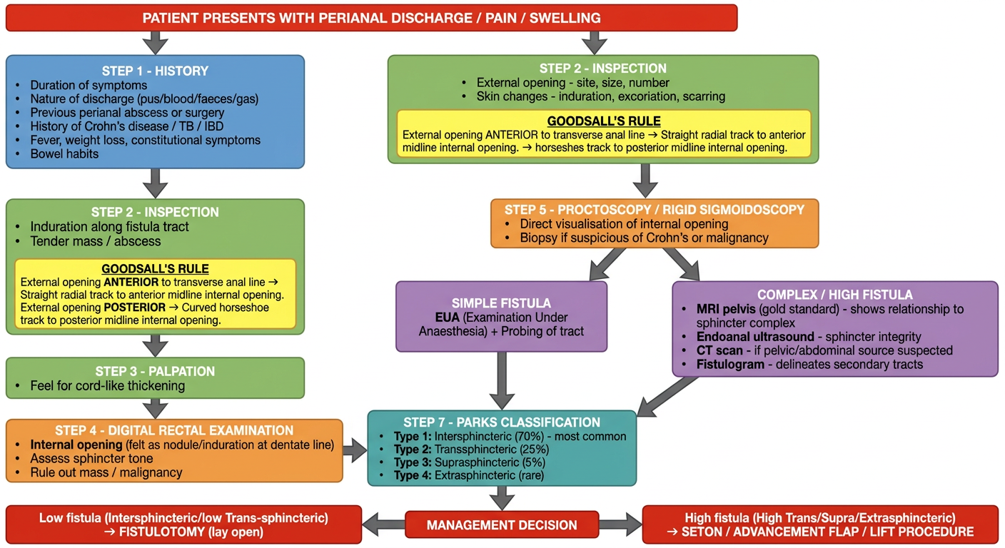
Quick Summary of the Flowchart Logic
- History - duration, discharge type, previous abscess/surgery, Crohn's/TB, constitutional symptoms
- Inspection - number and site of external openings, skin changes; apply Goodsall's Rule to predict where the internal opening lies
- Palpation - cord-like induration along the tract, tender mass
- Digital Rectal Examination - feel for internal opening at the dentate line, assess sphincter tone, exclude malignancy
- Proctoscopy - directly visualise the internal opening; methylene blue test to confirm
- Investigations branching by complexity:
- Simple fistula → EUA + probing
- Complex/high fistula → MRI pelvis (gold standard), endoanal USS, CT, fistulogram
- Parks Classification determines operative strategy
- Management decision - low fistula → fistulotomy; high fistula → seton / advancement flap / LIFT