
Why thyroid moves with deglutination
thyroid gland pretracheal fascia anatomy neck swallowing
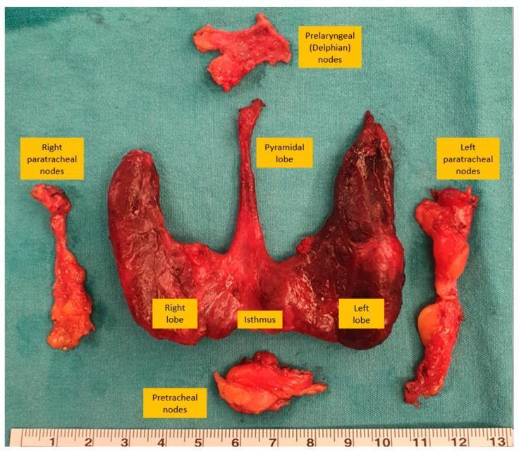
This clinical photograph shows a gross surgical specimen from a bilateral total thyroidectomy and central compartment (Level VI) neck dissection, arranged anatomically on a green sterile field with a centimeter scale. The main thyroid specimen consists of the right and left lobes connected by a central isthmus, with a distinct, elongated pyramidal lobe extending superiorly from the isthmus. Surrounding the gland are four excised lymph node groups: the prelaryngeal (Delphian) nodes located superiorly, the pretracheal nodes positioned inferiorly, and the right and left paratracheal nodes arranged laterally to their respective thyroid lobes. The thyroid tissue appears fleshy and deep red, while the associated lymph node packets appear as lighter, yellowish-pink fibrofatty tissue masses. This image serves as an educational reference for surgical anatomy and the extent of central neck dissection in the management of thyroid malignancies.
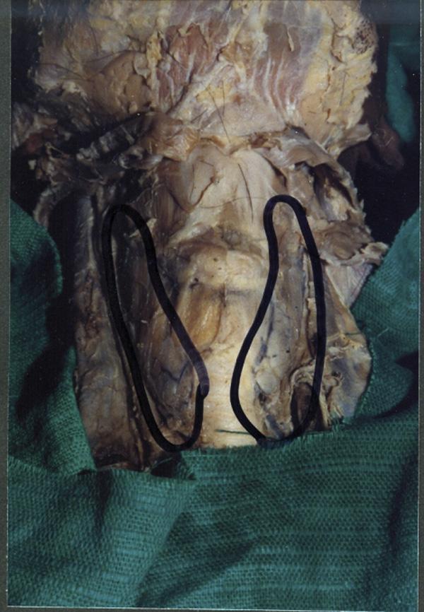
This clinical photograph of a gross anatomical specimen shows a midline dissection of the human neck, highlighting a congenital variation of the thyroid gland. The image depicts the thyroid gland consisting of two distinct, independent lateral lobes (outlined in black) positioned on either side of the trachea. Notably, there is a complete absence (agenesis) of the thyroid isthmus, which normally connects the two lobes across the second to fourth tracheal rings. In the area where the isthmus typically resides, only a thin, translucent layer of pre-tracheal fascia is visible covering the anterior surface of the trachea. The specimen demonstrates the anatomical relationship between the glandular lobes, the respiratory pathway, and the surrounding connective tissue structures. This visual is significant for medical education in endocrinology, anatomy, and surgery, as it illustrates a rare developmental anomaly where the median diverticulum of the thyroglossal duct failed to form the glandular bridge between the bifurcated lobes.
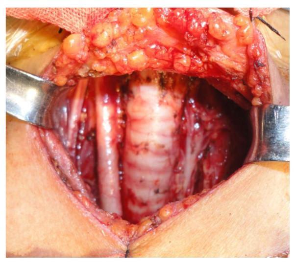
This clinical photograph displays an intraoperative view of a total thyroidectomy combined with level III-IV and VI neck dissection. The surgical field is exposed using metal retractors, revealing central neck anatomy. Centrally, the trachea is clearly identified by its characteristic horizontal cartilaginous rings. The thyroid gland has been removed, providing visibility of the pretracheal and paratracheal regions. Lateral to the trachea, neurovascular structures are visible, including the carotid sheath contents. Evidence of surgical intervention includes cauterized tissue edges, small ligated vessels, and visible subcutaneous adipose tissue in the superior flap. The image demonstrates the surgical bed after completion of the thyroidectomy and lymph node dissection, emphasizing the clear identification of the trachea and the preservation of adjacent vital structures. This visual is representative of surgical oncology and endocrine surgery, specifically for the management of papillary thyroid microcarcinoma requiring central and lateral compartment neck dissection.
Why the Thyroid Gland Moves with Deglutition (Swallowing)
The Core Anatomical Reason
Anatomy in Detail
- Begins superiorly at the hyoid bone
- Encloses the infrahyoid muscles anteriorly
- Wraps around the thyroid gland on all sides
- Continues inferiorly into the upper thoracic cavity
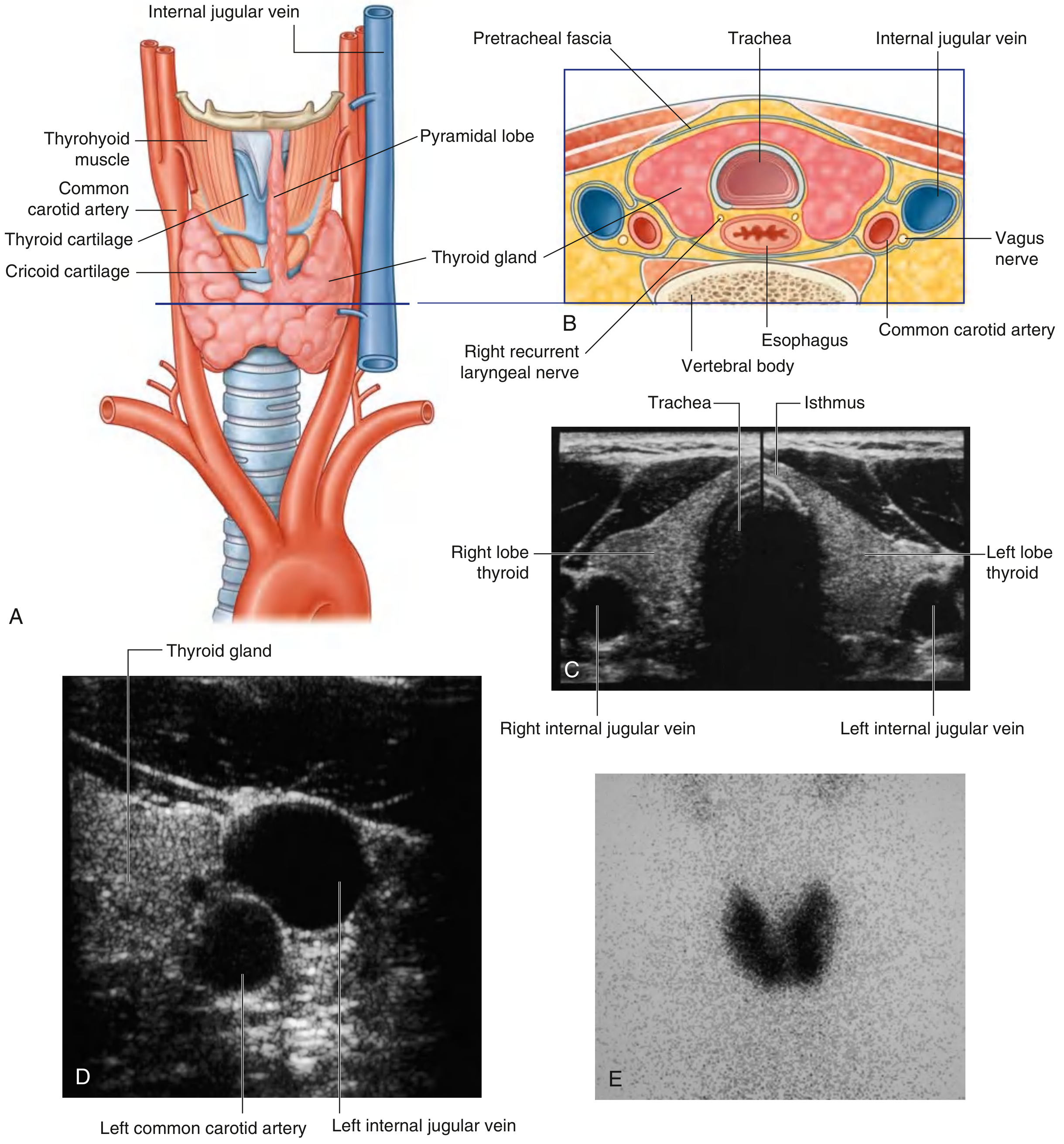
- Its two lateral lobes cover the anterolateral surfaces of the trachea, the cricoid cartilage, and the lower part of the thyroid cartilage
- The isthmus crosses the anterior surface of the 2nd and 3rd tracheal cartilages
"The swellings which are adherent to the larynx and trachea move upwards on swallowing, e.g. thyroid swelling, thyroglossal cyst and subhyoid bursitis." - S. Das, A Manual on Clinical Surgery
Clinical Importance
| Swelling | Moves with Swallowing? | Moves with Tongue Protrusion? |
|---|---|---|
| Thyroid gland / goitre | Yes (upward) | No |
| Thyroglossal cyst | Yes (upward) | Yes (pathognomonic - connected to foramen cecum via thyroglossal duct) |
| Cervical lymph nodes | No (unless fixed to larynx/trachea) | No |
| Subhyoid bursitis | Yes | No |
| Branchial cyst | No | No |
Diagram
Summary
- It is enclosed by the pretracheal fascia along with the larynx and trachea
- The larynx and trachea are pulled upward during swallowing
- The fascial binding transmits this movement directly to the thyroid gland
- Gray's Anatomy for Students, pp. 1142, 1157
- S. Das: A Manual on Clinical Surgery, p. 370