
History taking of RTA case comes with the 5th metatarsal fracture
Reading File
A 5th metatarsal fracture from an RTA (Road Traffic Accident) is a common orthopedic presentation. Before going to the books, here's a quick overview:
History taking should cover: mechanism of injury (direct blow vs. inversion), pain location/severity, ability to weight bear, previous foot injuries, and relevant PMH/medications. The 5th metatarsal has three fracture zones with very different management implications.
Searching sources...
Finding Sources
Finding Sources
Searching the Web
5th metatarsal fracture history taking RTA clinical assessment zones
Reading File
Reading File
Reading File
Loading Image
Reading File
Here is a complete, structured history-taking framework for an RTA patient presenting with a 5th metatarsal fracture:
History Taking: RTA Case with 5th Metatarsal Fracture
1. Presenting Complaint
- Pain on the lateral aspect of the foot/ankle following a road traffic accident
- Inability or difficulty bearing weight
- Swelling and bruising over the outer foot
2. History of Presenting Illness (HPI)
Mechanism of Injury
This is the most critical part - it predicts fracture zone and type:
| Mechanism | Likely Fracture |
|---|---|
| Forced inversion of foot (ankle rolls inward) | Zone 1 - Avulsion fracture (Pseudo-Jones) |
| Strong abduction force to forefoot (bending moment at base) | Zone 2 - True Jones fracture |
| Direct blow / crush (foot run over, pedal impact) | Diaphyseal/shaft fracture (Zone 3 or distal) |
| Stress over time (repeated loading) | Zone 3 stress fracture |
In an RTA, ask specifically:
- Was the foot on the brake/clutch pedal at time of impact?
- Was there a direct crush (motorcycle fall, wheel over foot)?
- Did the foot roll inward on impact?
- Was there a twisting component?
Pain
- Location: Lateral border of foot, base of the 5th metatarsal, or along the shaft?
- Onset: Immediate after injury
- Character: Sharp, throbbing
- Severity: VAS/NRS score (0-10)
- Radiation: Up the leg or into the toes?
- Aggravating factors: Weight bearing, movement
- Relieving factors: Rest, elevation, analgesia
- Associated "snap" or "pop" heard at the time?
Weight-Bearing Status
- Can the patient walk at all? (Ottawa Foot Rules: inability to weight-bear = X-ray indicated)
- Did they walk after the injury?
3. Associated Injuries (Especially Important in RTA)
RTA is a high-energy mechanism - always screen for:
- Ankle injuries (ligament tears, malleolar fractures - inversion is the same mechanism)
- Other metatarsal fractures (Lisfranc pattern if multiple metatarsals involved)
- Calcaneus and talus fractures
- Knee and leg injuries (dashboard injury pattern)
- Pelvic/spinal injuries in high-speed RTA
- Open wounds or abrasions over the fracture site (open fracture risk)
- Neurovascular symptoms: Numbness or tingling in the toes?
4. Past Medical History
- Previous injury to the same foot (prior fractures, non-union risk if prior zone 2/3)
- Diabetes mellitus (impaired bone healing, neuropathy)
- Osteoporosis or metabolic bone disease (lower-energy fractures possible)
- Peripheral vascular disease (affects healing)
- Inflammatory arthritis (rheumatoid, gout)
- Neuropathy (may mask pain, increases non-union risk)
- Corticosteroid use (bone fragility)
5. Drug History
- NSAIDs (can impair fracture healing - Cox-2 inhibitors with particular concern)
- Steroids (bone quality)
- Bisphosphonates (atypical fracture pattern risk)
- Anticoagulants (haematoma, surgical planning)
- Diabetic medications
- Allergies (especially to latex, anaesthetic agents if surgery likely)
6. Social History
- Occupation: Manual labour vs. sedentary (affects weight-bearing advice and time off work; average 22 days off work post-injury)
- Dominant side / Affected foot: Right vs. left
- Driving status: Especially relevant if right foot - affects fitness to drive
- Sports/physical activity level: Athlete vs. non-athlete (directly impacts management - athletes with Zone 2 fractures are often offered surgical fixation)
- Smoking: Significantly impairs fracture healing and increases non-union risk
- Alcohol use: Nutritional status, compliance with treatment
- Living situation: Stairs at home? Anyone to help with mobility?
- BMI / Body weight: Increased load on fracture site with weight bearing
7. Functional History
- Pre-injury mobility: Was the patient walking normally before the accident?
- Pre-injury foot problems: Any previous foot pain, deformity, or flat feet (pes planus) or high arch (cavovarus - associated with Zone 3 stress fractures)?
8. System Review (Relevant)
- Fever (rule out open fracture/infection)
- Bladder/bowel changes (if high-energy RTA with spinal concern)
- Chest pain/SOB (rib fractures, pneumothorax in polytrauma)
Key Classification to Know (Lawrence & Botte Zones)
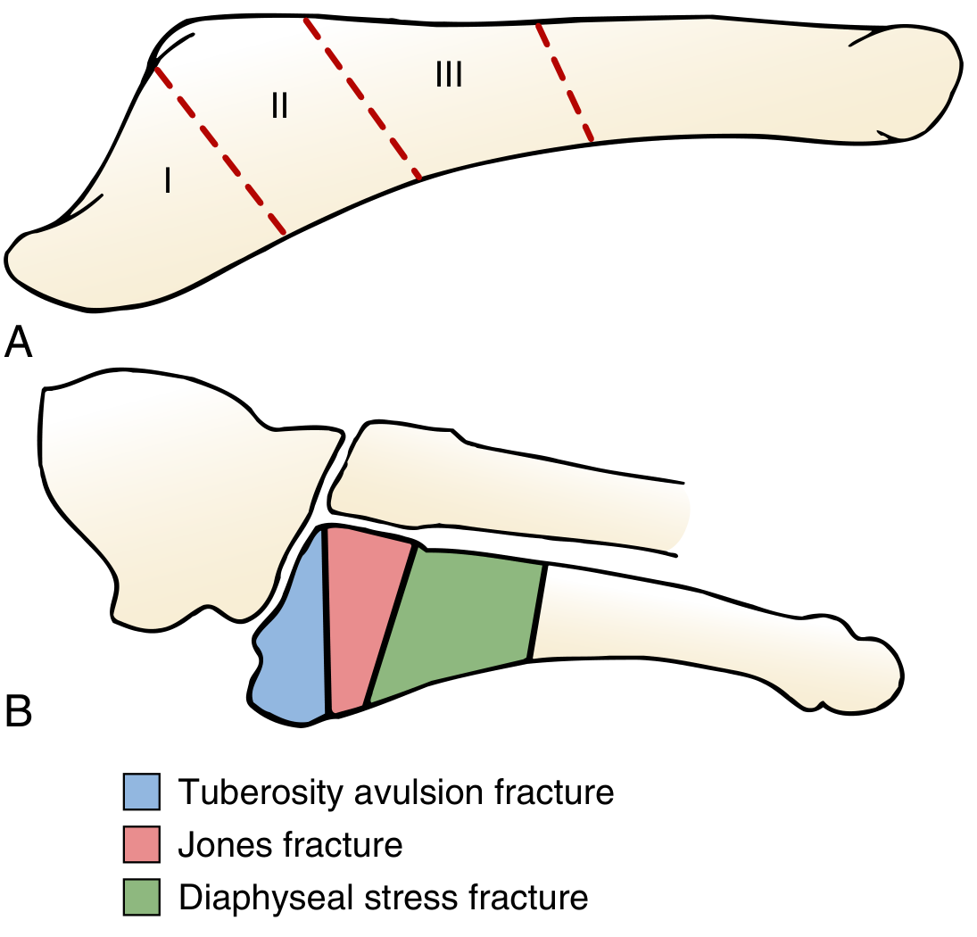
(From Miller's Review of Orthopaedics, 9th Ed, Fig. 6.108)
| Zone | Location | Fracture Type | In RTA? |
|---|---|---|---|
| Zone 1 | Proximal tuberosity | Avulsion (Pseudo-Jones) | Common - inversion |
| Zone 2 | Metaphyseal-diaphyseal junction, includes 4th-5th intermetatarsal joint | True Jones fracture | Less common in RTA |
| Zone 3 | Proximal diaphysis | Stress/diaphyseal | Rare - direct blow in RTA |
Examiner Tip: Ottawa Foot Rules (for X-ray decision)
X-ray of the foot is indicated if there is pain in the midfoot zone AND any of:
- Bone tenderness at the base of the 5th metatarsal, OR
- Bone tenderness at the navicular, OR
- Inability to bear weight immediately or in the ED (4 steps)
Sources:
- Miller's Review of Orthopaedics, 9th Ed, Ch. 12 (5th Metatarsal Base Fractures)
- Campbell's Operative Orthopaedics, 15th Ed, Ch. 93 (Metatarsals)
- Tintinalli's Emergency Medicine, Comprehensive Study (Foot and Toe Injuries)
- StatPearls - 5th Metatarsal Fracture (Smidt et al., 2023)
Blood transfusion notes
Reading File
Blood transfusion is a broad, high-yield topic covering blood products, indications, compatibility testing, reactions, and complications. Let me pull comprehensive notes from the textbook library.
Searching sources...
Finding Sources
Finding Sources
Reading File
Reading File
Reading File
Reading File
Reading File
Finding Sources
Reading File
Reading File
Finding Sources
Reading File
Finding Sources
Reading File
Finding Sources
Reading File
Reading File
I now have comprehensive data from multiple authoritative sources. Let me compile the full blood transfusion notes.
Blood Transfusion - Complete Notes
1. Definition & Overview
Blood transfusion is the process of transferring blood or blood components from a donor to a recipient (allogeneic transfusion) to restore oxygen-carrying capacity, volume, coagulation factors, or platelets. It is one of the most common medical interventions in modern healthcare.
- Tietz Textbook of Laboratory Medicine, 7th Ed: "Blood transfusion is one of the most common medical interventions in modern health care... the sole purpose of pRBC transfusion should be to increase a patient's oxygen-carrying capacity and consequently improve tissue oxygenation."
2. Blood Products
2a. Packed Red Blood Cells (pRBCs)
| Feature | Detail |
|---|---|
| Content | Red cells in ~100 mL plasma + additive solution |
| Volume | ~250-350 mL per unit |
| Haematocrit | ~55-65% |
| Effect | 1 unit raises Hb by ~1 g/dL (adults); 7 mL/kg WB raises Hb by 1 g/dL (children) |
| Shelf life | 35-42 days (4°C) |
| Indication | Anaemia, acute blood loss, impaired erythropoiesis |
Transfusion Thresholds (evidence-based):
- Stable, no CAD: Hb < 7 g/dL (restrictive strategy)
- Stable with CAD/cardiac risk: Hb < 8-9 g/dL (liberal strategy)
- Acute GI bleed without CAD: Transfuse to Hb ≥ 8 g/dL
- ICU/critical patients: Hb threshold 7 g/dL (TRICC trial)
- Normal Hb reference: 12-16 g/dL (females), 13.5-18 g/dL (males)
Restrictive strategy (Hb threshold 7-8 g/dL) in GUB associated with lower all-cause mortality and rebleeding vs. liberal strategy (Goldman-Cecil / Rosen's EM).
2b. Platelets
| Feature | Detail |
|---|---|
| Source | Pooled (from 4-6 whole blood donations) or apheresis (single donor) |
| Volume | ~250-300 mL (pool) / ~200-250 mL (apheresis) |
| Storage | 20-24°C, continuous agitation, 5-7 days |
| Effect | 1 pool raises platelet count ~30,000-50,000/μL |
| Indications | Thrombocytopenia with bleeding, pre-procedure prophylaxis |
Transfusion Thresholds:
- Platelet count < 10,000/μL - prophylactic in stable patient
- Platelet count < 50,000/μL - for procedures/surgery, or in active GI bleed (ACG guideline)
- Platelet count < 100,000/μL - neurosurgery or ophthalmic surgery
HLA-compatible platelets for refractory (alloimmunized) patients via:
- HLA matching (highest compatibility, limited donor pool)
- HLA antigen avoidance (larger donor pool)
- Platelet crossmatching (fastest, used when others unavailable)
2c. Fresh Frozen Plasma (FFP)
| Feature | Detail |
|---|---|
| Content | All clotting factors, fibrinogen, albumin |
| Volume | ~200-250 mL per unit |
| Storage | -18°C or below, 1 year |
| Thawing | 30-37 min at 37°C |
| Indication | Coagulopathy (liver disease, DIC, warfarin reversal, massive transfusion) |
| Dose | 10-15 mL/kg |
Use with caution in portal hypertension - overtransfusion worsens portal pressure.
2d. Cryoprecipitate
| Content | Fibrinogen, Factor VIII, vWF, Factor XIII, fibronectin |
|---|---|
| Volume | ~10-20 mL per unit |
| Indication | Hypofibrinogenaemia (fibrinogen < 1.5 g/L), haemophilia A, vWD, DIC |
| Dose | 1 unit per 5-10 kg body weight |
2e. Whole Blood
- Appropriate for: acute blood loss replacement, volume expansion in septicaemia with coagulation disorder
- Contains all components - cells, plasma, clotting factors
- Freshest available preferred for clotting factor supplementation
- Resurgent interest in trauma and military settings (damage control resuscitation)
2f. Granulocytes (Rarely Used)
- For severe prolonged neutropenia (ANC < 0.5 × 10⁹/L) with life-threatening infection refractory to antimicrobials
- Overall efficacy unproven (RING trial was underpowered)
- Risk of pulmonary toxicity and CMV transmission
3. Pre-Transfusion Testing
Step 1 - ABO & Rh Typing
- ABO group: Forward typing (patient's cells + anti-A/anti-B serum) + Reverse typing (patient's serum + known A/B cells)
- Rh typing: D antigen status (positive/negative)
Step 2 - Antibody Screen
- Detects unexpected red cell alloantibodies in recipient's serum
- Performed against panel of reagent RBCs with known antigen profiles
Step 3 - Crossmatch
| Type | Method | When Used |
|---|---|---|
| Electronic crossmatch | Computer verification of ABO/Rh compatibility | No alloantibodies, 2 prior ABO typings on record |
| Immediate spin crossmatch | Mix donor cells + recipient serum, spin, check agglutination | Low risk recipients |
| Full (antiglobulin) crossmatch | + incubation + antihuman globulin phase | Alloantibodies detected on screen |
Emergency situations: Group O Rh-negative pRBCs (universal donor) given before typing.
4. Massive Transfusion Protocol (MTP)
Definition: Transfusion of ≥1 circulating blood volume within 24 hours (approx. 10 units pRBCs in adults)
Damage Control Resuscitation Ratio (1:1:1):
1 unit pRBC : 1 unit FFP : 1 unit Platelets
- Aims to recapitulate whole blood composition
- Prevents dilutional coagulopathy and the "lethal triad" (hypothermia + acidosis + coagulopathy)
- Triggered by active, life-threatening haemorrhage
Tranexamic Acid (TXA) in Trauma:
- Antifibrinolytic - reduces mortality from haemorrhage
- Dose: 1 g IV over 10 min, then 1 g over 8 hours
- Must be given within 3 hours of injury (ideally within 1 hour)
- Given to all trauma patients with SBP < 110 mmHg or HR > 110 bpm
(Bailey & Love's Surgery, 28th Ed; Goldman-Cecil Medicine)
5. Transfusion Reactions
Incidence Summary Table
| Reaction | Risk per Unit Transfused |
|---|---|
| Febrile non-haemolytic (FNHTR) | ~1 : 1,100 |
| Allergic | ~1 : 1,200 (severe: 1 : 15,500) |
| TACO | ~1 : 9,000 |
| Delayed haemolytic | ~1 : 32,000 |
| Hypotensive | ~1 : 32,000 |
| Acute haemolytic (AHTR) | ~1 : 110,000 |
| TRALI | ~1 : 140,000 |
| TA-GvHD | < 1 : 10,000,000 |
(Goldman-Cecil Medicine, 2-Volume Set)
5a. Acute Haemolytic Transfusion Reaction (AHTR)
Cause: ABO incompatibility (usually clerical error - wrong patient/wrong unit)
Mechanism: Pre-formed recipient IgM antibodies → complement activation → intravascular haemolysis
Symptoms (onset within minutes to hours):
- Fever, rigors/chills, anxiety
- Chest pain, back pain, flank pain (classic "back pain")
- Nausea, vomiting, dyspnea
- Haemoglobinuria (red/cola-coloured urine)
- Diffuse bleeding (DIC), oliguria/anuria
- In anaesthetized patient: unexplained hypotension and haemoglobinuria
Diagnosis:
- ↓ Hb, ↓ haptoglobin, ↑ LDH, ↑ bilirubin, haemoglobinuria
- Positive Direct Antiglobulin Test (DAT)
- Incompatible crossmatch
Management:
- STOP the transfusion immediately
- Send unit back to blood bank; send patient blood for repeat crossmatch + DAT
- IV saline to maintain BP and urine output (>1 mL/kg/hr)
- Furosemide to increase renal blood flow
- Treat DIC with plasma, cryoprecipitate, platelets, ± heparin
- Monitor renal function
Prognosis: ~50% of ABO-incompatible transfusions have no adverse effect; 5% are fatal
5b. Febrile Non-Haemolytic Transfusion Reaction (FNHTR)
Cause:
- RBC units: donor leukocytes react with recipient HLA/granulocyte antibodies
- Platelet units: donor cytokines accumulated during storage
Diagnosis criteria (NHSN):
- During or <4 hours after transfusion
- Fever ≥38°C AND ≥1°C rise from pre-transfusion baseline, OR chills/rigors
- No other cause identified
Management:
- Stop transfusion; rule out AHTR (most important step)
- Antipyretics (paracetamol)
- Can restart at slower rate once AHTR excluded
Prevention:
- Prestorage leukoreduction (universal now in most countries) - greatly reduces incidence
- Premedication (paracetamol ± antihistamine) only if prior FNHTR history
5c. Allergic Transfusion Reaction
Cause: Reaction to donor plasma proteins
Special case: Anti-IgA antibodies in IgA-deficient recipients → anaphylaxis
| Severity | Symptoms | Management |
|---|---|---|
| Mild | Urticaria, pruritus, flushing | Stop transfusion, antihistamine, restart when resolved |
| Severe/Anaphylaxis | Stridor, wheezing, hypotension, shock | Stop, IV epinephrine, H1-antagonist, hydrocortisone |
IgA-deficient patients: Use washed pRBCs/platelets or plasma from IgA-deficient donors
5d. Transfusion-Associated Circulatory Overload (TACO)
Risk factors: Age >70, positive fluid balance >3 L, multiple units within 6 hours, cardiac disease
Symptoms: Dyspnoea, tachypnoea, ↓SpO₂, orthopnoea, hypertension, headache (onset at end of transfusion or up to 12 hours post)
Diagnosis: Clinical fluid overload + ↑BNP or NT-proBNP
Management: Stop transfusion, sit upright, O₂, furosemide
5e. Transfusion-Related Acute Lung Injury (TRALI)
Cause: Donor anti-HLA or anti-HNA antibodies → neutrophil/pulmonary endothelial activation → non-cardiogenic pulmonary oedema
Onset: Within 6 hours of cessation of transfusion
Features: Hypoxia, bilateral infiltrates on CXR, no fluid overload (contrasts with TACO)
Most commonly associated with platelet transfusions
Management: Stop transfusion, supportive (O₂, ventilation if needed); no diuretics
Prevention: Use of male-only / HLA-antibody-negative / nulliparous female plasma donors
5f. Transfusion-Associated GvHD (TA-GvHD)
Cause: Donor T-lymphocytes engraft and attack host tissues in immunosuppressed recipients
Prevention: Gamma-irradiation of cellular blood products for at-risk patients
Mortality: Very high (~90%)
6. Infectious Disease Risks
| Pathogen | Window Period | Residual Risk (per unit, USA) |
|---|---|---|
| HIV (MP-NAT) | 9 days | ~1 : 1,800,000 |
| HCV (MP-NAT) | 7 days | ~1 : 1,600,000 |
| HBV (NAT) | 15-50 days | ~1 : 1,500,000 |
| HBsAg | 30-38 days | ~1 : 300,000 |
| HTLV | 80 days | ~1 : 3,300,000 |
| Syphilis | - | 1 case reported in USA in 50 years |
(Goldman-Cecil Medicine, 2-Volume Set)
7. Special Situations
Autologous Transfusion
- Patient's own blood collected pre-operatively and reinfused
- Types: Pre-operative donation, intraoperative cell salvage, acute normovolemic haemodilution
- Eliminates alloimmunization and infectious risks
Transfusion in Jehovah's Witnesses
- May refuse blood products (religious grounds)
- Respect autonomy in competent adults
- For emergency/incompetent patients: follow local legal/ethical frameworks + hospital ethics board
Paediatric Transfusion Formula
- Blood volume in infancy = 80 mL/kg
- 7 mL whole blood/kg raises Hb by ~1 g/dL
- Do NOT use peristaltic pump (haemolysis risk)
8. Key Mnemonics
Transfusion Reactions - FATBACK
- Febrile non-haemolytic
- Allergic/Anaphylactic
- TACO (circulatory overload)
- Bacterial contamination (septic reaction)
- Acute haemolytic
- Cold transfusion
- Killer - TRALI
First step in ANY transfusion reaction = STOP the transfusion
Sources:
- Tietz Textbook of Laboratory Medicine, 7th Ed (Blood Transfusion Practices)
- Goldman-Cecil Medicine, International Ed (Transfusion Reactions + Adverse Events Tables)
- Bailey & Love's Surgery, 28th Ed (MTP, TXA in trauma)
- Rosen's Emergency Medicine (Blood Product Transfusion, MTP)
- Tintinalli's Emergency Medicine (Transfusion thresholds in GI bleed)
- Pye's Surgical Handicraft, 22nd Ed (Paediatric transfusion, rates)
This is a shared conversation. Sign in to Orris to start your own chat.