
Ventricular Muscle Action Potential, Conducting System of Heart, Type of Heart Block, List the factors affecting venous return.
I'll read the medical library skill first, then research all four topics in parallel.
Reading File
Now I'll search for all four topics in parallel:
Finding Sources
Finding Sources
Finding Sources
Finding Sources
Finding Sources
Finding Sources
Finding Sources
Reading File
Good results. Now let me read the key sections in parallel:
Reading File
Reading File
Reading File
Excellent data gathered. Now let me get the remaining details on action potential phases (3, 4) and heart block types, plus factors affecting venous return:
Reading File
Reading File
Reading File
Now let me get the AV block types description and factors affecting venous return:
Reading File
Reading File
Now let me get the complete AV block classification and factors affecting venous return in one final read:
Reading File
Reading File
Searching Images
ventricular muscle action potential phases diagram
Searching Images
conducting system of the heart diagram SA node AV node bundle of His Purkinje
I now have comprehensive content from the textbooks for all four topics. Let me fetch the key action potential diagram from Costanzo:
Loading Image
Loading Image
I now have all the content needed for a thorough, well-cited answer. Let me compose the full response.
1. Ventricular Muscle Action Potential
The ventricular muscle action potential has 5 phases (0-4) and is characterized by a distinctive long plateau, which accounts for its prolonged duration (~200-300 ms) and long refractory period - a feature that prevents tetanic contractions of cardiac muscle.
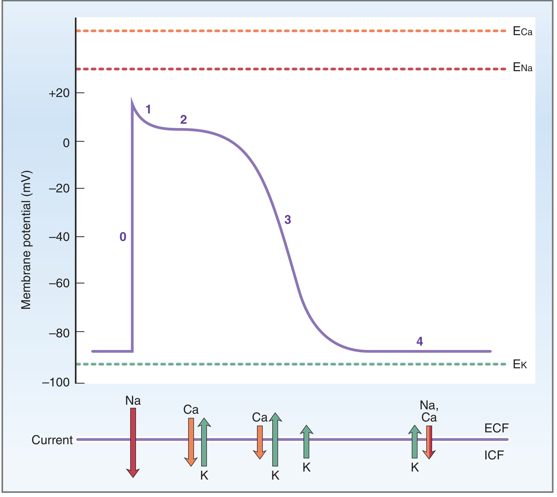
Phase 4 - Resting Membrane Potential
- Value: approximately -90 mV
- Maintained primarily by a high K+ conductance (gK); the membrane is most permeable to K+ at rest
- K+ moves out down its concentration gradient, leaving net negative charge inside
- Unlike SA/AV nodal cells, ventricular muscle shows no spontaneous depolarization (no "funny current" / If) - it is a stable flat line
Phase 0 - Upstroke (Rapid Depolarization)
- Triggered when an arriving impulse depolarizes the membrane to threshold (~-70 mV)
- Sudden opening of fast voltage-gated Na+ channels (activation gates open rapidly)
- Large inward Na+ current (INa) rapidly drives membrane toward ENa (+65 mV)
- Peak of upstroke reaches approximately +20 mV
- Na+ channels then close as inactivation gates respond to depolarization
- Rate of rise = dV/dT; greatest when resting membrane potential is most negative (-90 mV), because more Na+ channels have open inactivation gates
Phase 1 - Initial (Brief) Repolarization
- Short, transient dip after the upstroke peak
- Two mechanisms:
- Closure of Na+ channel inactivation gates → inward Na+ current ceases
- Transient outward K+ current (Ito) - at peak upstroke, large electrochemical driving force pushes K+ out of cell
Phase 2 - Plateau
- Duration: 150-200 ms - unique to cardiac muscle (absent in nerve/skeletal muscle)
- Maintained by a balance of inward and outward currents (net current ≈ 0):
- Inward: L-type Ca2+ channels open → slow inward Ca2+ current (ICa-L) - also called "slow inward current"
- Blocked by: nifedipine, diltiazem, verapamil
- Outward: sustained K+ current (IK), driven by electrochemical gradient
- Inward: L-type Ca2+ channels open → slow inward Ca2+ current (ICa-L) - also called "slow inward current"
- The Ca2+ influx during Phase 2 triggers Ca2+-induced Ca2+ release from the sarcoplasmic reticulum (CICR) - essential for excitation-contraction coupling
- Physiological importance: the plateau creates a long absolute refractory period, preventing re-stimulation of the ventricle before it has relaxed - the heart cannot tetanize
Phase 3 - Rapid Repolarization
- Inward Ca2+ current decreases (gCa falls as L-type channels inactivate)
- Simultaneously, K+ conductance increases further (IK rises) → large outward K+ current
- Net outward current drives membrane potential back toward resting level (~-90 mV)
- As repolarization proceeds, driving force on K+ decreases (approaching EK), so IK gradually lessens
Phase 4 - Resting (Restored)
- Membrane returns to -90 mV
- Na+/K+ ATPase restores ionic gradients; Na+/Ca2+ exchanger removes Ca2+ that entered during Phase 2
| Phase | Name | Key Ion(s) | Direction |
|---|---|---|---|
| 0 | Upstroke | Na+ | Inward (fast) |
| 1 | Initial repolarization | K+ (Ito), Na+ off | Outward |
| 2 | Plateau | Ca2+ in, K+ out | Balanced |
| 3 | Rapid repolarization | K+ (IK) ↑, Ca2+ off | Net outward |
| 4 | Resting | K+ (IKr) | Stable |
Source: Costanzo Physiology, 7th Ed.
2. Conducting System of the Heart
The conducting system is a network of specialized cardiac muscle cells that generate and propagate impulses in a coordinated sequence to produce rhythmic, efficient contractions. These cells differ histologically and functionally from the working myocardium.
Components (in order of impulse travel)
1. Sinoatrial (SA) Node - "Pacemaker"
- Location: beneath the epicardium on the posterior wall of the right atrium, near the opening of the superior vena cava, in the sulcus terminalis cordis (Keith-Flack node)
- Size: ~10 mm long, spindle-shaped
- Intrinsic rate: 60-80 impulses/minute (fastest of all pacemaker cells)
- Generates spontaneous action potentials due to the "funny current" (If) causing slow Phase 4 depolarization
- Sets the normal heart rate (sinoatrial nodal rhythm)
2. Internodal Pathways (Atrial Conduction)
- Impulse spreads from the SA node through the working myocardium of the right atrium to the AV node via preferential intra-atrial pathways with higher conduction velocity
- Simultaneously activates both atria
3. Atrioventricular (AV) Node - "Gatekeeper"
- Location: at the atrioventricular septum in the interatrial septum (Koch's triangle), between the coronary sinus opening and septal cusp of the tricuspid valve (Aschoff-Tawara node)
- Size: ~5 mm long
- Intrinsic rate: 50-60 impulses/minute (overridden by SA node normally)
- Key functional property: AV nodal delay (approx. 0.1 s) - slows conduction to allow ventricular filling before contraction
- Only normal electrical connection between atria and ventricles (the fibrous annulus electrically insulates the rest)
- Action potential: relies on L-type Ca2+ channels (ICa-L) for Phase 0 (no fast Na+ channels); slow upstroke; Phase 4 depolarization present
4. Bundle of His (AV Bundle)
- Extends from the AV node through the central fibrous body (cardiac skeleton) into the muscular interventricular septum
- Penetrating bundle traverses the annulus fibrosis; insulated from surrounding myocardium
- Blood supply: AV nodal artery (from RCA in 80-90%) and first septal perforator of LAD
5. Right Bundle Branch (RBB)
- Curves down the right side of the interventricular septum, enters the septomarginal trabecula (moderator band), reaches the anterior papillary muscle
- Peripheral branches form a subendocardial plexus, terminating at papillary muscles and ventricular myocardium near the apex
6. Left Bundle Branch (LBB)
- Fans out as flat broad bundles along the left side of the interventricular septum
- Divides into anterior fascicle and posterior fascicle, proceeding toward the papillary muscle bases
- Blood supply: dual (from LAD and posterior descending artery branches)
7. Purkinje Fibers
- Terminal subendocardial network from both bundle branches
- Fastest conduction in the heart: 1-3 m/s
- Action potential: rapid upstroke (fast Na+ channels), prolonged plateau, modest Phase 4 automaticity
- Transmit impulses rapidly to ventricular myocardium from apex toward base
Hierarchy of Pacemakers
| Site | Intrinsic Rate |
|---|---|
| SA node | 60-100 bpm |
| AV node | 50-60 bpm |
| Bundle of His / Purkinje | 25-45 bpm |
The SA node normally dominates because it fires fastest. Each lower site acts as a backup if the site above it fails.
Source: Color Atlas of Human Anatomy Vol. 2 - Internal Organs; Harrison's Principles of Internal Medicine 22E
3. Types of Heart Block
Heart block (AV block) refers to impaired conduction from atria to ventricles through the AV node or His-Purkinje system. It is classified by ECG appearance into three degrees.
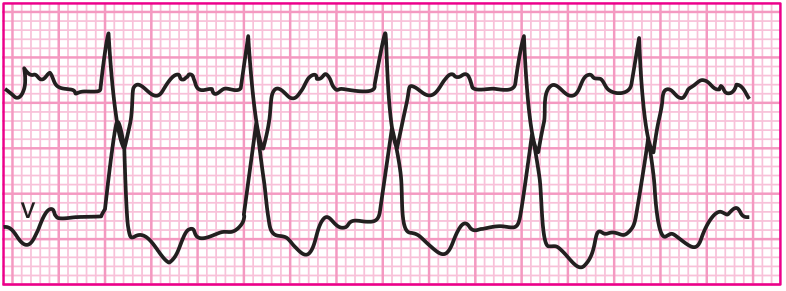
First-Degree AV Block
- Definition: All atrial impulses are conducted to the ventricle, but with abnormal delay
- ECG: PR interval > 200 ms (fixed prolongation), every P wave followed by a QRS
- Location: Delay usually within the AV node (may also be in atria, His bundle, or Purkinje)
- Significance: Generally benign; does not typically warrant pacing
- Note: "block" is a misnomer - conduction is slowed, not interrupted
Second-Degree AV Block
Intermittent failure of conduction between atrium and ventricle. There are two subtypes:
Type I (Mobitz I / Wenckebach):
- ECG: Progressive lengthening of the PR interval with each beat, until one P wave is not conducted ("dropped" QRS - the "dropped beat")
- Classic feature: PR interval before the dropped beat is longest; first PR after is shortest; RR intervals show progressive shortening
- Location: Typically within the AV node
- Significance: Usually hemodynamically stable; does not typically require pacing in the absence of symptoms
- Common in athletes (high vagal tone) or inferior MI
Type II (Mobitz II):
- ECG: Fixed PR interval (no progressive prolongation) preceding a suddenly non-conducted P wave
- Usually associated with QRS widening (bundle branch block pattern)
- Location: Infranodal (His bundle or bundle branches)
- Significance: More serious - risk of sudden death; permanent pacing required even without symptoms due to unpredictable complete block
- In 2:1 AV block, Type I vs II cannot be distinguished on ECG alone; if PR < 160 ms + wide QRS → infranodal (Type II) more likely
Third-Degree AV Block (Complete Heart Block)
- Definition: Complete interruption of conduction between atria and ventricles (AV dissociation)
- ECG: P waves and QRS complexes are independent, with ventricular rate slower than atrial rate
- Ventricular rhythm maintained by an escape pacemaker:
- Narrow QRS escape rhythm → block in AV node/proximal His (rate ~50-60 bpm; more stable)
- Wide QRS escape rhythm → block in distal His/bundle branches (rate ~25-45 bpm; unreliable)
- Significance: Life-threatening; permanent pacing is required
Summary Table
| Type | PR Interval | QRS after P | Location | Urgency |
|---|---|---|---|---|
| 1st degree | >200 ms, fixed | All conducted | AV node | Benign |
| 2nd degree Mobitz I | Progressive lengthening | Periodically dropped | AV node | Usually watch |
| 2nd degree Mobitz II | Fixed (no change) | Suddenly dropped | Infranodal | Pace even if asymptomatic |
| 3rd degree | AV dissociation | Independent of P waves | AV node or infranodal | Emergency pacing |
Source: Harrison's Principles of Internal Medicine, 22E; Miller's Anesthesia, 10E
4. Factors Affecting Venous Return
Venous return (VR) is the volume of blood flowing from the systemic circulation back to the right heart per unit time. In steady state, venous return = cardiac output (right heart input must equal its output). VR is driven by the pressure gradient between the peripheral venous system and the right atrium.
The key equation: VR = (MSFP - RAP) / Resistance to venous return
Where MSFP = Mean Systemic Filling Pressure; RAP = Right Atrial Pressure
1. Mean Systemic Filling Pressure (MSFP)
- The equilibrium pressure in the vascular system when flow is zero
- Increased by: Blood volume transfusion; venoconstriction (sympathetic tone increases venomotor tone, squeezing the unstressed volume into stressed volume)
- Decreased by: Hemorrhage/hypovolemia; venodilation
- A higher MSFP increases the driving pressure gradient for venous return → increases VR
2. Blood Volume
- Direct effect on MSFP: more blood → greater stressed volume → higher MSFP → higher VR
- Volume expansion (e.g., IV fluids, transfusion): shifts vascular function curve right → increases VR
- Volume depletion (e.g., hemorrhage, dehydration): shifts curve left → decreases VR
3. Right Atrial Pressure (RAP / CVP)
- Inverse relationship with VR: as RAP rises, the pressure gradient (MSFP - RAP) falls → VR decreases
- Reduced RAP (e.g., reduced cardiac filling) → increased gradient → increased VR
- Very negative RAP causes venous collapse (veins buckle), creating a "waterfall effect" that limits further increase in VR (the flat "knee" of the vascular function curve)
4. Venomotor Tone (Venoconstriction / Venodilation)
- Most blood volume (~70%) resides in veins; venomotor tone controls how much is in the "unstressed" vs. "stressed" compartment
- Venoconstriction (sympathetic stimulation, epinephrine): decreases venous compliance → shifts blood from unstressed to stressed volume → acts like a transfusion → raises MSFP → increases VR
- Venodilation (nitrates, heat): acts like a blood loss → reduces MSFP → decreases VR
5. Total Peripheral Resistance (TPR) / Arteriolar Tone
- Arterioles contain little of the total blood volume, so they don't affect MSFP much
- However, arteriolar resistance affects the slope of the vascular function curve:
- Arteriolar dilation → blood redistributes from arterial to venous side → raises capillary venous pressure (CVP) → increases VR (steeper vascular function curve)
- Arteriolar constriction → lowers CVP → decreases VR (flatter curve)
6. Skeletal Muscle Pump
- Rhythmic contraction of skeletal muscles compresses veins, propelling blood toward the heart
- Venous valves prevent backflow
- Major factor during exercise - dramatically increases VR and cardiac output
7. Respiratory Pump (Thoracic Pump)
- During inspiration: intrathoracic pressure falls → right atrial pressure decreases → pressure gradient increases → VR increases
- During expiration: reverse effect
- Positive pressure ventilation (IPPV) impairs this mechanism → reduces VR
8. Gravity / Body Position
- Standing: hydrostatic pressure increases venous pressure in lower limbs → venous pooling → reduces effective VR (can cause orthostatic hypotension)
- Lying flat or raising legs: reverses pooling → increases VR
- Trendelenburg position increases VR
9. Heart Rate and Cardiac Contractility (Indirect)
- A more vigorously contracting heart empties more completely → reduces RAP → increases the MSFP-RAP gradient → "sucks" more venous return
- Increased cardiac output lowers RAP, which in turn augments VR (Frank-Starling mechanism working in reverse to maintain steady state)
10. Intrapericardial Pressure / Cardiac Tamponade
- Elevated pericardial pressure compresses the heart, raises RAP → reduces VR → reduces cardiac output
Summary Table - Factors Affecting Venous Return
| Factor | Change | Effect on VR |
|---|---|---|
| Blood volume ↑ | More stressed volume | ↑ VR |
| Venoconstriction | ↑ MSFP | ↑ VR |
| Venodilation | ↓ MSFP | ↓ VR |
| RAP ↑ (e.g., RHF) | ↓ Pressure gradient | ↓ VR |
| RAP ↓ (cardiac suction) | ↑ Pressure gradient | ↑ VR |
| Arteriolar dilation | ↑ CVP, steeper curve | ↑ VR |
| Arteriolar constriction | ↓ CVP, flatter curve | ↓ VR |
| Skeletal muscle pump | Mechanical compression | ↑ VR |
| Inspiration (IPPB) | ↓ Intrathoracic pressure | ↑ VR |
| IPPV / PEEP | ↑ Intrathoracic pressure | ↓ VR |
| Standing/gravity | Venous pooling | ↓ VR |
| Lying/leg elevation | ↓ Pooling | ↑ VR |
| Cardiac tamponade | ↑ RAP | ↓ VR |
Source: Costanzo Physiology, 7th Ed.; Medical Physiology (Boron & Boulpaep)
This is a shared conversation. Sign in to Orris to start your own chat.