
Create a 30–35 slide postgraduate-level PowerPoint presentation on “Interstitial Lung Disease (ILD): Classification and HRCT Patterns” for an MD Radiodiagnosis resident academic seminar using standard textbooks
UIP usual interstitial pneumonia HRCT honeycombing subpleural
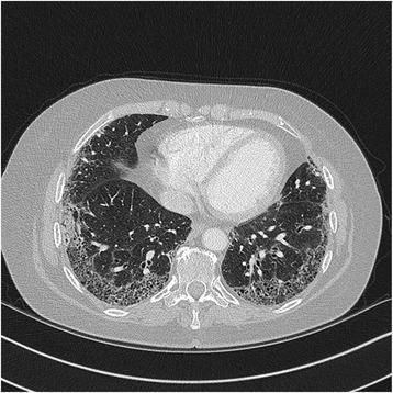
High-resolution computed tomography (HRCT) axial section of the chest demonstrating advanced interstitial lung disease. The image shows extensive reticular opacities with a predominant peripheral and subpleural distribution. There is evidence of architectural distortion characterized by early honeycombing, visible as small, clustered cystic spaces in the subpleural regions of both lower lobes. These findings are consistent with a Usual Interstitial Pneumonia (UIP) pattern, often associated with Idiopathic Pulmonary Fibrosis (IPF). The central lung parenchyma remains relatively spared compared to the dense fibrotic changes at the periphery. The imaging highlights the characteristic superior-to-inferior gradient of UIP, where fibrotic destruction is most pronounced in the lung bases. This diagnostic image serves as an educational tool for identifying the hallmarks of chronic fibrosing interstitial pneumonia, including subpleural reticulation, traction bronchiectasis, and honeycombing.
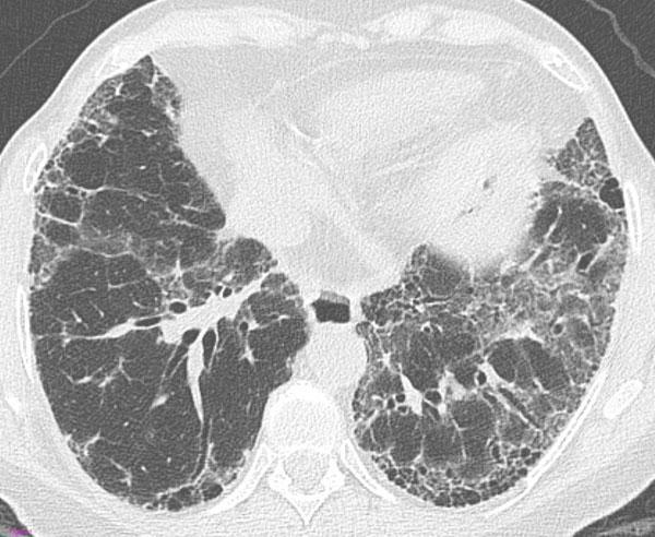
This transverse High-Resolution Computed Tomography (HRCT) axial scan of the thorax demonstrates a 'definite Usual Interstitial Pneumonia (UIP)' pattern, a hallmark of Idiopathic Pulmonary Fibrosis (IPF). The image shows bilateral, predominantly subpleural and basal lung involvement. Key diagnostic features include honeycombing—manifesting as clustered cystic airspaces with thick walls—which is particularly evident in the subpleural region of the left lung. Associated findings include a prominent reticular pattern, traction bronchiectasis (dilatation of bronchi within the fibrotic areas), and mild ground-glass opacities. The distribution is peripheral and basal-predominant, which is characteristic of the ATS/ERS/JRS/ALAT guidelines for diagnosing UIP. This imaging is crucial for medical students and radiologists to distinguish between different patterns of interstitial lung disease (ILD), specifically identifying the architectural distortion and honeycombing required for a definite diagnosis of UIP versus non-specific interstitial pneumonia (NSIP).
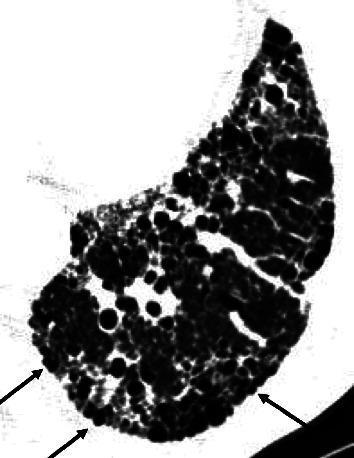
High-resolution computed tomography (HRCT) axial view of the lung parenchyma demonstrating classic honeycombing patterns associated with Usual Interstitial Pneumonia (UIP). The image shows clustered, subpleural cystic airspaces with thick, well-defined walls. These cysts appear as rounded or oval lucencies (dark regions) interspersed with bright, high-density fibrotic tissue. A key diagnostic feature highlighted by black arrows is the outward protuberance of these honeycomb cysts toward the extrapulmonary region, distorting the pleural surface. The distribution is predominantly peripheral and subpleural, creating a heterogeneous architectural distortion typical of end-stage pulmonary fibrosis. This visual serves as an educational tool to differentiate honeycombing in UIP from the more uniform traction bronchiolectasis seen in Nonspecific Interstitial Pneumonia (NSIP), where such pleural protuberance is typically absent.
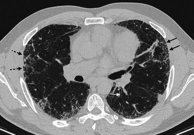
This axial High-Resolution Computed Tomography (HRCT) scan of the thorax demonstrates characteristic findings of a Usual Interstitial Pneumonia (UIP) pattern. The image shows a predominantly subpleural and peripheral distribution of lung abnormalities in both hemispheres. Key diagnostic features include honeycombing, represented by clusters of subpleural cystic airspaces (indicated by black solid arrows), and a fine reticular pattern (indicated by black dotted arrows) consisting of intersecting linear opacities. Additionally, there is evidence of architectural distortion and traction bronchiectasis (indicated by a white arrow), characterized by the irregular dilatation of small bronchi within the areas of fibrosis. These findings are hallmarks of idiopathic pulmonary fibrosis (IPF) and are critical for differentiating UIP from other interstitial lung diseases like Non-Specific Interstitial Pneumonia (NSIP). The image serves as an educational reference for identifying progressive fibrosing interstitial lung disease through imaging markers such as basal predominance, reticulation, and subpleural honeycombing.
NSIP non-specific interstitial pneumonia ground glass subpleural sparing HRCT
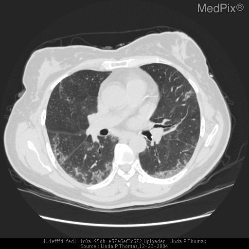
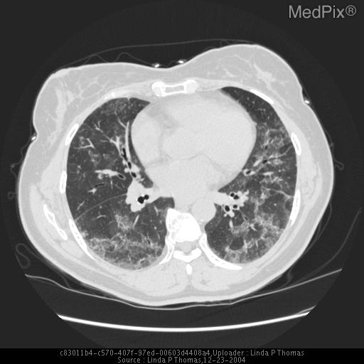
sarcoidosis perilymphatic nodules HRCT CT scan
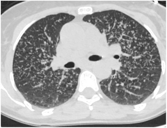
This diagnostic axial CT scan of the chest demonstrates non-fibrotic pulmonary sarcoidosis. The primary finding is a diffuse, bilateral distribution of small interstitial nodules. These nodules exhibit a characteristic perilymphatic pattern, localized prominently along the subpleural surfaces, interlobular septa, and the bronchovascular bundles. There is an absence of architectural distortion, honeycombing, or traction bronchiectasis, which distinguishes this as a non-fibrotic phase of the disease. The lung parenchyma between the nodules remains relatively clear, with no significant ground-glass opacities or consolidations visible. The central airways and hilar regions are depicted at this level, providing context for the nodular distribution relative to the major pulmonary vessels and bronchi. This image serves as a classic educational example of how lymphatic-related lung diseases like sarcoidosis manifest on high-resolution computed tomography (HRCT).
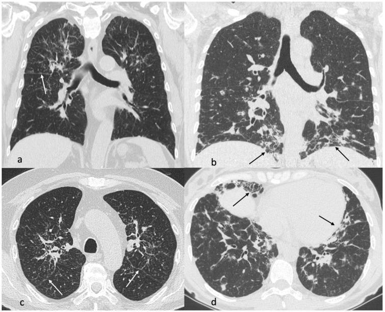
This diagnostic image provides a comparative CT scan analysis of sarcoidosis and Granulomatous-Lymphocytic Interstitial Lung Disease (GLILD). Images (a) and (c) depict thoracic CT views of a 68-year-old male with sarcoidosis, showing characteristic perilymphatic nodules and periscissural involvement (white arrows) concentrated in the mid-to-upper lung fields, alongside perihilar fibrosis. In contrast, images (b) and (d) present CT findings of a 38-year-old female with Common Variable Immunodeficiency (CVID) and GLILD, where the pathology is predominantly located in the lower lung fields. These findings include dense consolidations, multiple nodules, and prominent reticulations (black arrows) with associated fibrotic changes. The comparison highlights key differential diagnostic features: the upper lobe/perihilar distribution of sarcoidosis versus the lower lobe/reticular predominance of GLILD. The imaging modality is High-Resolution Computed Tomography (HRCT) in both axial and coronal planes, serving as a critical tool for identifying patterns of interstitial lung disease and guiding clinical management in pulmonology and immunology.
hypersensitivity pneumonitis centrilobular nodules mosaic attenuation CT chest
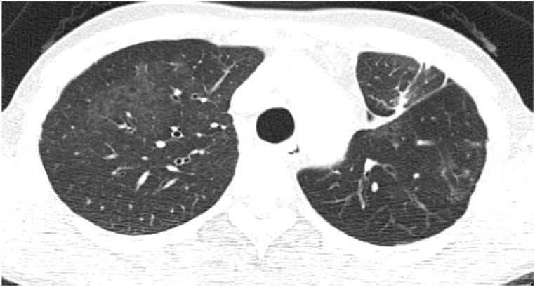
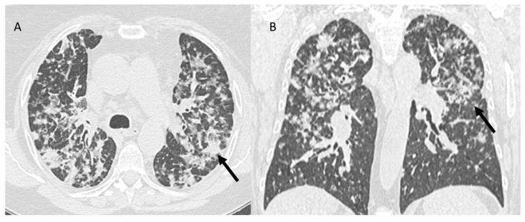
This diagnostic axial chest CT scan demonstrates diffuse pulmonary parenchymal abnormalities characteristic of hypersensitivity pneumonitis, specifically 'humidifier lung.' The image reveals bilateral, though asymmetric, mosaic attenuation. The left lung exhibits extensive, ill-defined centrilobular ground-glass nodules and hazy increased density. Within these regions of ground-glass opacity, small, lucent, cystic-like structures are visible, representing dilated bronchioles or air trapping. In contrast, the right lung shows a more heterogeneous pattern, with patches of normal-attenuating lung interspersed with areas of subtly increased density. The distribution of these findings is predominantly centrilobular, typical of small airway involvement and granulomatous inflammation following organic antigen exposure. This CT illustrates key features used in differentiating acute or subacute hypersensitivity pneumonitis from other interstitial lung diseases, highlighting the importance of recognizing mosaic perfusion and ground-glass patterns in the clinical context of recurrent pneumonia.
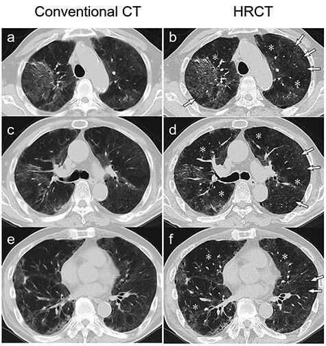
This diagnostic image provides a side-by-side comparison between conventional 5-mm-thick axial CT scans (panels a, c, e) and 2-mm-thick high-resolution CT (HRCT) scans (panels b, d, f) of the chest. The imaging captures three anatomical levels: the aortic arch, the tracheal carina, and the aortic valve. Both modalities demonstrate diffuse, bilateral ground-glass opacities (GGOs) with a predominant peripheral distribution. However, the HRCT images offer superior detail for characterizing parenchymal lung disease. Specifically, the HRCT scans reveal ill-defined centrilobular nodules (indicated by arrows) and distinct areas of mosaic attenuation (indicated by asterisks), characterized by alternating regions of varying lung density. These visual markers are key diagnostic features of hypersensitivity pneumonitis (HP), such as humidifier lung, and serve as important radiographic findings to differentiate HP from other interstitial lung diseases or viral pneumonias like COVID-19.
cryptogenic organizing pneumonia COP peribronchovascular consolidation CT
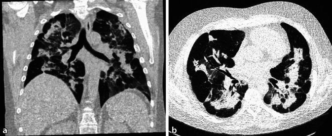
This diagnostic imaging set consists of two non-contrast CT scans of the chest: a coronal reconstruction (a) and a transverse/axial slice (b). The images demonstrate extensive, symmetrical, bilateral lung abnormalities characterized by mixed ground-glass opacities and areas of consolidation. A hallmark feature of the distribution is the predominant involvement of the central, peribronchovascular zones with notable subpleural sparing, where the peripheral lung parenchyma adjacent to the pleura remains clear. There is also evidence of bronchial wall thickening. These findings are consistent with diffuse parenchymal lung disease processes, such as cryptogenic organizing pneumonia (COP) or eosinophilic pneumonia, characterized by peribronchovascular infiltrates. The image serves as an educational example of central lung consolidation patterns and the significance of subpleural sparing in narrowing differential diagnoses for interstitial or inflammatory lung diseases.
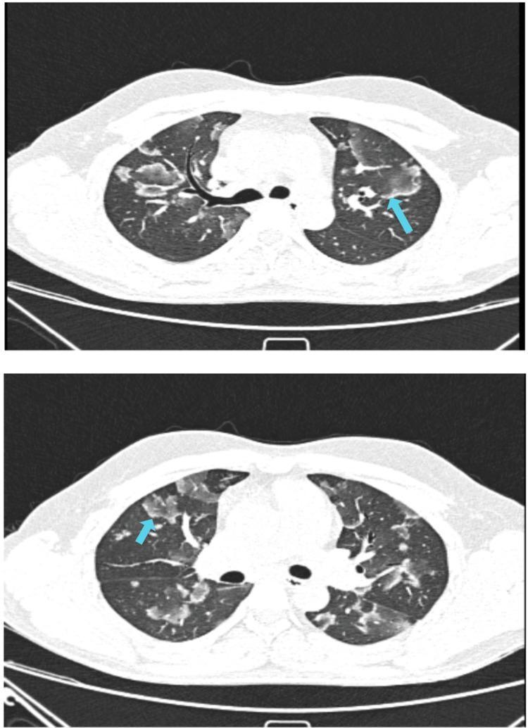
This diagnostic image consists of two axial non-contrast CT sections of the thorax in lung window setting, demonstrating bilateral pulmonary abnormalities characteristic of Cryptogenic Organizing Pneumonia (COP). The primary findings are multifocal ground-glass opacities (GGO) and areas of consolidation. Notably, both sections display the 'reverse halo sign' (or 'atoll sign'), indicated by blue arrows, where a central area of ground-glass attenuation is surrounded by a crescentic or ring-like rim of denser consolidation. These lesions are distributed throughout the lung parenchyma with a predominantly subpleural and peribronchovascular orientation. The image illustrates a classic radiological manifestation of interstitial lung disease (ILD), specifically organizing pneumonia, highlighting the distinction between partial alveolar filling (GGO) and complete airspace replacement (consolidation). This material is suitable for advanced medical education in radiology and pulmonology focusing on the differential diagnosis of diffuse parenchymal lung diseases.
mkdir -p /tmp/workspace/ILD-presentation && ls /tmp/workspace/ILD-presentation/
Continue
Continue
Make ppt for me