Scapula
Reading File
Finding Sources
Searching Images
scapula anatomy diagram bones
Reading File
Reading File
Searching Images
scapula costal dorsal surface anatomy labeled
Reading File
Loading Image
Finding Sources
Reading File
Reading File
Loading Image
Loading Image
Loading Image
Reading File
Loading Image
I now have excellent material for a comprehensive scapula overview. Here it is:
The Scapula
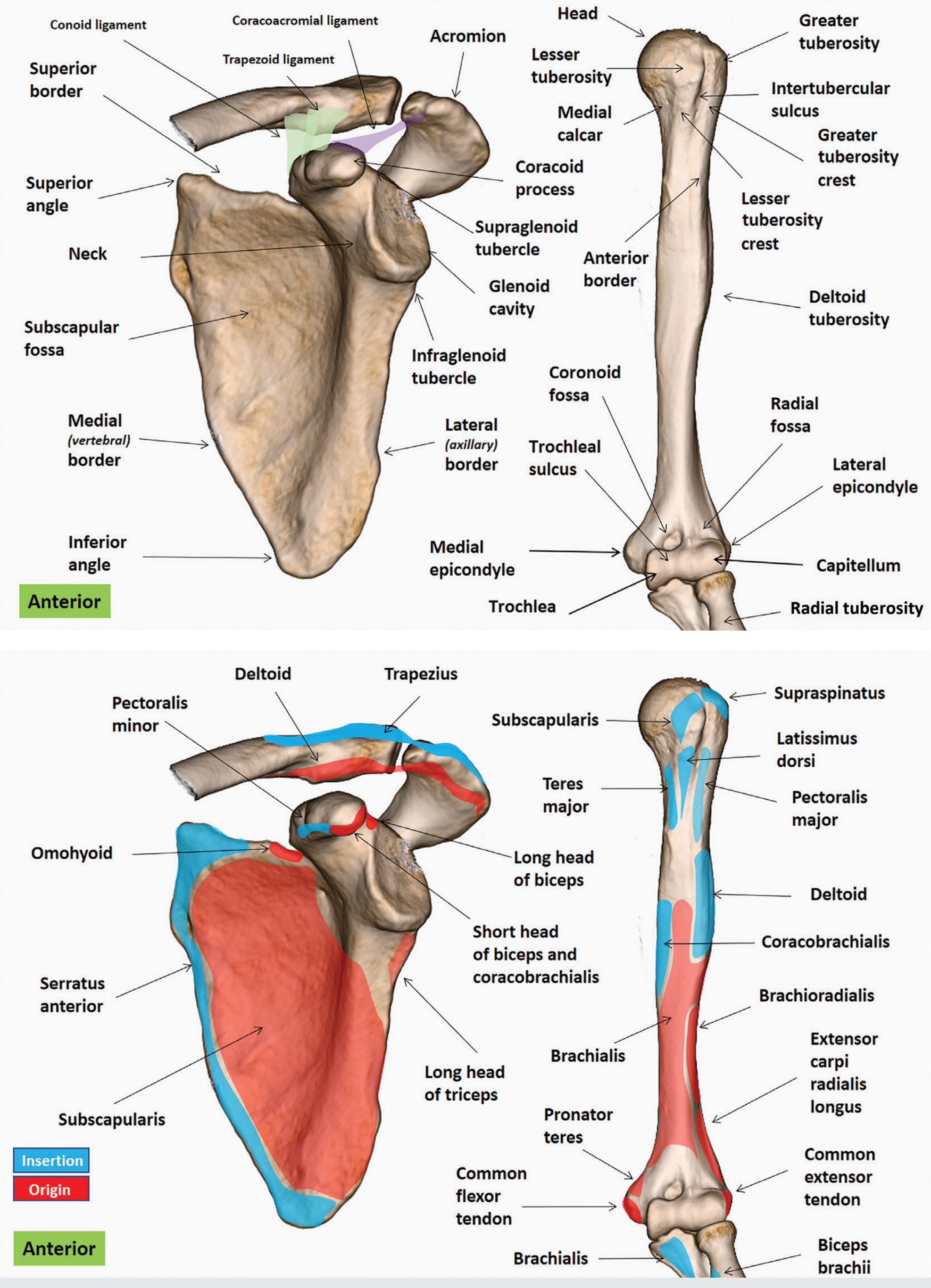
The scapula is a large, flat, triangular bone on the posterolateral aspect of the thoracic wall, overlying ribs 2–7. It connects to the humerus at the glenohumeral joint and to the sternum indirectly via the clavicle.
Surfaces (2)
Costal (anterior/ventral) surface
Dominated by the subscapular fossa — a broad concavity where the subscapularis muscle originates. Oblique ridges give attachment to the fibrous intramuscular septa of subscapularis. A narrow strip along the medial edge is the attachment of serratus anterior (which keeps the scapula pressed against the thorax).
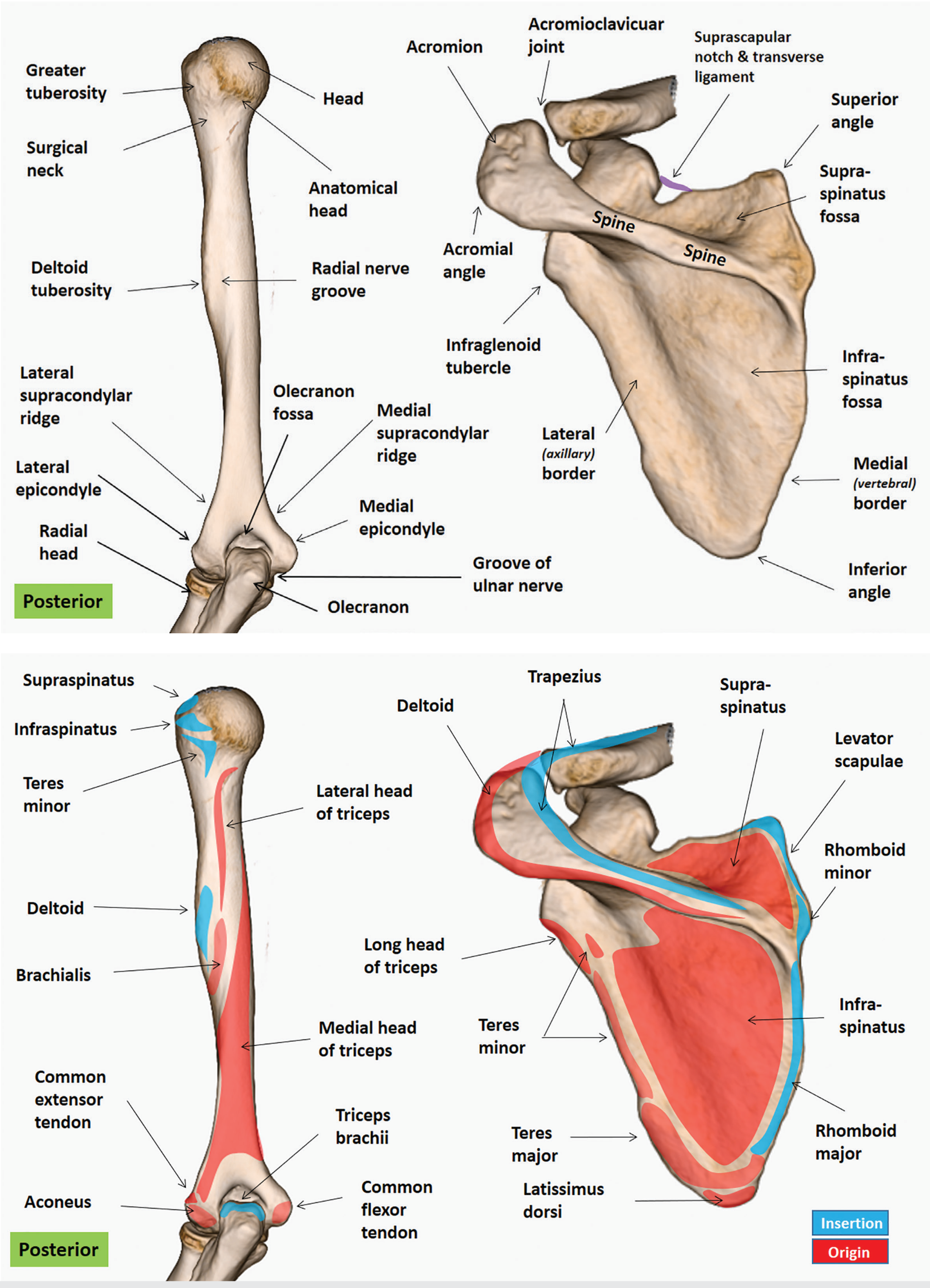
Dorsal (posterior) surface
Divided into two asymmetric regions by the spine of the scapula:
- Supraspinous fossa (smaller, superior) — origin of supraspinatus
- Infraspinous fossa (larger, inferior) — origin of infraspinatus (medially), teres minor (superolateral), and teres major (inferolateral) The two fossae communicate via the spinoglenoid notch between the dorsal scapular neck and the lateral border of the spine.
Borders (3)
| Border | Course | Key Attachments |
|---|---|---|
| Superior | Concave; from superior angle to coracoid base | Inferior belly of omohyoid; contains the suprascapular notch (bridged by the transverse scapular ligament, through which the suprascapular nerve passes) |
| Medial (vertebral) | Longest; from superior to inferior angle | Levator scapulae (upper), rhomboid minor (mid), rhomboid major (inferior) |
| Lateral (axillary) | From inferior glenoid margin to inferior angle | Infraglenoid tubercle at its superior end; separates subscapularis, teres minor, and teres major |
Angles (3)
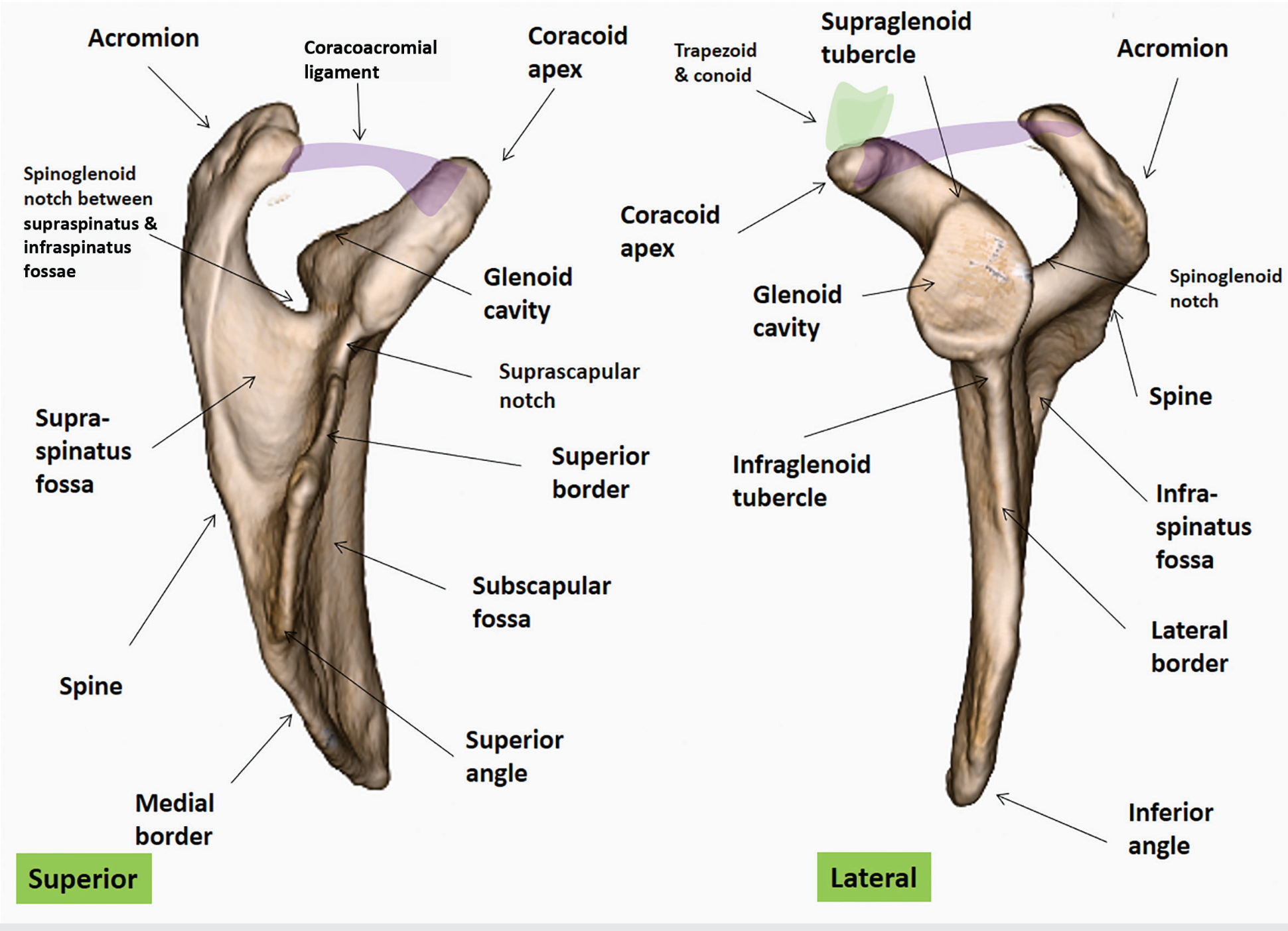
- Lateral angle — marked by the glenoid cavity, a shallow comma-shaped socket that articulates with the humeral head (glenohumeral joint). Articular surface = ~6–8 cm², about one-third to one-fourth the size of the humeral head; retroverted ~4–8°.
- Superior angle — at the junction of the superior and medial borders
- Inferior angle — palpable landmark at the bottom of the bone, at ~T7–T8; the reference point clinically for scapular winging
Processes (3)
Spine
A triangular, plate-like posterior projection. Broadens and elevates laterally to become the acromion. Trapezius inserts on its superior surface; deltoid originates from its inferior surface.
Acromion
The lateral continuation of the spine, forming the roof of the glenohumeral joint. Articulates with the clavicle at the acromioclavicular (AC) joint. Can be flat (type 1) or curved (type 2). Os acromiale — failure of the acromial ossification center to fuse by age 25 (7–15% of individuals); clinically associated with shoulder impingement.
Coracoid process
A hook-like projection arising from the superior border of the neck that curves anteriorly and laterally over the glenoid. Attachment site:
- Apex: short head of biceps brachii + coracobrachialis
- Medial surface: pectoralis minor
- Base (lateral): coracohumeral ligament
- Superior surface: conoid and trapezoid ligaments (coracoclavicular ligaments connecting to clavicle) The coracoacromial ligament spans from the coracoid to the acromion, completing the coracoacromial arch above the rotator cuff.
Glenoid Landmarks
| Landmark | Location | Attachment |
|---|---|---|
| Supraglenoid tubercle | Just superior to glenoid | Long head of biceps brachii (intra-articular) |
| Infraglenoid tubercle | Inferior to glenoid, on scapular neck | Long head of triceps brachii (extra-articular) |
| Glenoid notch | Ventral margin of glenoid | Present in ~55% of scapulas |
Muscular Attachments — Summary
Origins from scapula: subscapularis, supraspinatus, infraspinatus, teres minor, teres major, long heads of biceps and triceps, short head of biceps, coracobrachialis, pectoralis minor, omohyoid, serratus anterior (insertion)
Insertions onto scapula: trapezius (spine/acromion), levator scapulae (medial border), rhomboid major & minor (medial border), serratus anterior (medial border, costal surface)
Nerves of the Scapular Region
| Nerve | Origin | Key point |
|---|---|---|
| Suprascapular nerve | C5–C6 (upper trunk) | Passes through the suprascapular notch under the transverse scapular ligament; supplies supraspinatus and infraspinatus |
| Long thoracic nerve | C5–C7 (roots) | Runs on external surface of serratus anterior along lateral chest wall; vulnerable in axillary dissection (e.g. mastectomy) |
| Dorsal scapular nerve | C5 root | Supplies rhomboids and levator scapulae |
| Axillary nerve | C5–C6 | Passes through quadrilateral space; supplies deltoid and teres minor |
Winged Scapula
Damage to the long thoracic nerve paralyses serratus anterior, causing the medial border and inferior angle to protrude posteriorly — classic "winging." This is accentuated when the patient pushes against a wall or raises their arms. It is the most common complication of axillary lymph node dissection (e.g. radical mastectomy).
Joints
| Joint | Bones | Type |
|---|---|---|
| Glenohumeral | Glenoid cavity + humeral head | Synovial ball-and-socket; most mobile joint in body |
| Acromioclavicular (AC) | Acromion + lateral clavicle | Synovial plane |
| Scapulothoracic | Scapula + thoracic wall | Physiological joint (gliding); not a true synovial joint |
Illustrated Anatomy
Anterior view — bony landmarks & muscle attachments:
Posterior/lateral views — spine, fossae, processes:
Posterior muscle attachments:
Sources: Gray's Anatomy for Students; Imaging Anatomy: Bones, Joints, Vessels and Nerves, Vol. 3
This is a shared conversation. Sign in to Orris to start your own chat.