

IMG_4555.jpeg
Analyze the ecg.What’s the diagnosis
Reading File
The ECG image is visible. Let me analyze it carefully and search for relevant clinical context simultaneously.
Finding Sources
Finding Sources
Loading Image
I have a clear view of the ECG. Here is my full systematic analysis:
ECG Analysis - Abdul Motin, Male, 51 Years
Patient & Technical Details
- Name: Abdul Motin | Age: 51 years | Sex: Male
- Date/Time: 25-05-2026, 9:32:14 AM
- Machine: GE MAC2000
- Settings: 25 mm/s paper speed, 10 mm/mV gain, 12-lead standard
Measured Parameters (from machine)
| Parameter | Value | Normal |
|---|---|---|
| Heart Rate | 83 bpm | 60-100 bpm ✓ |
| QRS duration | 78 ms | <120 ms ✓ |
| PR interval | 152 ms | 120-200 ms ✓ |
| QT / QTc (Bazett) | 382 / 448 ms | QTc borderline (≤440 ms in males) |
| RR / PP | 722 / 722 ms | Regular ✓ |
| P / QRS / T axis | 54 / 36 / 23 degrees | Normal axis ✓ |
Systematic Lead-by-Lead Analysis
Rhythm:
- Regular rhythm, rate 83 bpm. P waves present and upright in leads I and II - Normal Sinus Rhythm
P waves:
- Normal morphology, normal axis (+54°), duration appears normal. No evidence of atrial enlargement.
PR interval: 152 ms - normal, no heart block.
QRS complexes:
- Duration 78 ms - narrow, no bundle branch block
- Normal axis +36°
- No pathological Q waves identifiable in the visible leads
- No left or right ventricular hypertrophy pattern
ST segments / T waves (the key finding):
- There are nonspecific ST-T wave changes visible across multiple leads
- The T waves appear flattened or with mild morphological changes, consistent with a nonspecific ST-T abnormality
- No clear ST elevation or horizontal/downsloping ST depression that would indicate STEMI or NSTEMI
- No deep symmetric T-wave inversions
QTc: 448 ms - borderline prolonged (upper limit of normal for males is 440 ms; some guidelines use 450 ms). This warrants attention.
Machine Interpretation (confirmed on strip)
Normal sinus rhythm Nonspecific ST abnormality Abnormal ECG
Clinical Interpretation
Primary Diagnosis: Abnormal ECG with Nonspecific ST-T Wave Changes
The machine (GE MAC2000 auto-analysis) and the visual findings agree on:
- Normal sinus rhythm at 83 bpm - no arrhythmia
- Nonspecific ST-T abnormality - this is a non-diagnostic finding that can be caused by multiple conditions:
| Possible Cause | Notes |
|---|---|
| Myocardial ischemia / ACS | Must be ruled out first given age 51, male |
| Hypertensive heart disease | Very common in this age group |
| Electrolyte imbalance (hypokalemia, hypomagnesemia) | Flattened T waves |
| Left ventricular hypertrophy with strain | Review voltage criteria |
| Digoxin or other drug effect | Check medication history |
| Early repolarization variant | Benign, but diagnosis of exclusion |
| Cardiomyopathy | Less common |
- Borderline QTc of 448 ms - check for medications that prolong QT, electrolyte disturbances
What This ECG Does NOT Show
- No ST elevation (no acute STEMI pattern)
- No new LBBB
- No pathological Q waves suggesting prior infarct
- No complete heart block or significant conduction abnormality
- No atrial fibrillation or flutter
Clinical Recommendations
Given this is a 51-year-old male with an abnormal ECG (nonspecific ST-T changes + borderline QTc):
- Correlate clinically - Is the patient symptomatic? Chest pain, dyspnoea, palpitations?
- Troponin I/T - to exclude acute myocardial injury
- Electrolytes (K+, Mg2+, Ca2+) - for QTc and T-wave changes
- Previous ECG comparison - Are these changes new or old? Nonspecific ST changes have far greater significance if new.
- 2D Echocardiogram - assess wall motion, LV function, LVH
- Stress test / Holter if ischemia is clinically suspected
- Review medications for QT-prolonging drugs
Important: This ECG analysis is for educational/informational purposes. Clinical decisions must be made by the treating physician with full clinical context, history, and examination findings. An "Abnormal ECG" with nonspecific changes in a 51-year-old male requires active clinical evaluation to rule out ischemic heart disease.
He is hypertensive,he has sudden chest pain,sweating
Finding Sources
Finding Sources
Reading File
Reading File
Loading Image
⚠️ High Suspicion: ACUTE CORONARY SYNDROME (ACS) - Likely NSTEMI / Unstable Angina
With the clinical context now provided - 51-year-old hypertensive male, sudden chest pain, diaphoresis (sweating) - this ECG takes on much greater urgency.
Clinical Picture Synthesis
| Finding | Significance |
|---|---|
| Age 51, Male | High-risk demographic for ACS |
| Hypertension | Major CAD risk factor |
| Sudden chest pain | Classic ischemic symptom |
| Diaphoresis (sweating) | Autonomic activation - hallmark of myocardial infarction |
| Nonspecific ST-T changes on ECG | Consistent with NSTEMI / UA |
| Borderline QTc 448 ms | Reflects myocardial stress/ischemia |
| No ST elevation | Rules out STEMI on this tracing - but NSTEMI cannot be excluded |
Sudden chest pain + sweating + abnormal ECG in a hypertensive 51-year-old male = NSTEMI / Unstable Angina until proven otherwise.
Why NOT STEMI (yet)?
- No ST elevation seen on this ECG
- No new LBBB
- However: a normal or nonspecific ECG does NOT exclude ACS - up to 6% of evolving ACS cases have a normal initial ECG. Serial ECGs are mandatory. (Sabiston Textbook of Surgery)
Differential Diagnoses to Consider
- NSTEMI - most likely given triad of symptoms + ECG changes
- Unstable Angina - if troponins return negative
- Aortic Dissection - must be considered in a hypertensive patient with sudden severe chest pain (requires CXR + CT aorta to exclude)
- STEMI equivalent - repeat ECG in 15-30 min, early changes may evolve
TIMI Risk Score (for NSTEMI/UA)
This patient scores high based on: age >45, hypertension as CAD risk factor, chest pain, ST-T changes on ECG = Moderate-High Risk → indicating Routine Invasive Strategy is appropriate.
Immediate Management Protocol
Following the ACS pathway (Washington Manual of Medical Therapeutics):
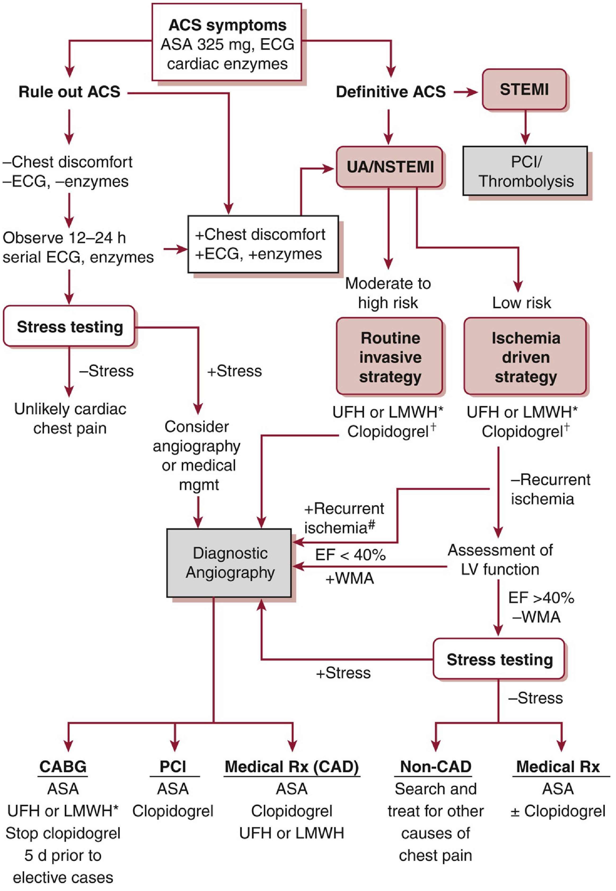
Right now - ACUTE ACTIONS:
| Priority | Action |
|---|---|
| 🔴 IMMEDIATE | IV access + continuous cardiac monitoring + pulse oximetry |
| 🔴 IMMEDIATE | 12-lead ECG repeat in 15-30 min; compare for dynamic changes |
| 🔴 IMMEDIATE | Aspirin 325 mg (loading dose, chew and swallow) |
| 🔴 IMMEDIATE | Sublingual nitroglycerin if BP allows (SBP >90 mmHg) |
| 🔴 IMMEDIATE | Troponin I or T (high-sensitivity if available) - draw now and at 3-6 hours |
| 🟠 URGENT | Dual antiplatelet therapy - add Clopidogrel 300-600 mg OR Ticagrelor 180 mg loading |
| 🟠 URGENT | Anticoagulation - LMWH (Enoxaparin) or UFH |
| 🟠 URGENT | Beta-blocker (oral, if no contraindication - HR >60, no acute HF, no bronchospasm) |
| 🟡 SOON | CXR - assess cardiac size, pulmonary congestion, mediastinum width |
| 🟡 SOON | Electrolytes, RFT, CBC, blood glucose, lipid profile |
| 🟡 SOON | 2D Echo - wall motion abnormalities |
| 🟡 SOON | Cardiology consultation for consideration of coronary angiography |
⚠️ Do NOT Miss
- If sweating + pain is very severe and blood pressure is markedly elevated or unequal in both arms → rule out Aortic Dissection (CT aortogram) before giving anticoagulants
- Monitor for arrhythmias - ischemia can precipitate VT/VF
- Do not discharge this patient without ruling out ACS with serial ECGs + troponins
This patient requires emergency hospital admission and cardiology evaluation immediately. This ECG analysis is clinical decision support - the treating physician must manage the patient based on full examination and investigations.
This is a shared conversation. Sign in to Orris to start your own chat.