
squamous cell carcinoma keratin pearl histology oral
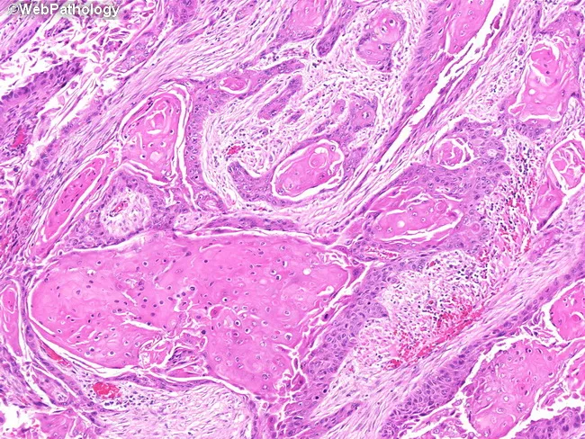
High-magnification histopathology image of oral mucosa showing keratinizing, well-differentiated squamous cell carcinoma with classic keratin pearl formation. The neoplasm displays nests and cords of malignant squamous cells invading the fibrous lamina propria, with abundant eosinophilic cytoplasm and intercellular bridges. Keratinization is evident both centrally within tumor nests and at the periphery, producing concentric keratin pearls of pale eosinophilic keratin. Tumor cells are relatively well differentiated, with recognizable squamous maturation and occasional mitotic figures. The adjacent mucosa demonstrates diverse epithelial alterations, including squamous metaplasia (keratinizing and nonkeratinizing variants), dysplasia, and areas consistent with squamous cell carcinoma in situ, reflecting field cancerization around the invasive focus. Inflammatory infiltrate and edema are present in the superficial lamina propria, while deeper stroma shows variable desmoplastic response. The surface epithelium may show thinning or ulceration over the lesion. No clear vascular invasion is evident at this magnification, though deeper planes could reveal invasion into connective tissue and perineural spread. This image provides valuable diagnostic features for distinguishing keratinizing SCC from nonkeratinizing variants and other mucosal neoplasms, and highlights the relevance of adjacent dysplasia/CIS as indicators of tumor field effect and oncologic margins. It serves as an educational reference for pathology, histology, and clinical tumor biology.
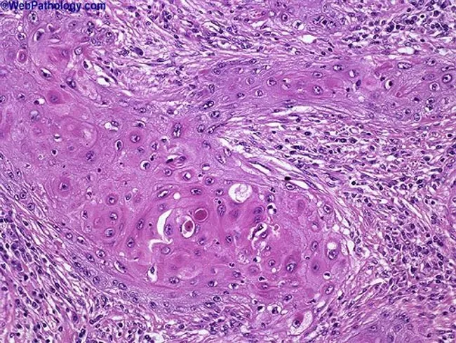
This histopathology image depicts a cutaneous squamous cell carcinoma on routine hematoxylin and eosin stained section. The biopsy shows nests of malignant squamous cells with keratinization and abundant keratin pearls, indicating squamous differentiation. The tumor exhibits moderate differentiation, evidenced by intercellular bridges, eosinophilic cytoplasm, nuclear pleomorphism, hyperchromasia, and mitotic activity. Invasion into the surrounding dermal stroma is evident, with desmoplastic-type fibrous response in some areas. The epidermis overlying the lesion shows dysplastic changes, and keratin pearl formation within tumor nests is a hallmark of keratinizing SCC. The architecture includes irregular cords and nests of polygonal cells, occasional central keratinization, and keratinized pearls of varying sizes. Clinically, these features suggest a malignant cutaneous neoplasm with potential for local invasion; staging would consider depth of invasion and perineural or lymphovascular involvement. This image is relevant for diagnostic pathology, histology education, and tumor biology research, illustrating characteristic squamous differentiation, keratin production, and invasion patterns. Differential considerations include well-differentiated keratinizing SCC versus moderately-to-poorly differentiated forms, verrucous carcinoma, and basal cell carcinoma with squamous features. The visual cues—keratin pearls, keratinization, and cohesive epidermal-derived tumor islands—assist in confirming diagnosis and guiding surgical management. Correlation with clinical data will inform prognosis and adjuvant therapy decisions and multidisciplinary care planning.
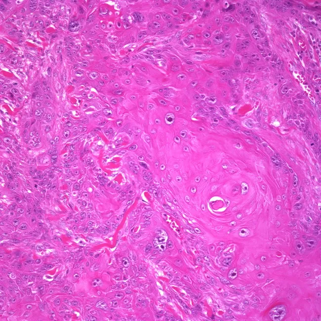
High-power light microscopic histology of vulvar tissue demonstrates invasive, well-differentiated keratinizing squamous cell carcinoma centered on the labium majus. The specimen shows malignant squamous cells arranged in nests and radiating cords with orderly keratinization and conspicuous keratin pearl formation within desmoplastic stroma. Individual tumor cells exhibit abundant eosinophilic cytoplasm, polygonal outlines, intercellular bridges, hyperchromatic rounded to angulated nuclei, and mitotic activity is present but not excessive in this field. Invasion through the squamous epithelium into the underlying dermis is evident, with variable stromal reaction and a lymphocytic inflammatory component. The keratinizing phenotype and absence of conspicuous koilocytosis or basaloid features favor a traditional keratinizing vulvar squamous cell carcinoma as opposed to HPV-associated basaloid or warty subtypes. The appearance aligns with a histologic diagnosis of invasive vulvar SCC; depth of invasion and perineural or lymphovascular invasion risk require correlation with surgical margins and regional nodal status. Clinically, this entity is associated with vulvar mass and potential regional metastasis; management typically involves radical vulvectomy with bilateral inguinal lymph node dissection or equivalent radiotherapeutic strategies depending on staging. This image supports diagnostic confirmation, guides surgical planning, and informs prognosis and follow-up strategies. Accurate staging and multidisciplinary coordination remain critical for optimal outcomes globally.
Answer: B) Squamous Cell Carcinoma
Key Histological Findings in the Image
- Keratin pearls - the most prominent finding: concentric whorls of laminated, pale eosinophilic keratin arranged in onion-skin layers within tumor nests
- Nests and cords of malignant squamous cells invading the fibrous stroma
- Desmoplastic stromal response - reactive fibrous tissue surrounding tumor islands
- Nuclear pleomorphism and hyperchromasia within the squamous cell nests
- Stromal inflammatory infiltrate
Why Not the Other Options?
| Option | Why Excluded |
|---|---|
| A) Basal cell carcinoma | BCC shows basaloid cells in palisading arrangement at the periphery of tumor nests, with retraction artifact and mucin stroma - NO keratin pearls |
| C) Molluscum contagiosum | Shows epidermal lobules with large intracytoplasmic Henderson-Patterson (molluscum) bodies - entirely different picture |
Clinical Correlation
- Elderly man - SCC is far more common in older age groups
- Chronic verrucous lesion over the cheek - the cheek is a classic "sun terrace" location for SCC, especially verrucous SCC, which presents as a slowly growing exophytic/cauliflower-like lesion
- Verrucous SCC is a well-recognized low-grade variant of SCC that commonly affects the oral cavity and facial skin, and histologically still demonstrates keratinization and keratin pearl formation