

Summarise assessment of pulmonary circulation tests for seminar presentation with all important points in understanding way easy to understand
Assessment of Pulmonary Circulation - Seminar Summary
Why Is It Important?
"Assessment of pulmonary function is incomplete and unsatisfactory without the study of pulmonary circulation."
1. Measuring Pulmonary Blood Flow & Pressure
How?
- Done in ICU using invasive monitoring
- Tool used: Swan-Ganz catheter (flow-directed pulmonary arterial catheter)
- Measures:
- Pulmonary arterial pressure (PAP)
- Pulmonary capillary wedge pressure (PCW)
- Cardiac output (CO) - by thermodilution method
Key Formula - Pulmonary Vascular Resistance (PVR):
PVR = 80 × (PAP - PCW) / CO
| Term | Meaning |
|---|---|
| PAP | Mean pulmonary arterial pressure (mm Hg) |
| PCW | Pulmonary capillary wedge pressure (mm Hg) |
| CO | Cardiac output (L/min) |
2. Disturbances in Pulmonary Circulation (PVR Increases By 4 Mechanisms)
| # | Mechanism | What Happens |
|---|---|---|
| 1 | Vasoconstriction | Arteriolar spasm in response to alveolar hypoxia |
| 2 | Thromboembolism | Intraluminal clots reduce the cross-sectional area of vessels |
| 3 | Vascular hypertrophy | Smooth muscle proliferation narrows vessel lumen |
| 4 | Pulmonary injury | Destruction of small vessels (scarring, alveolar wall loss) |
Increased PVR --> increased pulmonary artery pressure --> decreased right ventricular output
Conditions That Increase PVR:
- Heart diseases - e.g., mitral stenosis raises left atrial pressure
- Lung diseases - COPD, interstitial lung disease, chest wall diseases (kyphoscoliosis), obesity hypoventilation, sleep apnea
- Diseases of pulmonary vessels - recurrent pulmonary embolism, scleroderma
3. Respiratory Causes of Hypoxemia
- Non-respiratory causes: anemia, carbon monoxide poisoning, decreased inspired O₂ (high altitude)
- Respiratory dysfunction is the most common cause in adults
5 Respiratory Causes of Hypoxemia:
- Regional hypoventilation
- Large blood clot (occlusion of major artery)
- Shunts
- Generalized hypoventilation
- Diffusion block
4. Alveolar-Arterial O₂ Gradient (A-aO₂ Gradient)
- Normal arterial PaO₂ = 85-95 mm Hg (Hb is ~98% saturated)
- Normal A-aO₂ gradient = 5-15 mm Hg
5 Causes of Increased A-aO₂ Gradient:
A) Regional Hypoventilation (most common)
- A local ventilation-perfusion (V/Q) mismatch - some lung areas are hypoventilated but still perfused
- Blood passes un-oxygenated areas → increases venous admixture
- Result: High A-aO₂ gradient, Low PO₂, Low O₂ content
B) Large Blood Clot
- A major artery in the lung is occluded → greater cardiac output is diverted to the remaining lung
- That remaining lung gets over-perfused relative to ventilation
- Result: Venous admixture increases, V/Q ratio falls
C) Shunts
- Blood bypasses the lungs entirely:
- Right-to-left cardiac shunt (e.g., Tetralogy of Fallot)
- Intrapulmonary shunt (e.g., foreign body or tumor obstructing a bronchus)
- Key feature: Breathing 100% O₂ does NOT correct low arterial PO₂ (because shunted blood never contacts alveolar air)
- Result: High A-aO₂ gradient, Low PO₂, Low O₂
D) Generalized Hypoventilation
- Occurs when overall alveolar ventilation is globally reduced (not just regional):
- COPD (emphysema)
- Head injury or drug overdose (morphine) - depresses respiratory center
- As ventilation falls → arterial PCO₂ rises + arterial pH falls
- KEY distinguishing feature: A-aO₂ gradient is NORMAL (unlike other causes)
- Treatment: Mechanical ventilation with room air
E) Diffusion Block (least common)
- Occurs when the diffusion distance across the alveolar-capillary membrane increases OR its permeability decreases
- Example: Pulmonary edema
- Features: Low PaO₂, High A-aO₂ gradient, High PaCO₂
5. Quick Comparison Table for Seminar
| Cause | A-aO₂ Gradient | PaO₂ | PaCO₂ | Corrected by 100% O₂? |
|---|---|---|---|---|
| Regional hypoventilation | High | Low | Variable | Yes |
| Large blood clot | High | Low | Variable | Partially |
| Shunt | High | Low | Low | No |
| Generalized hypoventilation | Normal | Low | High | Yes |
| Diffusion block | High | Low | High | Yes |
Memory Tips for Exams
- PVR formula: "80 times pressure drop divided by flow" = 80 × (PAP - PCW) / CO
- Shunt = the only cause where 100% O₂ fails to correct hypoxemia
- Generalized hypoventilation = only cause with normal A-aO₂ gradient
- Diffusion block = low permeability or long distance (think pulmonary edema)
- 4 mechanisms of increased PVR: Vasoconstriction, Thromboembolism, Hypertrophy, Injury (mnemonic: VTHI)
Summarise assessment of pulmonary circulation tests for seminar presentation with all important points in understanding way easy to understand
🫁 Assessment of Pulmonary Circulation - Seminar Summary
Why Study Pulmonary Circulation?
"Assessment of pulmonary function is incomplete and unsatisfactory without the study of pulmonary circulation."
- The pulmonary vasculature accommodates 5 L/min of right ventricular output
- Vessels are thin-walled and offer less resistance than systemic vessels
- Normal mean pulmonary artery pressure = 15 mm Hg
- In upright posture: pressure is lowest at apex, highest at lung bases
PART 1 - Measuring Pulmonary Blood Flow and Pressure
How Is It Done?
- Performed in ICU using invasive monitoring
- Tool: Swan-Ganz catheter (flow-directed pulmonary arterial catheter)
What Does It Measure?
- Pulmonary arterial pressure (PAP)
- Pulmonary capillary wedge pressure (PCW)
- Cardiac output (CO) - by thermodilution method
Key Formula - Pulmonary Vascular Resistance (PVR):
PVR = 80 × (PAP - PCW) / CO
| Term | Full Form | Unit |
|---|---|---|
| PAP | Mean pulmonary arterial pressure | mm Hg |
| PCW | Pulmonary capillary wedge pressure | mm Hg |
| CO | Cardiac output | L/min |
Normal PVR = 50-150 dynes.s/cm³
PART 2 - Disturbances in Pulmonary Circulation
PVR Increases by 4 Mechanisms:
| # | Mechanism | Simple Explanation |
|---|---|---|
| 1 | Pulmonary Vasoconstriction | Alveolar hypoxia causes arteriolar spasm → narrows vessels |
| 2 | Pulmonary Thromboembolism | Clots inside vessels reduce the cross-sectional area |
| 3 | Vascular Hypertrophy | Smooth muscle thickens the vessel wall → narrows lumen |
| 4 | Pulmonary Injury | Scarring or alveolar wall destruction → loss of vascular bed |
Increased PVR → Increased pulmonary artery pressure → Decreased right ventricular output
Conditions That Increase PVR:
- Conditions that raise left atrial pressure (e.g., mitral stenosis)
- COPD
- Interstitial lung disease
- Chest wall diseases (e.g., kyphoscoliosis)
- Obesity hypoventilation
- Sleep apnea syndrome
- Recurrent pulmonary embolism
- Scleroderma (occludes small pulmonary arteries and arterioles)
PART 3 - Respiratory Causes of Hypoxemia
- Non-respiratory causes: anemia, carbon monoxide poisoning, decreased inspired O₂ tension (e.g., high altitude)
- Respiratory dysfunction = most common cause of hypoxemia in adults
5 Respiratory Causes:
- Regional hypoventilation
- Occlusion of a major artery (large blood clot)
- Shunts
- Generalized hypoventilation
- Diffusion block
PART 4 - Alveolar-Arterial Oxygen Gradient (A-aO₂ Gradient)
Normal Physiology:
- Hb is 100% saturated with O₂ as blood passes pulmonary capillaries
- End-capillary PO₂ = alveolar PO₂
- Blood in pulmonary veins returning to left atrium has slightly lower PO₂
- Normal PaO₂ in systemic arterial blood = 85-95 mm Hg (Hb ~98% saturated)
- Normal A-aO₂ gradient = 5-15 mm Hg
PART 5 - The 5 Causes Explained Simply
1. Regional Hypoventilation (Most Common)
- A local area of the lung is underventilated but still perfused
- Blood passes through un-oxygenated alveoli → venous admixture increases
- Only a small amount of venous admixture is normal
- Patients with abnormally low V/Q ratio:
| Finding | Value |
|---|---|
| A-aO₂ gradient | HIGH |
| PO₂ | LOW |
| O₂ content | LOW |
2. Large Blood Clot (Occlusion of Major Artery)
- A major artery is blocked → blood is diverted to the remaining lung
- That part gets over-perfused relative to ventilation → V/Q ratio falls
- Result: venous admixture increases
3. Shunts
- Blood bypasses ventilated alveoli entirely:
- Right-to-left cardiac shunt
- Intrapulmonary shunt (foreign body or tumor obstructing airway)
- Shunted blood never contacts alveolar air
- Critical point: Breathing 100% O₂ does NOT correct the low PaO₂ (unique to shunts!)
| Finding | Value |
|---|---|
| A-aO₂ gradient | HIGH |
| PO₂ | LOW |
| O₂ | LOW |
| Response to 100% O₂ | No correction |
4. Generalized Hypoventilation
- Overall alveolar ventilation is globally reduced (not just one area)
- Causes:
- COPD (emphysema)
- Head injury or drug overdose (morphine) depressing the respiratory center
- As ventilation falls → PaCO₂ rises → arterial pH falls
A-aO₂ gradient is NORMAL (unlike all other causes!)
- Best treated by mechanical ventilation breathing room air
5. Diffusion Block (Least Common)
- Alveolar-capillary membrane has increased thickness or decreased permeability
- Classic example: Pulmonary edema
| Finding | Value |
|---|---|
| PaO₂ | LOW |
| A-aO₂ gradient | HIGH |
| PaCO₂ | HIGH |
PART 6 - Master Comparison Table (Most Important for Exam!)
| Cause | A-aO₂ Gradient | PaO₂ | PaCO₂ | 100% O₂ Corrects? |
|---|---|---|---|---|
| Regional hypoventilation | HIGH | Low | Variable | Yes |
| Large blood clot | HIGH | Low | Variable | Partially |
| Shunt | HIGH | Low | Low | NO |
| Generalized hypoventilation | NORMAL | Low | HIGH | Yes |
| Diffusion block | HIGH | Low | High | Yes |
Quick Memory Tips
| Point | Memory Aid |
|---|---|
| PVR formula | "80 times pressure drop divided by flow" |
| 4 mechanisms raising PVR | Vasoconstriction, Thromboembolism, Hypertrophy, Injury = VTHI |
| Shunt hallmark | Only cause where 100% O₂ fails to correct hypoxemia |
| Generalized hypoventilation hallmark | Only cause with normal A-aO₂ gradient |
| Diffusion block hallmark | Pulmonary edema - thick membrane, high PaCO₂ |
| Swan-Ganz catheter | Used to measure PAP, PCW, CO directly in ICU |
Pulmonary circulation introduction
pulmonary circulation diagram heart lungs blood flow
Summary : This figure illustrates three different configurations of ventricular assist devices (VADs) used to support heart function: left VAD (A), right VAD (B), and biventricular assist device (C). The diagram shows the anatomical connections and blood flow pathways for each configuration, including the heart chambers, lungs, and systemic circulation.
Summary : This is a comprehensive medical illustration summarizing the pathophysiology, clinical features, and complications of portal hypertension in liver disease, integrating vascular, hemodynamic, and systemic effects with annotated diagrams, flowcharts, and explanatory boxes.
pulmonary circulation zones West zone lung apex base blood flow
🫁 Pulmonary Circulation - Introduction
1. What Is the Pulmonary Circulation?
| System | Type | Supplied by | Purpose |
|---|---|---|---|
| Pulmonary circulation | Low-pressure, HIGH-flow | Pulmonary artery (from right ventricle) | Gas exchange at alveoli |
| Bronchial circulation | High-pressure, LOW-flow | Bronchial arteries (from aorta) | Nutrition of lung tissue itself |
The pulmonary circulation is the one responsible for oxygenating blood - it carries deoxygenated blood to the alveoli and returns oxygenated blood to the left heart. Bronchial circulation accounts for only 1-2% of cardiac output and supplies the airways, connective tissue, and lung support structures.
2. Anatomical Path of Blood Flow
Right Ventricle
      ↓
Pulmonary Artery (only 5 cm long, then divides)
      ↓
Right + Left Pulmonary Arteries → enter each lung at the hilum
      ↓
Branch alongside bronchi at every airway generation
      ↓
Pulmonary Arterioles → Alveolar Capillaries (GAS EXCHANGE)
      ↓
Pulmonary Venules → Pulmonary Veins
      ↓
Left Atrium → Left Ventricle → Systemic Circulation
The pulmonary artery branches travel alongside airways at every generation - this close arrangement ensures ventilation and perfusion are always matched. Pulmonary veins, unlike arteries, travel away from the airways - found in the surrounding connective tissue peripherally.
3. Structure of Pulmonary Vessels - Why They Are Different
| Feature | Pulmonary Vessels | Systemic Vessels |
|---|---|---|
| Wall thickness | Thin (1/3 of aortic wall) | Thick |
| Diameter | Larger for same generation | Smaller |
| Compliance | High (~7 mL/mm Hg) | Lower |
| Resistance | Low (5-10x less) | High |
| Pressure | Low (15 mm Hg mean) | High (95 mm Hg mean) |
- High compliance allows pulmonary arteries to accommodate the full stroke volume of the right ventricle
- Low pressure protects the thin alveolar-capillary membrane from rupture, keeping it thin enough for efficient gas diffusion
- Low resistance means the right ventricle does much less work than the left ventricle
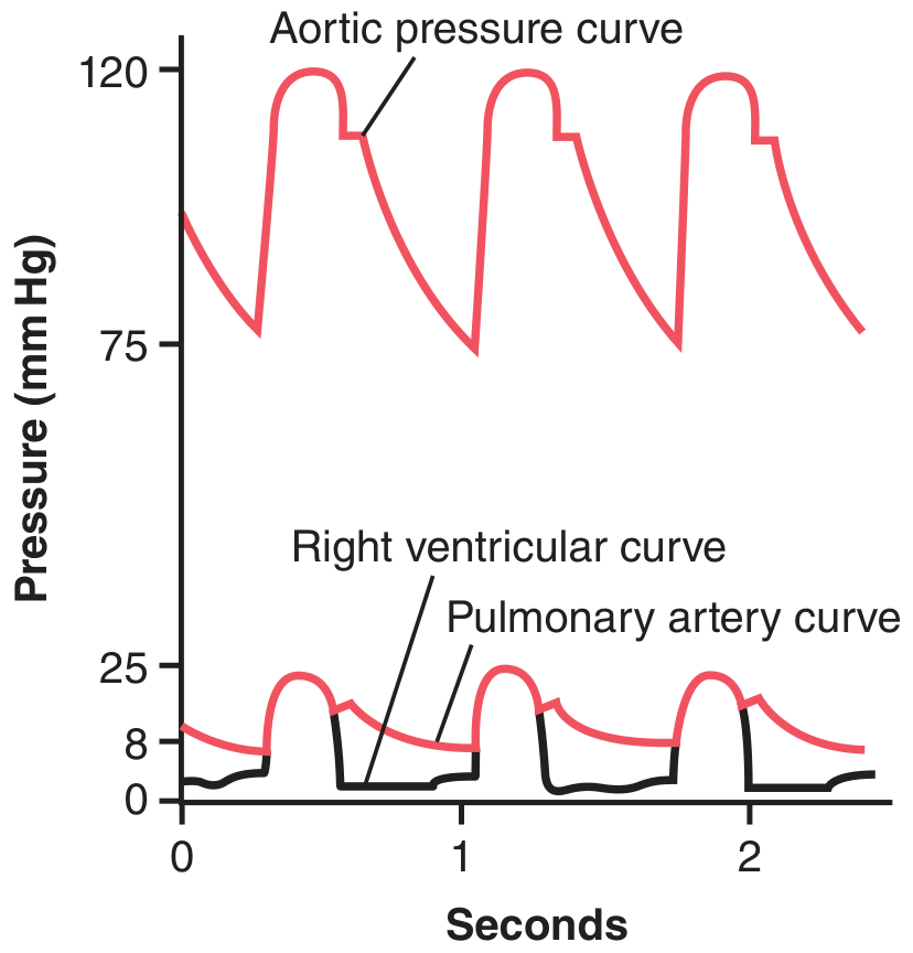
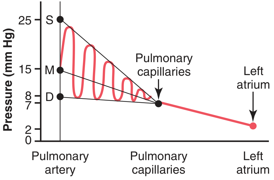
4. Pressures in the Pulmonary Circulation
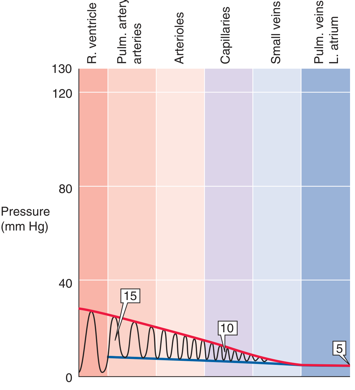
Normal Pressure Values:
| Location | Pressure |
|---|---|
| Right ventricle - systolic | 25 mm Hg |
| Right ventricle - diastolic | 0-1 mm Hg |
| Pulmonary artery - systolic | 25 mm Hg |
| Pulmonary artery - diastolic | 8 mm Hg |
| Mean pulmonary artery pressure | 15 mm Hg |
| Pulmonary capillaries | ~7 mm Hg |
| Pulmonary wedge pressure | ~5 mm Hg |
| Left atrium | ~2 mm Hg |
5. Blood Volume of the Lungs
- Total blood in lungs at any time = ~450 mL (~9% of total blood volume)
- In capillaries = ~70 mL
- Rest equally divided between arteries and veins
The Lungs Act as a Blood Reservoir!
- Under stress, up to 250 mL can be shifted from lungs to systemic circulation (e.g., when blowing hard against resistance)
- In hemorrhage, pulmonary blood can shift into systemic vessels as compensation
- In left heart failure or mitral stenosis, blood dams up in lungs - pulmonary blood volume can double, causing pulmonary edema
6. Blood Flow Through the Lungs
- Pulmonary blood flow = cardiac output (the entire output of the right ventricle)
- Normally ~5 L/min
- Pulmonary vessels are distensible - they widen with rising pressure and narrow with falling pressure
- This passive distension allows the lung to handle large increases in cardiac output (e.g., during exercise) without major pressure rises
7. Regulation of Pulmonary Blood Flow - Hypoxic Vasoconstriction
| Situation | Systemic Vessels | Pulmonary Vessels |
|---|---|---|
| Low O₂ (hypoxia) | Vasodilate (bring more blood) | Vasoconstrict (divert blood away) |
| Normal O₂ | Normal tone | Normal tone |
Why Does the Lung Vasoconstrict During Hypoxia?
- O₂-sensitive K⁺ channels in smooth muscle are inhibited
- Cell membrane depolarizes
- Voltage-gated Ca²⁺ channels open → Ca²⁺ influx
- Vasoconstriction occurs
- Release of vasoconstrictors (endothelin, reactive oxygen species)
- Decreased release of vasodilators (nitric oxide)
8. Effect of Gravity on Pulmonary Blood Flow
West's Zones of the Lung:
| Zone | Location | Pressures | Blood Flow |
|---|---|---|---|
| Zone I | Apex | Alveolar P > Arterial P | No flow (only in mechanical ventilation) |
| Zone II | Middle | Arterial P > Alveolar P > Venous P | Intermittent flow (systole only - "waterfall" effect) |
| Zone III | Base | Arterial P > Venous P > Alveolar P | Continuous flow (throughout cardiac cycle) |
In normal standing humans, Zone I barely exists - most of the lung is Zone II and III. In positive pressure ventilation, Zone I expands and creates dead space.
9. Summary - Key Points at a Glance
| Feature | Value/Concept |
|---|---|
| Blood flow | = Cardiac output (~5 L/min) |
| Mean PA pressure | 15 mm Hg |
| PA systolic/diastolic | 25/8 mm Hg |
| Capillary pressure | ~7 mm Hg |
| Total lung blood volume | ~450 mL |
| Resistance vs systemic | 5-10x lower |
| Unique regulation | Hypoxic vasoconstriction |
| Gravity effect | Zonal blood flow distribution (West's zones) |
Memory Tricks
- "Low pressure, HIGH flow" - pulmonary = the exact opposite of what you'd expect
- Pulmonary hypoxia = vasoconstriction (remember: it's the OPPOSITE of systemic)
- Bronchial = nutrition (1-2%) | Pulmonary = gas exchange (99%)
- Apex = less blood, Base = more blood (gravity wins in a low-pressure system)
- Wedge pressure ≈ Left atrial pressure (used clinically to estimate left heart filling)