
Anatomy of mouth /oral cavity
Reading File
Now let me search the medical library for oral cavity anatomy content.
Finding Sources
Finding Sources
Gray's Anatomy for Students is the best source here. Let me read that section plus the Cummings Otolaryngology content, and also search for specific substructures in parallel.
Reading File
Reading File
Finding Sources
Gray's Anatomy is giving excellent content. Let me read more of it plus search for specific structures (tongue, palate, teeth, salivary glands).
Reading File
Finding Sources
Finding Sources
Excellent. Now let me read the tongue section and the palate sections in parallel.
Reading File
Reading File
I have excellent content. Now let me fetch the key anatomical images and read more about muscles of tongue, salivary glands, and teeth.
Loading Image
Loading Image
Loading Image
Reading File
I now have very rich content from Gray's Anatomy for Students. Let me get the salivary glands and teeth/floor of mouth content, plus one more key image.
Finding Sources
Finding Sources
Reading File
Reading File
I now have comprehensive content from Gray's Anatomy for Students. Let me fetch one more important image (the salivary glands/floor of mouth) and compile the full answer.
Loading Image
I now have all the content needed for a thorough, well-illustrated answer. Here is the complete anatomy of the mouth/oral cavity:
Anatomy of the Mouth / Oral Cavity
Source: Gray's Anatomy for Students
Overview
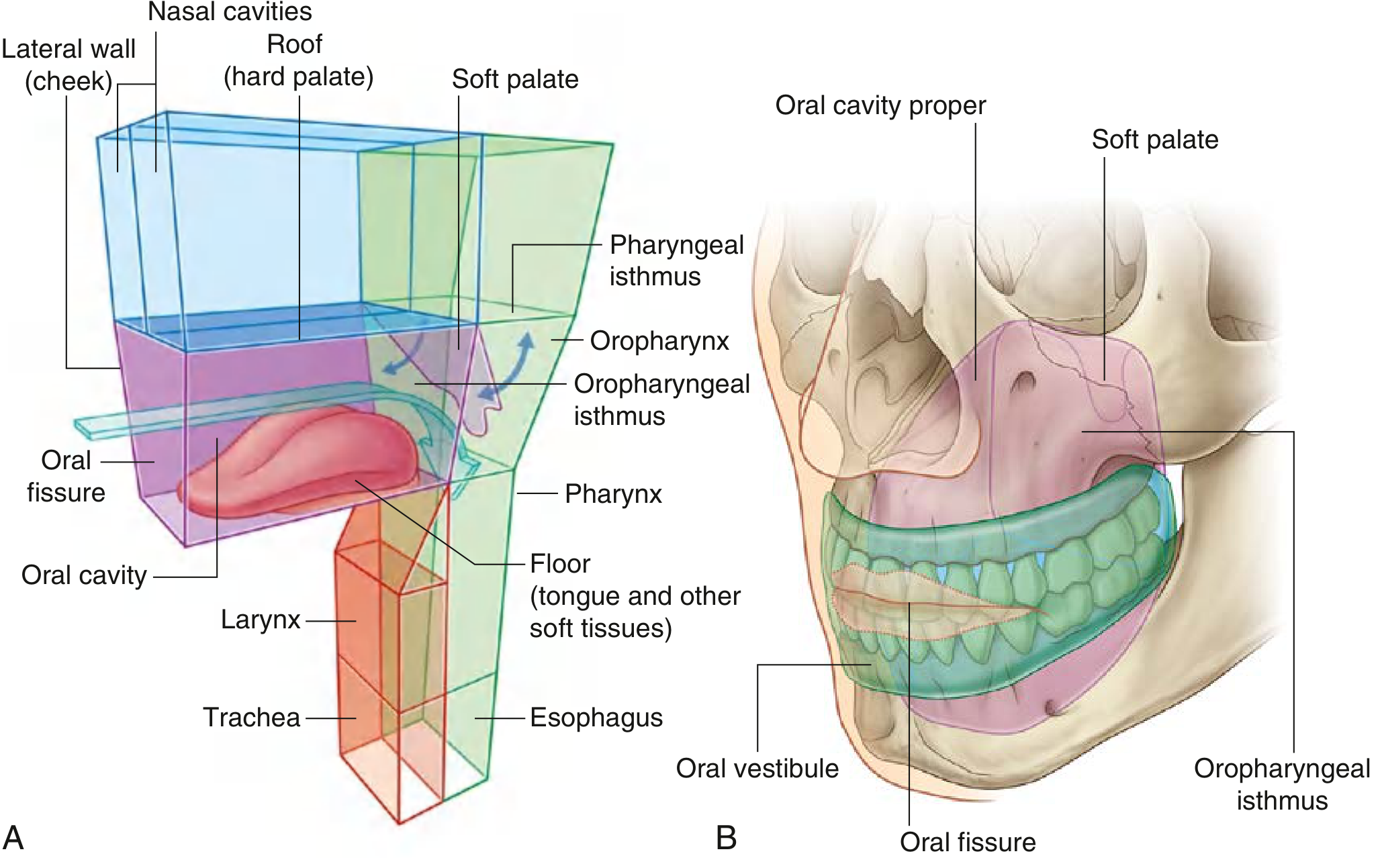
The oral cavity is inferior to the nasal cavities. It opens onto the face through the oral fissure anteriorly and communicates with the pharynx posteriorly through the oropharyngeal isthmus. It has four walls:
- Roof - hard and soft palates
- Floor - muscular diaphragm (mylohyoid) and tongue
- Lateral walls - cheeks (buccinator muscle)
- Anterior opening - oral fissure, bordered by the lips
Two Regions
The upper and lower dental arches (teeth + alveolar bone) divide the oral cavity into two regions:
- Oral vestibule - horseshoe-shaped space between the dental arches and the inner surfaces of the cheeks and lips.
- Oral cavity proper - the inner space enclosed by the dental arches.
Skeletal Framework
The bones contributing to the oral cavity include:
- Paired: maxillae, palatine bones, temporal bones
- Unpaired: mandible, sphenoid, hyoid
The two maxillae form most of the roof via their alveolar processes (carrying upper teeth) and palatine processes (forming the anterior 3/4 of the hard palate). The palatine bones (horizontal plates) complete the posterior 1/4 of the hard palate.
The mandible forms the lower jaw; it carries the lower dental arch and articulates with the temporal bone at the temporomandibular joint (TMJ).
Roof - The Palate
Hard Palate
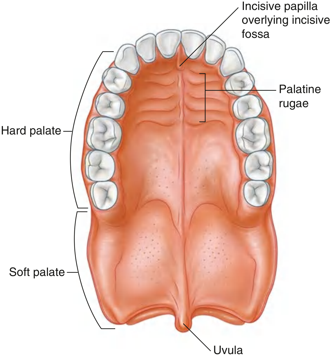
- Bony plate covered by mucosa above (respiratory) and below (oral).
- Anterior 3/4 formed by the palatine processes of the maxillae; posterior 1/4 by the horizontal plates of the palatine bones.
- Mucosal features: transverse palatine rugae, a median palatine raphe, and anteriorly an incisive papilla overlying the incisive fossa (transmits nasopalatine nerve and greater palatine vessels).
Soft Palate
- A muscular, mobile extension of the hard palate posteriorly.
- Ends in the uvula - a small, tear-shaped muscular projection.
- Acts as a valve: depressed to close the oropharyngeal isthmus; elevated to seal off the nasopharynx from the oropharynx during swallowing.
- Formed by 5 paired muscles:
| Muscle | Action |
|---|---|
| Tensor veli palatini | Tenses soft palate; opens auditory tube |
| Levator veli palatini | Elevates soft palate |
| Palatoglossus | Lowers soft palate; narrows oropharyngeal isthmus |
| Palatopharyngeus | Lowers soft palate; narrows pharyngeal inlet |
| Musculus uvulae | Shortens/elevates uvula |
All soft palate muscles are innervated by the vagus nerve [X] via the pharyngeal plexus, except tensor veli palatini (innervated by the mandibular nerve [V3] via the nerve to medial pterygoid).
Floor of the Oral Cavity
The floor is primarily formed by the mylohyoid muscle on each side, which creates a muscular diaphragm stretching between the two sides of the mandible and the hyoid bone. Above the mylohyoid sits the tongue, sublingual glands, and the deep part of the submandibular gland.
A large triangular oropharyngeal aperture is formed between the free posterior border of the mylohyoid, the superior constrictor, and the middle constrictor muscles - this is the gateway through which muscles, vessels, nerves, and lymphatics reach the floor of the oral cavity from the neck.
The Tongue
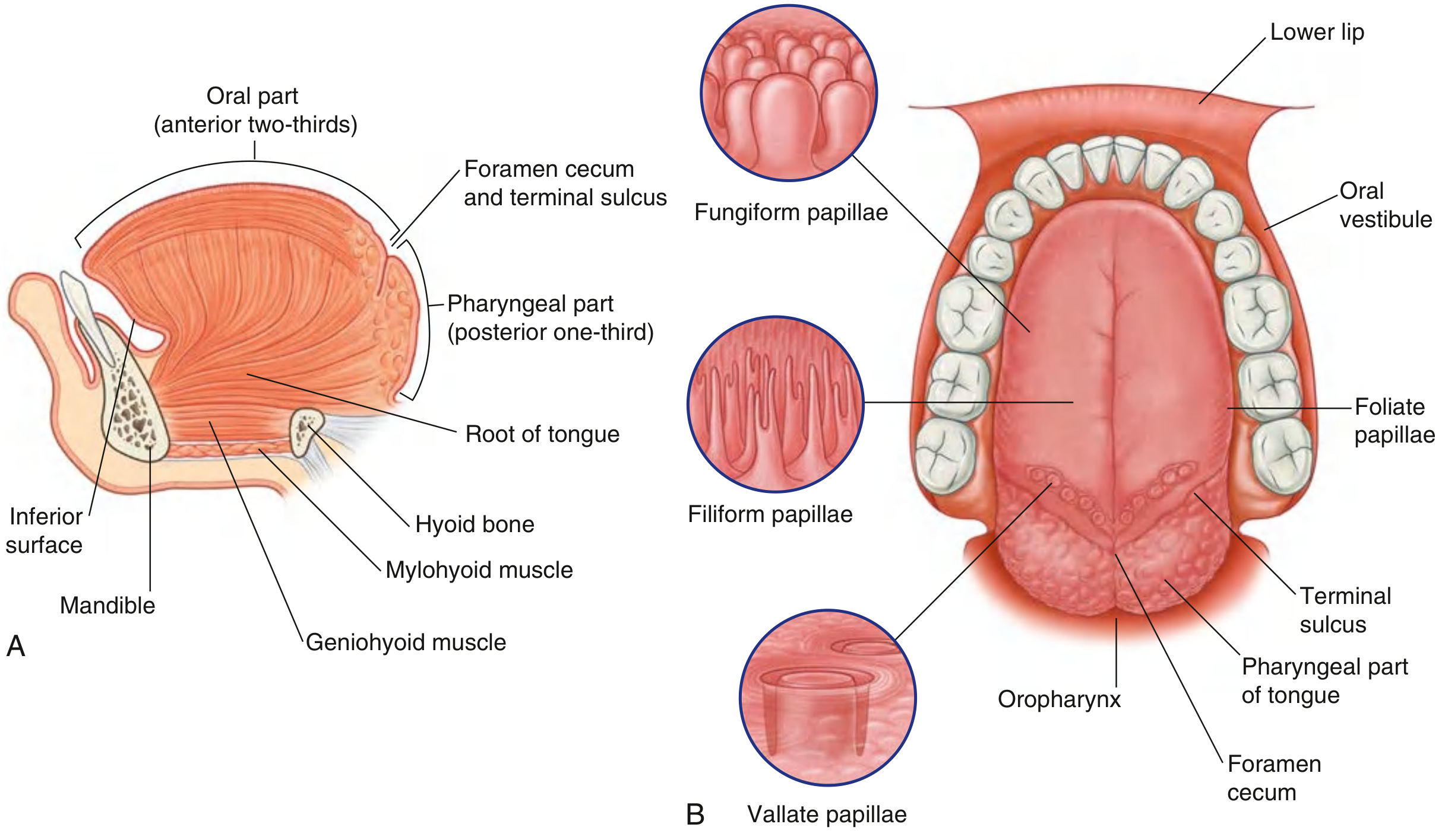
The tongue is a muscular structure forming part of the oral floor and the anterior wall of the oropharynx.
Parts
- Oral (anterior 2/3) - in the oral cavity, horizontal surface bearing papillae; apex points forward behind the incisor teeth.
- Pharyngeal (posterior 1/3) - curves inferiorly into the vertical plane; covered by irregular mucosa with lingual tonsil (lymphoid nodules).
- Separated by the V-shaped terminal sulcus, at the apex of which is the foramen cecum (embryonic thyroglossal duct origin).
- The tongue is divided into left and right halves by a median fibrous septum.
Papillae (on dorsal surface of oral part)
| Papillae | Features |
|---|---|
| Filiform | Most numerous; small, cone-shaped; no taste buds; give tongue its rough texture |
| Fungiform | Rounded, larger; concentrated at tongue margins; have taste buds |
| Vallate (circumvallate) | Largest; only 8-12 in a V-line just anterior to terminal sulcus; have taste buds in sulcus walls |
| Foliate | Linear folds on lateral tongue margin near sulcus; have taste buds |
Inferior Surface
- No papillae.
- A midline frenulum connects the tongue to the floor of the oral cavity.
- On each side of the frenulum: a lingual vein, and laterally a fimbriated (plica fimbriata) fold.
Muscles of the Tongue
All tongue muscles are paired (divided by the median septum) and innervated by the hypoglossal nerve [XII], except palatoglossus (vagus nerve [X]).
Intrinsic muscles (alter tongue shape):
| Muscle | Function |
|---|---|
| Superior longitudinal | Shortens tongue; curls apex upward |
| Inferior longitudinal | Shortens tongue; turns apex downward |
| Transverse | Narrows and elongates tongue |
| Vertical | Flattens and widens tongue |
Extrinsic muscles (move tongue as a whole):
| Muscle | Origin | Function |
|---|---|---|
| Genioglossus | Superior mental spines (mandible) | Protrudes tongue; depresses center |
| Hyoglossus | Greater horn of hyoid | Depresses tongue |
| Styloglossus | Styloid process | Retracts and elevates tongue |
| Palatoglossus | Palatine aponeurosis | Elevates root; narrows oropharyngeal isthmus |
Innervation of the Oral Cavity
General sensory innervation is via the trigeminal nerve [V]:
- Maxillary nerve [V2] - upper parts: palate, upper teeth, upper gums
- Mandibular nerve [V3] - lower parts: lower teeth, anterior 2/3 of tongue (general sensation via lingual nerve)
Taste:
- Anterior 2/3 of tongue - chorda tympani branch of facial nerve [VII]
- Posterior 1/3 of tongue - glossopharyngeal nerve [IX]
Autonomic:
- Parasympathetic to glands: facial nerve [VII] (via chorda tympani to submandibular ganglion for submandibular/sublingual; via lesser petrosal to otic ganglion for parotid)
- Sympathetic from T1 via the superior cervical ganglion, travelling on blood vessels and trigeminal branches
Salivary Glands
There are numerous small mucosal glands plus three large paired major salivary glands:
Parotid Gland (largest)
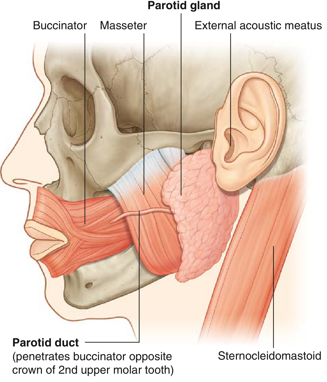
- Located outside the oral cavity in a triangular space bounded by the sternocleidomastoid (behind), the ramus of the mandible (in front), and the external acoustic meatus/zygomatic arch (superiorly).
- Contains the external carotid artery, retromandibular vein, and the facial nerve [VII].
- Parotid (Stensen's) duct passes anteriorly over the masseter, then pierces the buccinator to open opposite the crown of the 2nd upper molar tooth.
Submandibular Gland
- Hook-shaped; the large superficial part lies below the mylohyoid in the submandibular triangle; the deep part hooks around the posterior border of mylohyoid into the floor of the oral cavity.
- Submandibular (Wharton's) duct runs forward and opens on the sublingual caruncle beside the frenulum.
- The lingual nerve loops beneath the submandibular duct (crossing lateral then medial) before entering the tongue.
Sublingual Gland (smallest)
- Located on the floor of the oral cavity beneath the tongue mucosa, resting on the mylohyoid muscle.
- Drains via multiple small ducts (ducts of Rivinus), some joining Wharton's duct, others opening directly onto the sublingual fold.
Blood Supply
- Upper oral cavity: branches of the maxillary artery (greater palatine, posterior superior alveolar, infraorbital arteries)
- Lower oral cavity and tongue: lingual artery (branch of external carotid); inferior alveolar artery (branch of maxillary)
- Venous drainage follows the arterial supply into the internal jugular vein.
Lymphatic Drainage
- Tip of tongue and lower lip → submental nodes → deep cervical nodes
- Body of tongue, floor of mouth, cheeks → submandibular nodes → deep cervical nodes
- Posterior tongue → directly to deep cervical (jugulodigastric) nodes
- Upper lip and cheek → parotid and buccal nodes → submandibular nodes
Functions of the Oral Cavity
- Digestion - mechanical processing of food (mastication), lubrication and initial chemical digestion by saliva (amylase, lipase).
- Deglutition - tongue shapes a bolus and propels it through the oropharyngeal isthmus.
- Speech - the tongue, lips, teeth, and palate modify sounds produced by the larynx.
- Breathing - the oral cavity can substitute for the nasal airway (pharynx is a shared food-air pathway).
- Taste and sensation - taste buds on papillae and somatosensory receptors throughout the mucosa.
Gray's Anatomy for Students, pp. 1227-1248
This is a shared conversation. Sign in to Orris to start your own chat.