
Hey, Otis. I want you to teach me the clinical features. Of pneumonia Give me history points and, uh... Investigations. Treatment. Complications and its differential diagnosis and how to differentiate it from other its differentials.
Reading File
Now let me search the library across all the required topics in parallel.
Finding Sources
Finding Sources
Good - I have key sources. Now let me read the most comprehensive pneumonia sections in parallel - Goldman-Cecil, Rosen's EM, and Fishman's for clinical features, investigations, treatment, complications, and differential diagnosis.
Reading File
Reading File
Reading File
Excellent content found. Now let me read more - specifically the Goldman-Cecil chapter on CAP for treatment + complications, and continue reading the Rosen's section for treatment and differentials.
Reading File
Reading File
Very rich content. The Rosen's file has extensive coverage. Let me continue reading the Rosen's treatment section and also search for CAP treatment guidelines and complications.
Reading File
Finding Sources
Excellent - lots of content found in Rosen's (treatment section) and Fishman's. Let me now read:
- The Rosen's treatment and disposition section (continues from offset 3790)
- Fishman's CAP treatment section for IDSA/ATS guidelines
Reading File
Reading File
I have great content now. Let me get the treatment/antibiotic section from Rosen's and complications from Fishman's.
Reading File
Reading File
Now let me get the antibiotic treatment recommendations and complications from Rosen's management section.
Reading File
I now have comprehensive data covering all topics. Let me also quickly fetch the chest X-ray images from Rosen's to include visuals in the answer.
Loading Image
Loading Image
I now have everything I need to write a thorough, well-structured answer. Let me compile it all.
Pneumonia - Complete Clinical Guide
(Sources: Rosen's Emergency Medicine 9e, Fishman's Pulmonary Diseases & Disorders, Goldman-Cecil Medicine)
1. HISTORY POINTS
When taking a history from a patient with suspected pneumonia, the key components are:
Presenting Symptoms
- Cough - productive of purulent, rust-colored (pneumococcal), blood-streaked (streptococcal/TB), or currant jelly sputum (Klebsiella)
- Fever and chills - abrupt single shaking chill strongly suggests pneumococcal pneumonia
- Dyspnea / shortness of breath
- Pleuritic chest pain - sharp, worsened by breathing; suggests lobar pneumonia
- Hemoptysis - raises concern for MRSA, TB, or malignancy
Associated Symptoms to Ask About
- Headache, malaise, myalgias - viral prodrome or Mycoplasma
- Diarrhea + confusion + hyponatremia - classic for Legionella
- Non-productive cough + gradual onset + younger patient - Mycoplasma ("walking pneumonia")
- Confusion or acute change in mental status - especially in elderly; may be the only presenting symptom
- Abdominal or back pain - lower lobe pneumonia can present this way
Epidemiological History (Critical)
- Setting of acquisition: community-acquired (CAP), healthcare-associated, hospital-acquired, or ventilator-associated
- Recent hospitalization or long-term care facility (risk for resistant organisms: Klebsiella, Pseudomonas, MRSA)
- Geographic exposures: travel to endemic areas (histoplasmosis - Mississippi Valley; coccidioidomycosis - American Southwest)
- Animal exposures: birds (Chlamydophila psittaci), farm animals (Coxiella burnetii - Q fever), bats (histoplasma)
- Recent influenza illness - raises strong suspicion for secondary S. aureus/MRSA pneumonia
Host Factors (Risk History)
- Asplenia, sickle cell disease, HIV, multiple myeloma, agammaglobulinemia - increased risk of pneumococcal bacteremia
- COPD - colonized with S. pneumoniae, H. influenzae, Moraxella catarrhalis
- Cystic fibrosis - prone to Pseudomonas and S. aureus
- Immunosuppression (chemotherapy, transplant, HIV with CD4 <200) - adds CMV, PCP (P. jirovecii), Aspergillus, Cryptococcus
- IV drug use, alcoholism, undomiciled status - raises TB risk
- Recent antibiotic use (in last 3 months) - raises likelihood of drug-resistant organisms
- Swallowing dysfunction, impaired consciousness - aspiration risk
2. CLINICAL FEATURES (Examination)
Typical Pneumonia (Pyogenic Bacteria - e.g., S. pneumoniae)
- High fever (>38.5°C), tachycardia, tachypnoea
- Signs of consolidation: dullness to percussion, bronchial breathing, increased tactile fremitus
- Crackles (crepitations) over involved area
- Bronchophony, egophony, whispered pectoriloquy - (note: poor inter-rater reliability despite being "classic")
- Pleural rub if pleuritis is present
Atypical Pneumonia (Mycoplasma, Chlamydophila, Legionella)
- More gradual, "flu-like" onset
- Non-productive cough, lower degree of fever
- Absence of rigors, absent consolidation signs
- Extra-pulmonary features: rash (erythema multiforme in Mycoplasma), bullous myringitis, diarrhea (Legionella)
- Patient appears less toxic despite abnormal CXR
Important: The "typical vs atypical" distinction cannot reliably differentiate pathogens on clinical grounds alone and does NOT reliably guide treatment decisions. - Rosen's Emergency Medicine 9e
In Elderly / Debilitated Patients
- Classic symptoms may be absent
- May present with only acute confusion, functional decline, or sepsis without a clear respiratory syndrome
- More likely to have advanced disease at presentation
3. INVESTIGATIONS
Chest Imaging
Chest X-ray - Standard first-line imaging:
- Lobar consolidation + air bronchograms - typical pyogenic bacteria (S. pneumoniae, Klebsiella)
- Patchy/segmental infiltrates (bronchopneumonia) - S. pyogenes, Mycoplasma, viruses
- Dense lobar consolidate with bulging fissure ("lobar expansion") - classically Klebsiella
- Bilateral interstitial infiltrates - PCP, viral pneumonia
- Cavitation - Staphylococcus, Klebsiella, TB, anaerobes, MRSA
- Pleural effusion - common in pyogenic bacteria, Legionella, TB
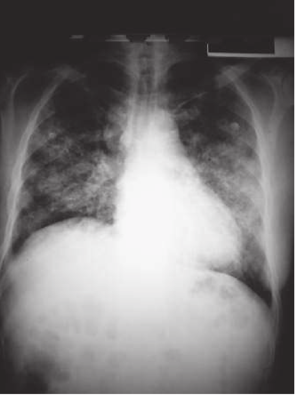
CT Chest - more sensitive than plain radiograph; detects infiltrates in ~33% of CXR-negative cases; indicated for elderly, immunocompromised, diagnostic uncertainty, or non-responding patients.
Lung Ultrasound - Highly sensitive and specific; consolidation appears as a hepatization pattern with air bronchograms and B-lines. Useful at bedside in ED.
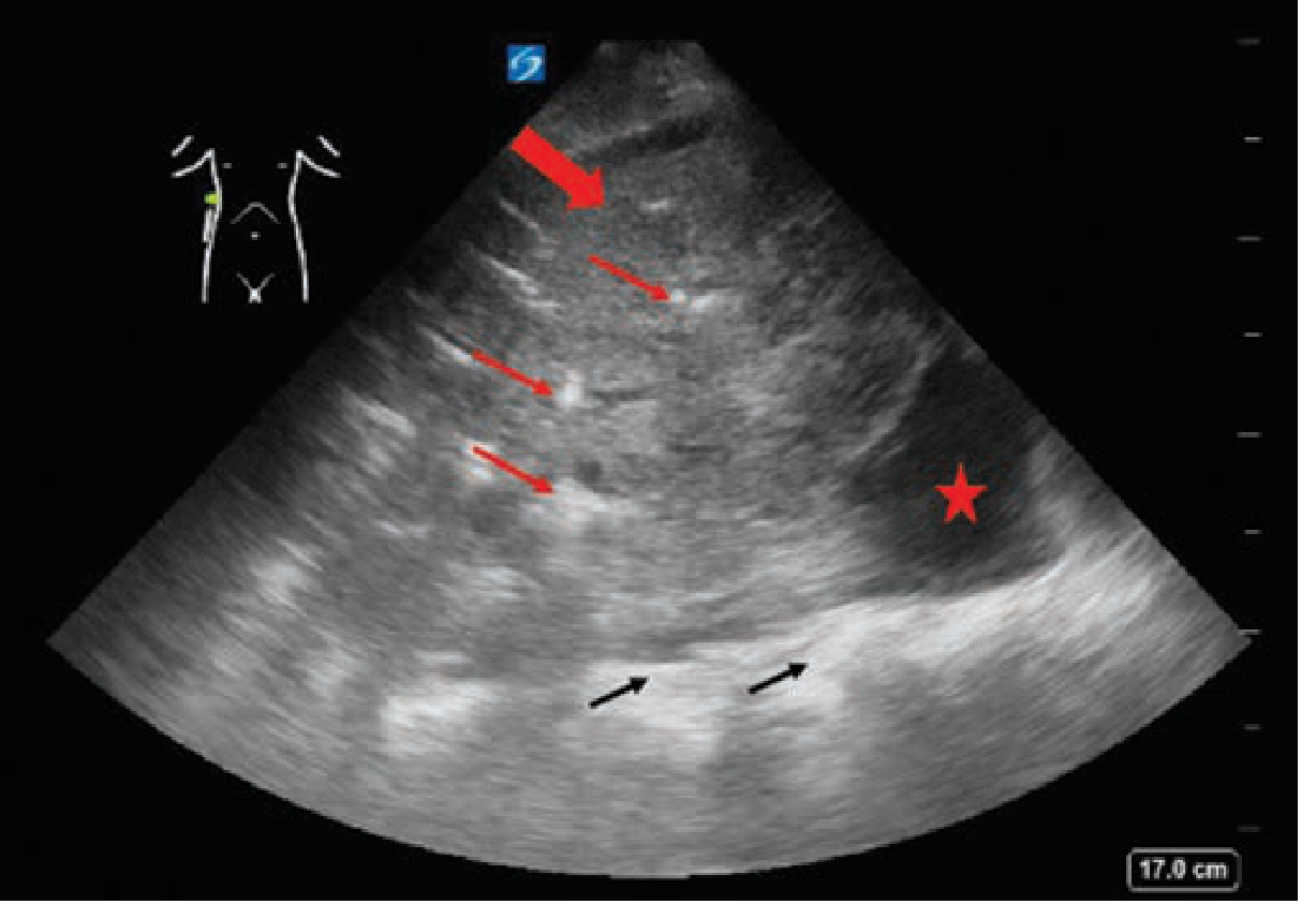
Blood Tests
- FBC (CBC) - WBC >15,000/mm³ suggests pyogenic bacterial cause; normal WBC more common with atypical/viral
- CRP, Procalcitonin - elevated in bacterial pneumonia; procalcitonin can help guide antibiotic duration
- Blood cultures - low yield (~5.8%) in immunocompetent outpatients; recommended for severe disease, ICU patients, suspected resistant organisms, or prior to antibiotics
- LFTs, U&E, Creatinine - for severity scoring and organ dysfunction
- Arterial Blood Gas (ABG) - if SpO2 <94% or severe disease; assess hypoxemia, hypocapnia (seen in PCP)
- LDH - elevated in PCP pneumonia; useful in HIV patients
Microbiology
- Sputum Gram stain + culture - recommended for severe disease or if resistant pathogens are suspected; requires <10 squamous epithelial cells and >25 WBCs per high-power field for an adequate specimen; obtain BEFORE antibiotics
- Urinary antigen tests:
- Legionella pneumophila serogroup 1 - sensitivity ~74%, specificity ~99%
- S. pneumoniae - sensitivity ~74%, specificity ~96%
- PCR/Multiplex panel (e.g., BioFire) - rapidly identifies bacteria and viruses; not affected by prior antibiotics; especially useful in outbreak settings
- Serology - C. pneumoniae, Legionella, fungi; useful retrospectively with paired titres, limited acute utility
Severity Scoring
CURB-65 (preferred for simplicity):
| Criterion | Points |
|---|---|
| Confusion | 1 |
| Urea (BUN) >20 mg/dL | 1 |
| Respiratory rate ≥30/min | 1 |
| BP <90 systolic or ≤60 diastolic | 1 |
| Age ≥65 | 1 |
- Score 0-1: Outpatient treatment
- Score 2: Consider hospital admission
- Score ≥3: Hospitalize; consider ICU
PSI (Pneumonia Severity Index) - 20-variable scoring system; Classes IV-V are high risk requiring admission; more precise but less practical at bedside.
4. TREATMENT
Site of Care Decision
Use CURB-65 or PSI, combined with clinical judgment. Factors beyond the score that favor admission include: inability to maintain oral intake, lack of home support, severe hypoxemia, and rapidly progressive disease.
Outpatient Treatment (CAP)
| Clinical Setting | Preferred Regimen |
|---|---|
| Previously healthy, no antibiotics in last 3 months | Amoxicillin 1g PO TID x 7 days OR Doxycycline 100mg PO BID x 7 days |
| Comorbidities OR recent antibiotic use | Amoxicillin-clavulanate 875/125mg TID + Azithromycin 500mg day 1 then 250mg days 2-5 |
| Alternative for comorbid patients | Levofloxacin 750mg PO daily x 5 days OR Moxifloxacin 400mg daily x 7-14 days |
Macrolide monotherapy is no longer recommended unless local pneumococcal resistance is <25%.
Inpatient (Non-ICU)
- Beta-lactam (ceftriaxone 1-2g IV daily) + macrolide (azithromycin)
- OR respiratory fluoroquinolone monotherapy (levofloxacin 750mg IV/PO daily)
ICU / Severe CAP
- Beta-lactam + macrolide (preferred combination)
- OR beta-lactam + respiratory fluoroquinolone
- Consider MRSA coverage (vancomycin, linezolid, ceftaroline) if: necrotizing pneumonia, preceding influenza, known MRSA contact, severe sepsis in young healthy adult
Special Pathogens
- Legionella: fluoroquinolone or azithromycin
- PCP (HIV/immunocompromised): TMP-SMX (co-trimoxazole) + corticosteroids if PaO2 <70 mmHg
- Aspiration pneumonia: only treat with antibiotics if signs of secondary bacterial infection develop >36 hours later; avoid prophylactic antibiotics
Duration
- Outpatient CAP: 5-7 days
- Hospitalized CAP: 5-7 days; no less than 5 days; can step down to oral once stable
- Reassess if no improvement in 48-72 hours
5. COMPLICATIONS
| Complication | Key Features |
|---|---|
| Parapneumonic effusion | Pleural fluid accumulates; send for pH, cell count, glucose, culture; pH <7.2 requires chest drain |
| Empyema | Frank pus in pleural space; common with S. pyogenes (30-40% of cases) and anaerobes; requires drainage + prolonged antibiotics |
| Lung abscess | Cavitation on CXR; anaerobes (aspiration), Klebsiella, MRSA; prolonged antibiotics needed |
| Bacteremia / Sepsis | More common with S. pneumoniae (especially in asplenic patients), S. pyogenes (10-15%), and gram-negatives |
| ARDS | Develops in severe pneumonia, particularly with Legionella, MRSA, and streptococcal toxic shock; 55% of streptococcal toxic shock patients |
| Respiratory failure | May require mechanical ventilation |
| Septic shock | Multi-organ dysfunction, metabolic acidosis; especially with gram-negatives and S. pyogenes |
| Organizing pneumonia | Incomplete resolution leading to fibrosis; seen with some viral and atypical pneumonias |
| Pericarditis / Myocarditis | Rare; seen with S. pneumoniae spread |
6. DIFFERENTIAL DIAGNOSIS - AND HOW TO DIFFERENTIATE
A. Pulmonary Tuberculosis (TB)
| Feature | Pneumonia | Tuberculosis |
|---|---|---|
| Onset | Acute (days) | Subacute-chronic (weeks-months) |
| Fever | Acute, high | Low-grade, evening |
| Cough | Purulent sputum | Chronic, blood-streaked |
| Night sweats | Uncommon | Classic feature |
| Weight loss | Mild if any | Prominent |
| CXR | Lower-lobe consolidation common | Upper lobe, cavitation, lymphadenopathy |
| Risk factors | Age, COPD, immunosuppression | Close contact, HIV, immigrant, homelessness |
| Response to antibiotics | Rapid (48-72h) | No response to standard antibiotics |
| Diagnosis | CXR, cultures | AFB smear, GeneXpert PCR, culture |
Always consider TB in any patient with HIV and respiratory complaints, and place them in respiratory isolation.
B. Congestive Heart Failure (CHF) - Acute Pulmonary Edema
| Feature | Pneumonia | CHF |
|---|---|---|
| Fever | Present | Absent (unless co-infection) |
| Cough | Purulent sputum | Pink frothy sputum |
| History | Respiratory infection prodrome | History of cardiac disease, orthopnoea, PND |
| JVP | Normal | Elevated |
| Oedema | Absent | Pedal oedema |
| CXR | Focal consolidation | Bilateral perihilar "bat-wing" opacification, Kerley B lines, cardiomegaly |
| BNP/proBNP | Normal | Markedly elevated |
| Response | Antibiotics | Diuretics, vasodilators |
| Lung US | Focal consolidation with air bronchograms | Bilateral diffuse B-lines |
C. Pulmonary Embolism (PE)
| Feature | Pneumonia | PE |
|---|---|---|
| Onset | Gradual | Sudden |
| Fever | Common | Usually absent (low-grade may occur) |
| Pleuritic chest pain | Present with pleuritis | Present |
| Haemoptysis | Uncommon (unless MRSA/TB) | Can occur (infarction) |
| DVT symptoms | Absent | Leg swelling/pain |
| CXR | Infiltrate / consolidation | Usually normal; Hampton hump, Westermark sign |
| CT | Consolidation | Filling defect in pulmonary arteries on CTPA |
| D-dimer | Can be elevated in both | Elevated in PE |
| Risk factors | Infection | Immobility, recent surgery, cancer, oral contraceptive |
D. Aspiration Pneumonitis vs Aspiration Pneumonia
| Feature | Aspiration Pneumonitis | Aspiration Pneumonia |
|---|---|---|
| Mechanism | Chemical injury from acidic gastric contents | Bacterial infection from aspiration of oropharyngeal flora |
| Onset | Immediate - within hours | Delayed >36 hours after aspiration |
| Sputum | Non-purulent, may clear | Purulent, foul-smelling |
| Fever | May appear early then resolve | Sustained, worsens |
| Antibiotics | Not recommended initially | Required |
| Location on CXR | Right lower lobe (RLL in supine) | Right lower lobe; can be bilateral |
E. Lung Cancer (or Post-obstructive Pneumonia)
| Feature | Pneumonia | Lung Cancer |
|---|---|---|
| Age | Any | Usually >40, smoker |
| Smoking history | Not required | Usually heavy smoker |
| Symptoms | Acute onset | Chronic cough, weight loss, haemoptysis |
| CXR resolution | Resolves in 4-6 weeks | Persistent mass/infiltrate |
| Key test | Follow-up CXR at 6 weeks | CT chest + bronchoscopy/biopsy |
Any pneumonia that fails to resolve on CXR after 6 weeks should prompt CT chest to rule out underlying malignancy.
F. Sarcoidosis / Hypersensitivity Pneumonitis
| Feature | Pneumonia | Sarcoidosis / HP |
|---|---|---|
| Onset | Acute | Subacute to chronic |
| Fever | High, acute | Low-grade or absent |
| Occupational exposure | Absent | Present in HP (farmer's lung, bird fancier's) |
| CXR | Lobar consolidation | Bilateral hilar lymphadenopathy (sarcoid); upper lobe reticulonodular (HP) |
| Response to antibiotics | Good | No response |
| Diagnosis | Culture, CXR | Bronchoalveolar lavage, biopsy |
Quick Memory Summary
PNEUMONIA
├── History: Cough + Fever + Purulent sputum + Pleuritic pain
│   Ask: Setting (CAP/HAP), Epidemiology, Host factors, Prodrome
├── Exam: Consolidation signs - Dull, ↑ Fremitus, Bronchial breath
├── Investigations:
│   ├── CXR (consolidation/infiltrate)
│   ├── FBC, U&E, CRP, Cultures
│   ├── Urine antigens (Legionella + Pneumococcal)
│   └── Severity: CURB-65
├── Treatment:
│   ├── Outpatient: Amoxicillin ± Doxycycline
│   ├── Inpatient: Ceftriaxone + Azithromycin (or Levofloxacin)
│   └── ICU: Add MRSA cover if indicated
├── Complications:
│   Effusion → Empyema → Abscess → Bacteremia → ARDS → MOF
└── Differentials:
    TB (subacute, upper lobe, night sweats)
    CHF (cardiomegaly, bat-wing, elevated BNP)
    PE (sudden, no fever, CTPA)
    Aspiration (timing, risk factors)
    Cancer (non-resolving infiltrate at 6 weeks)
Sources: Rosen's Emergency Medicine Concepts and Clinical Practice 9e (Ch. 62); Fishman's Pulmonary Diseases and Disorders 2-Volume Set (Ch. 128); Goldman-Cecil Medicine International Edition
This is a shared conversation. Sign in to Orris to start your own chat.