
Midbrain anatomy
Reading File
Searching sources...
Finding Sources
Finding Sources
The best sources are Gray's Anatomy for Students (EXTERNAL + INTERNAL MIDBRAIN sections), Localization in Clinical Neurology (Anatomy of the Mesencephalon), and Neuroscience: Exploring the Brain. Let me read them all in parallel.
Reading File
Reading File
Reading File
Now let me read the INTERNAL MIDBRAIN section from Gray's Anatomy and more of the Localization in Clinical Neurology text, plus fetch key cross-section images.
Reading File
Reading File
Loading Image
Excellent - I have very rich content from Gray's Anatomy for Students and Localization in Clinical Neurology. Let me fetch the rostral midbrain cross-section image and read a bit more about mesencephalic syndromes.
Loading Image
Loading Image
Reading File
I now have comprehensive material from multiple authoritative textbooks. Let me compile the full answer.
Midbrain (Mesencephalon) Anatomy
Overview
The midbrain is the shortest segment of the brainstem, approximately 2 cm in length. It connects the forebrain (diencephalon) superiorly to the pons inferiorly. Its rostral boundary is defined by the plane of the superior colliculi and mammillary bodies; its caudal boundary lies just caudal to the inferior colliculi.
The midbrain is divided into three fundamental regions along its dorsoventral axis:
- Tectum (dorsal) - the "roof"
- Tegmentum (central)
- Cerebral peduncles / Crus cerebri (ventral)
The cerebral aqueduct (of Sylvius) runs through the centre of the midbrain, connecting the third and fourth ventricles.
External Surface
Anterior (Ventral) Surface
- A deep midline depression called the interpeduncular fossa lies between the two crura cerebri (crus cerebri) - the prominent anterior bulges.
- CN III (oculomotor nerve) emerges from the medial aspect of each crus, from the interpeduncular fossa, and runs anteriorly between the posterior cerebral and superior cerebellar arteries.
Posterior (Dorsal) Surface - the Tectum
- The tectum bears the corpora quadrigemina - four rounded eminences arranged as two superior and two inferior colliculi.
- CN IV (trochlear nerve) emerges from the dorsal surface just below the inferior colliculi, decussates immediately, and wraps around the lateral aspect of the midbrain - it is the only cranial nerve to exit from the dorsal surface of the brainstem.
- Superior brachium and inferior brachium project anterolaterally from the respective colliculi toward the medial and lateral geniculate bodies of the thalamus.
(eFig. 9.25, Gray's Anatomy for Students)
Internal Structure - Cross Sections
Two key cross-sectional levels are recognised:
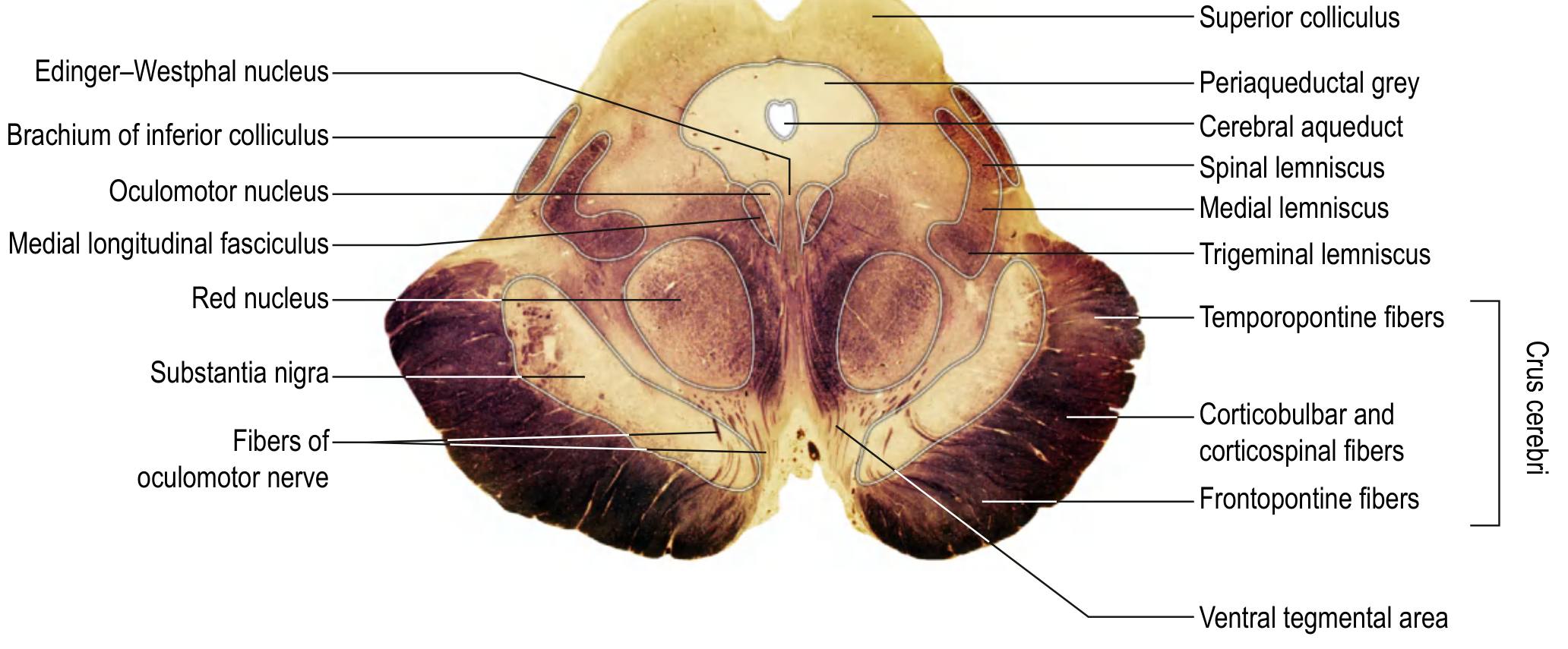
Rostral Midbrain (Level of Superior Colliculus)
Tectum: Superior colliculi - relay centres for visual reflexes (e.g., pupillary light reflex, optokinetic tracking).
Tegmentum contains:
| Structure | Location / Notes |
|---|---|
| Oculomotor nucleus (CN III) | Ventral to periaqueductal gray, posterior to MLF |
| Edinger-Westphal nucleus | Dorsal to CN III nucleus; preganglionic parasympathetic to pupil/ciliary muscle |
| Red nucleus | Large, spherical; source of rubrospinal tract; receives cerebellar and cortical input |
| Substantia nigra | Between tegmentum and crus cerebri; zona compacta (dopaminergic neurons, melanin-pigmented) + zona reticulata |
| Medial longitudinal fasciculus (MLF) | Near midline; coordinates conjugate gaze |
| Medial lemniscus | Ascending fine touch/proprioception |
| Spinothalamic tract (spinal lemniscus) | Ascending pain and temperature |
| Reticular formation | Arousal, autonomic modulation |
| Periaqueductal gray (PAG) | Surrounds aqueduct; pain modulation, defensive behaviors |
| Ventral tegmental area (VTA) | Source of mesolimbic/mesocortical dopamine pathways |
| Mesencephalic nucleus of CN V | Only sensory nucleus residing in the brainstem itself |
Cerebral peduncle (Crus cerebri) - medial to lateral:
- Medial 1/5 - frontopontine (corticopontine) fibers
- Middle 3/5 - corticospinal + corticobulbar fibers (somatotopically arranged: arm fibers medial, leg fibers lateral, trunk in between)
- Lateral 1/5 - temporopontine fibers
(Rostral midbrain section - Gray's Anatomy for Students, eFig. 9.28)
Caudal Midbrain (Level of Inferior Colliculus)
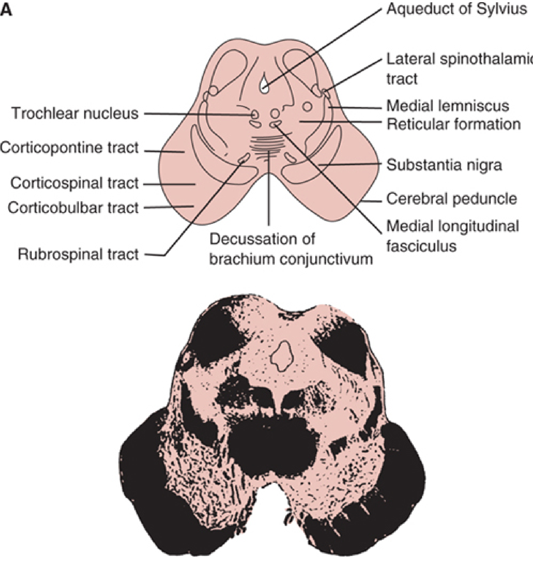
Tectum: Inferior colliculi - relay stations for auditory pathways, connected to medial geniculate body via inferior brachium.
Tegmentum contains:
- Trochlear nucleus (CN IV) - located in the ventral periaqueductal gray
- Decussation of the brachium conjunctivum (superior cerebellar peduncle decussates here on its way to the contralateral red nucleus and thalamus)
- Substantia nigra (present at both levels)
- Medial lemniscus, spinothalamic tract, MLF, reticular formation continue
(Caudal midbrain cross-section - Localization in Clinical Neurology, 8e, Fig. 15-5A)
Key Nuclei - Summary
| Nucleus | CN / Function | Location |
|---|---|---|
| Oculomotor (CN III) | Eye movement (superior, inferior, medial rectus; inferior oblique; levator palpebrae) | Rostral tegmentum, beneath superior colliculus |
| Edinger-Westphal (CN III para.) | Pupillary constriction, lens accommodation | Dorsal to CN III nucleus |
| Trochlear (CN IV) | Superior oblique muscle | Caudal tegmentum, beneath inferior colliculus |
| Red nucleus | Motor coordination (rubrospinal tract) | Tegmentum, rostral midbrain |
| Substantia nigra | Dopamine (nigrostriatal pathway) | Between tegmentum and crus cerebri |
| Mesencephalic nucleus CN V | Proprioception from jaw muscles | Lateral periaqueductal gray, full length of midbrain |
Important Tracts Passing Through the Midbrain
| Tract | Location | Function |
|---|---|---|
| Corticospinal/corticobulbar | Middle 3/5 crus cerebri | Voluntary motor |
| Corticopontine (fronto-, temporo-) | Medial + lateral 1/5 crus cerebri | Cortex → pons → cerebellum |
| Medial lemniscus | Tegmentum | Fine touch, vibration, proprioception (ascending) |
| Spinothalamic tract | Tegmentum (lateral) | Pain, temperature (ascending) |
| MLF | Paramedian tegmentum | Coordinates gaze (CN III/IV/VI) |
| Dentatorubrothalamic tract | Tegmentum | Cerebellum → red nucleus → thalamus |
| Posterior commissure | Roof, rostral midbrain | Connects superior colliculi; involved in pupillary light reflex |
Vascular Supply
The midbrain receives blood from multiple sources - Localization in Clinical Neurology, 8e:
- Paramedian vessels (from posterior cerebral artery origins): supply the medial peduncles and tegmentum, including CN III nucleus, red nucleus, and substantia nigra.
- Quadrigeminal arteries (from posterior cerebral arteries): supply the superior and inferior colliculi.
- Superior cerebellar arteries: branches to the cerebral peduncles and brachium conjunctivum.
- Posterior choroidal arteries: cerebral peduncles, lateral superior colliculi, thalamus.
- Anterior choroidal arteries (from internal carotid): contribute to cerebral peduncles.
Clinically Important Midbrain Syndromes
| Syndrome | Lesion Site | Features |
|---|---|---|
| Weber syndrome | Ventromedial midbrain (crus cerebri + CN III fascicle) | Ipsilateral CN III palsy (dilated pupil, ptosis, eye down-and-out) + contralateral hemiplegia |
| Benedikt syndrome | Midbrain tegmentum (red nucleus + CN III fascicle) | Ipsilateral CN III palsy + contralateral hemiataxia with intention tremor |
| Claude syndrome | Dorsal tegmentum (dorsal red nucleus + brachium conjunctivum + CN III) | Ipsilateral CN III palsy + contralateral cerebellar signs (ataxia, dysmetria) |
| Nothnagel syndrome | Dorsal midbrain | Unilateral or bilateral CN III palsy + contralateral cerebellar ataxia |
| Parinaud syndrome (dorsal midbrain / Sylvian aqueduct syndrome) | Rostral dorsal midbrain | Paralysis of upward gaze, large pupils with light-near dissociation, convergence-retraction nystagmus, Collier sign (lid retraction) |
(Localization in Clinical Neurology, 8e, p. 944-946 | Gray's Anatomy for Students, p. 1316-1318)
This is a shared conversation. Sign in to Orris to start your own chat.