
Left it fracture
Reading File
Left IT (iliotibial) band fracture likely refers to a left IT band syndrome or more probably a left tibial fracture. The query "Left it fracture" most likely means left tibia fracture. Let me search the library while also checking web sources.
Searching sources...
Finding Sources
Finding Sources
I need to clarify what "Left it fracture" means - this is ambiguous. Let me ask before spending more budget.
Asking for Preferences
Could you clarify what fracture you mean by 'Left it fracture'?
Left intertrochanteric (IT) hip fracture - a very common and clinically important injury, especially in elderly patients. Let me pull comprehensive information from the medical library.
Searching sources...
Finding Sources
Finding Sources
Searching PubMed
intertrochanteric hip fracture management
Good results. Now let me read the most relevant sections from the top orthopedic sources.
Reading File
Reading File
Reading File
Excellent - I now have rich content. Let me also fetch the key classification diagram images and read the Campbell's section for surgical details.
Loading Image
Finding Sources
Reading File
Reading File
I now have comprehensive information. Let me compose a thorough, well-structured answer on left intertrochanteric hip fractures.
Left Intertrochanteric (IT) Hip Fracture
Anatomy and Definition
An intertrochanteric fracture is an extracapsular hip fracture where the fracture line runs between the greater and lesser trochanters of the proximal femur. Because it lies outside the hip joint capsule, the blood supply to the femoral head is generally preserved - unlike intracapsular (femoral neck) fractures.
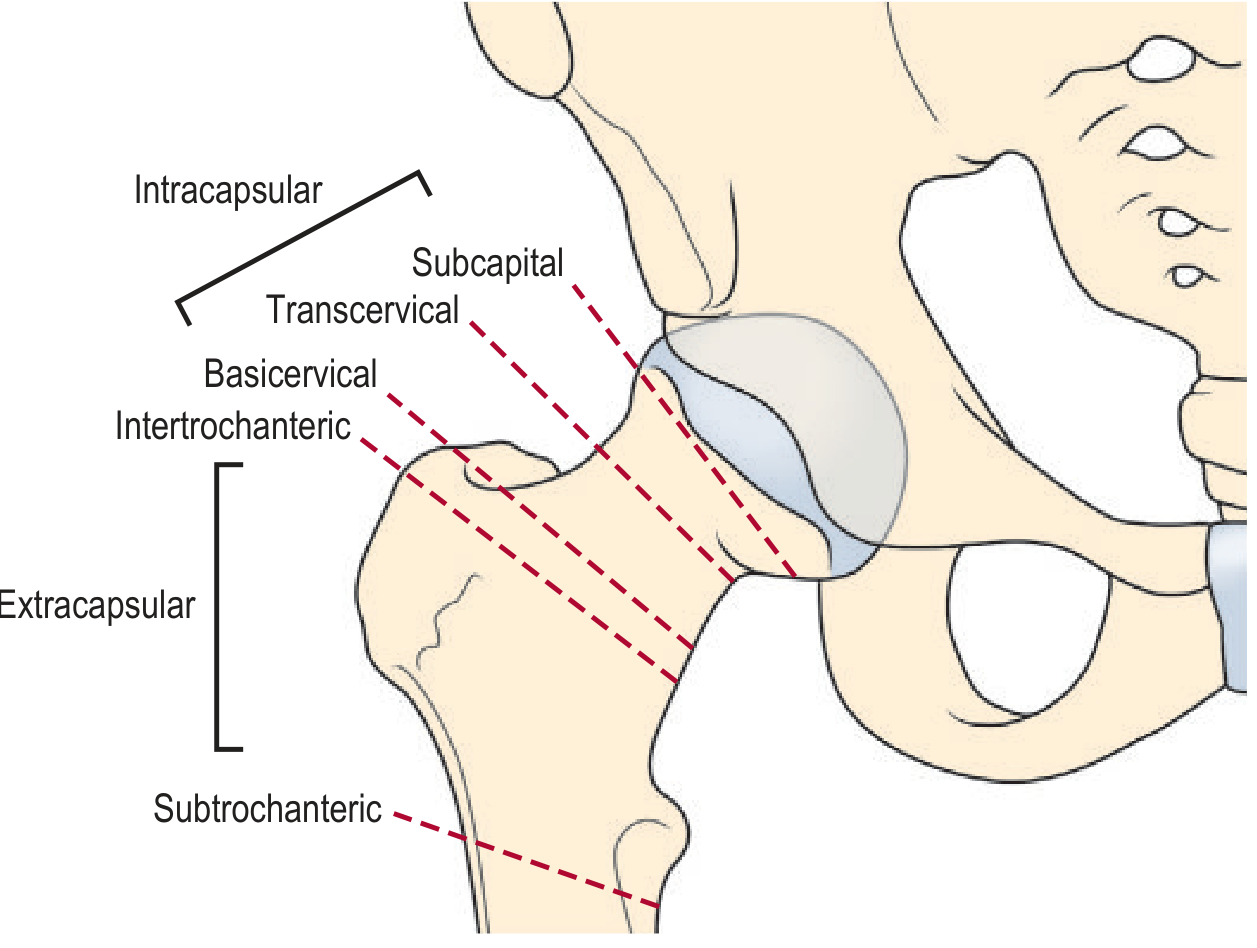
Epidemiology
- Most common in elderly patients (osteoporotic bone), typically after a low-energy fall
- Affects women more than men due to greater osteoporosis prevalence
- 30% mortality in the first year after injury
- Life expectancy returns to near-normal in patients who survive beyond one year
- Only ~50% of patients regain their pre-fracture level of ambulation
Classification
The most widely accepted system is the OTA/AO classification (incorporated into national hip fracture guidelines in Europe and the USA):
| Type | Description | Stability |
|---|---|---|
| 31-A1 | Simple 2-part fracture, intact lateral wall, fracture runs between trochanters | Relatively stable |
| 31-A2 | Comminuted (3-4 parts), lateral wall essentially intact, lesser trochanter detached | Less stable |
| 31-A3 | Reverse oblique or horizontal fracture line extending to lateral cortex | Most unstable |
The lateral wall integrity is a key surgical planning parameter - its compromise significantly affects implant choice.
An older but still-used system classifies by number of parts:
- 2-part: femoral head fragment + shaft fragment
- 3-part: one trochanter also fractured (greater or lesser)
- 4-part: both trochanters fractured - highest instability
(Rosen's Emergency Medicine, 8th ed.; Rockwood & Green's Fractures in Adults, 10th ed., 2025)
Clinical Presentation
- Pain in the hip/groin after a fall or trauma
- Leg shortened and externally rotated - due to the pull of the iliopsoas muscle on the lesser trochanter
- Inability to bear weight
- Associated injuries are frequently missed (distal radius, proximal humerus, ribs, spine) because the hip fracture draws attention away
Diagnosis
- AP pelvis + lateral hip X-ray - confirms diagnosis in most cases; include both hips on AP for comparison
- ~15% of fractures are subtle on plain film - look for interruption of trabecular lines or a sclerotic line
- MRI - gold standard for occult fractures (if X-ray inconclusive)
- CT - practical alternative when MRI is unavailable
- Consider internal rotation view if diagnosis remains uncertain
Management
Surgical (Definitive)
The vast majority require internal fixation. Surgery should be urgent (within 24-48 hours) but not emergent - the patient should be medically optimized first. Operating on the day of injury increases mortality risk; however, delays beyond 48 hours worsen 1-year mortality.
Implant Options:
| Implant | Indication |
|---|---|
| Dynamic Hip Screw (DHS) / Sliding hip screw + plate | Stable A1 fractures (intact lateral wall) |
| Cephalomedullary (intramedullary) nail (e.g., PFNA, InterTAN, Gamma nail) | Unstable A2/A3 fractures, compromised lateral wall, reverse oblique patterns |
A 2025 systematic review and meta-analysis (PMID 40130944) comparing intramedullary devices in the elderly confirms intramedullary nailing is the preferred approach for unstable patterns.
Hemiarthroplasty is a salvage option for severely comminuted/unstable fractures in elderly patients where internal fixation is unlikely to hold.
Pre-operative / Medical Management
- Skin or skeletal traction - not routinely recommended; comfort measures suffice
- IV fluids for resuscitation (dehydration is common)
- Adequate analgesia (nerve blocks are increasingly used)
- Multidisciplinary team optimization (cardiology, medicine) reduces 1-year mortality
- VTE prophylaxis: low molecular weight heparin or direct oral anticoagulants for 7-28 days
Post-operative
- Early mobilization is the goal - reduces pneumonia, DVT, pressure ulcers
- Weight bearing as tolerated from day 1 post-op in most cases
- Zoledronic acid (yearly IV infusion, started within 90 days of fracture repair) reduces new fractures and overall mortality
Complications
| Complication | Notes |
|---|---|
| Mortality | Up to 30% at 1 year |
| Implant failure / cut-out | More common with unstable fractures or poor reduction |
| Nonunion / malunion | Rare vs. femoral neck fractures (good blood supply) |
| Infection | Perioperative antibiotic prophylaxis required |
| DVT/PE | Mandatory anticoagulation prophylaxis |
| Functional decline | ~50% fail to return to baseline ambulation |
Key Differences from Femoral Neck Fracture
| Feature | Intertrochanteric | Femoral Neck |
|---|---|---|
| Location | Extracapsular | Intracapsular |
| Blood supply to head | Preserved | At risk (AVN risk) |
| Bone type | Cancellous (heals well) | Cortical (slower healing) |
| Primary treatment | Internal fixation | Arthroplasty (displaced) or fixation (undisplaced) |
| Nonunion rate | Low | Higher |
Sources:
- Rosen's Emergency Medicine, 10th ed.
- Rockwood and Green's Fractures in Adults, 10th ed., 2025
- Campbell's Operative Orthopaedics, 15th ed., 2026
- Grainger & Allison's Diagnostic Radiology
- Recent evidence: Intramedullary devices meta-analysis, JBJS Reviews, 2025 - PMID 40130944
This is a shared conversation. Sign in to Orris to start your own chat.