
Aneurysm and its types photos
Reading File
An aneurysm is a localized dilation or bulge in a blood vessel wall, typically an artery. The main types are saccular (berry), fusiform, pseudoaneurysm, mycotic, and dissecting aneurysms, each with distinct morphology and clinical significance.
Searching sources...
Finding Sources
Searching the Web
aneurysm types saccular fusiform dissecting diagram medical images
Finding Sources
Loading Image
Loading Image
Now let me also fetch the textbook content on aneurysm definitions and get textbook images if available.
Reading File
Loading Image
Now let me also fetch the textbook's dissecting aneurysm image:
Loading Image
Here is a complete visual and textbook-based guide to Aneurysm and its Types.
Aneurysm - Definition
A localized dilatation of a segment of the arterial wall caused by weakening of the vessel wall. Aneurysms are classified into True, False (Pseudoaneurysm), and Arteriovenous types - and further by shape.
- S Das A Manual on Clinical Surgery, 13th Ed.
Diagram 1 - Morphological Types Overview

Diagram 2 - True vs False vs Dissection (with vessel wall layers)
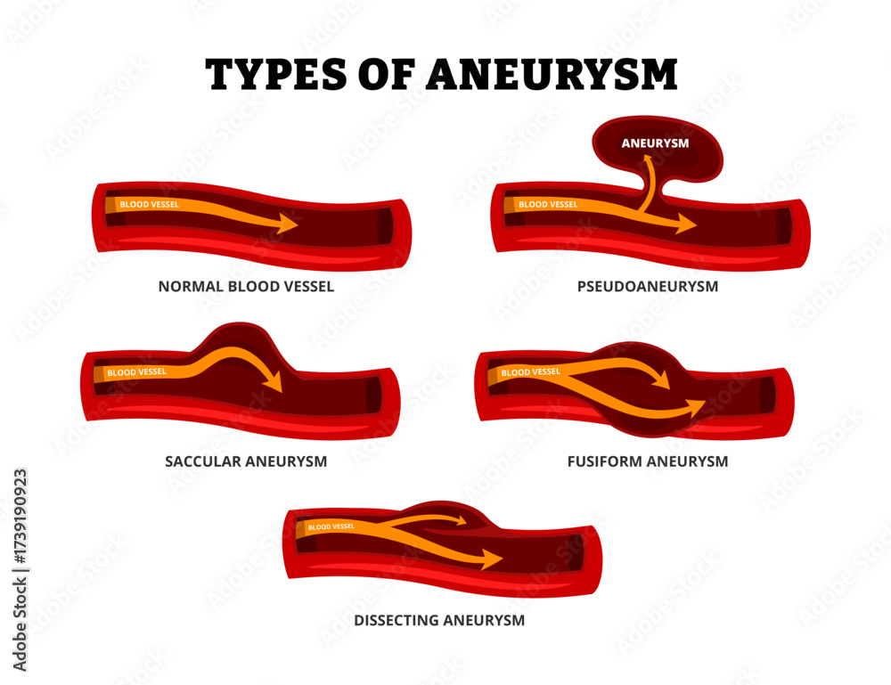
Diagram 3 - Fusiform, Pseudoaneurysm, Saccular, Dissecting, Ruptured

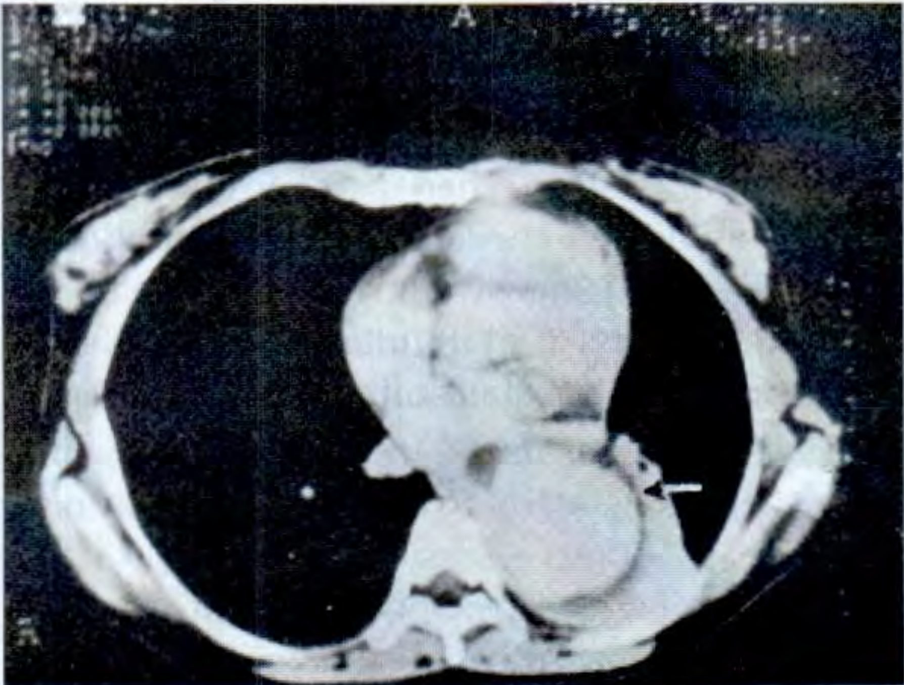
CT Scan - Dissecting Aneurysm of the Thoracic Aorta
(From S Das A Manual on Clinical Surgery - Fig 6.18)
Types Explained (Textbook Definitions)
1. True Aneurysm
Contains all three layers of the arterial wall (intima, media, adventitia). Subtypes by shape:
| Type | Description |
|---|---|
| Fusiform | Uniform expansion of the entire circumference of the arterial wall - the most common type. The vessel bulges symmetrically on all sides. |
| Saccular (Berry) | Expansion of only part of the circumference - forms a localized outpouching or "sac." Often traumatic; the classic form in the Circle of Willis is called a Berry aneurysm. |
| Dissecting | The tunica intima ruptures (usually beneath an atheromatous plaque) and blood is forced between the inner and outer layers of the tunica media, splitting the wall. |
2. False Aneurysm (Pseudoaneurysm)
- The wall of the sac is made of only a single layer of fibrous tissue (not true vessel wall layers).
- Blood leaks outside the vessel and is contained by surrounding connective tissue.
- Usually caused by trauma, infection, or iatrogenic injury.
3. Arteriovenous Aneurysm (AV Fistula)
- An abnormal communication between an artery and a vein, forming a pulsatile swelling.
- Can be congenital (cirsoid aneurysm) or acquired (traumatic).
4. Mycotic Aneurysm
- Caused by infection (bacterial, despite the misleading "mycotic" name).
- Weakening of the wall by an infected embolus or direct infection.
By Location
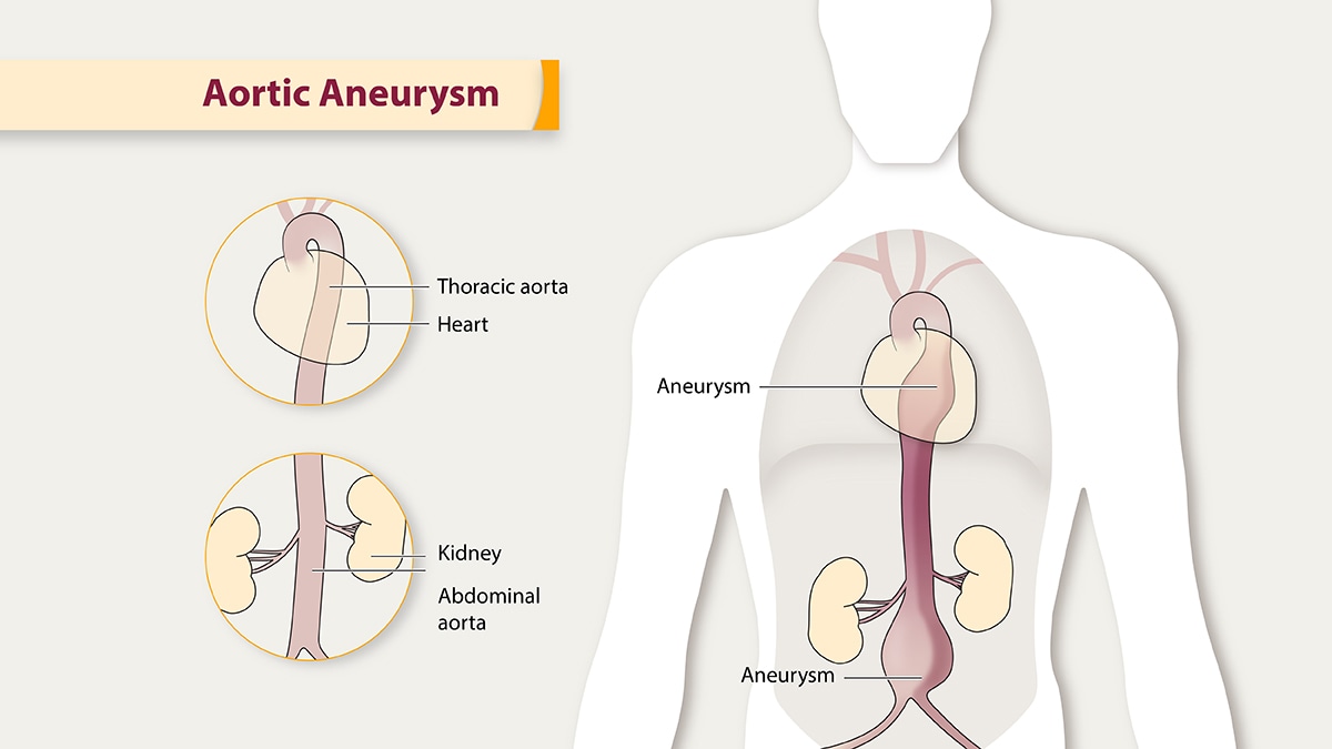
- Abdominal Aortic Aneurysm (AAA) - most common; fusiform type predominates.
- Intracranial (Cerebral) Aneurysm - saccular/berry type; typically at Circle of Willis branch points.
- Thoracic Aortic Aneurysm - often dissecting in hypertensive/Marfan patients.
- Peripheral - popliteal and femoral arteries most common peripherally.
Sources: S Das A Manual on Clinical Surgery, 13th Ed. | Fuster & Hurst's The Heart, 15th Ed. | Robbins & Cotran Pathologic Basis of Disease | Sabiston Textbook of Surgery
By location diagrams
Searching for aneurysm-by-location diagrams from the textbook library and web sources.
Searching sources...
Finding Sources
Searching the Web
aneurysm by location diagram aortic cerebral popliteal femoral medical illustration
Finding Sources
Loading Image
Loading Image
Loading Image
Loading Image
Searching the Web
abdominal aortic aneurysm thoracic aortic aneurysm location diagram body illustration
Loading Image
Loading Image
Finding Sources
Loading Image
Searching the Web
aneurysm sites body human full anatomy diagram brain aorta peripheral splenic renal
Loading Image
Loading Image
Here is a complete visual guide to Aneurysms by Location.
1. Aneurysms in the Body - Overview (Saccular, Fusiform, Pseudoaneurysm at different sites)

2. Aortic Aneurysm - Thoracic vs Abdominal Locations (CDC)
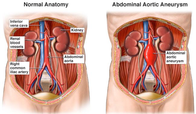
3. Abdominal Aortic Aneurysm (AAA) - Detailed Anatomy
Comparison of normal abdominal aorta vs AAA, showing relationship to kidneys, iliac arteries, and inferior vena cava:
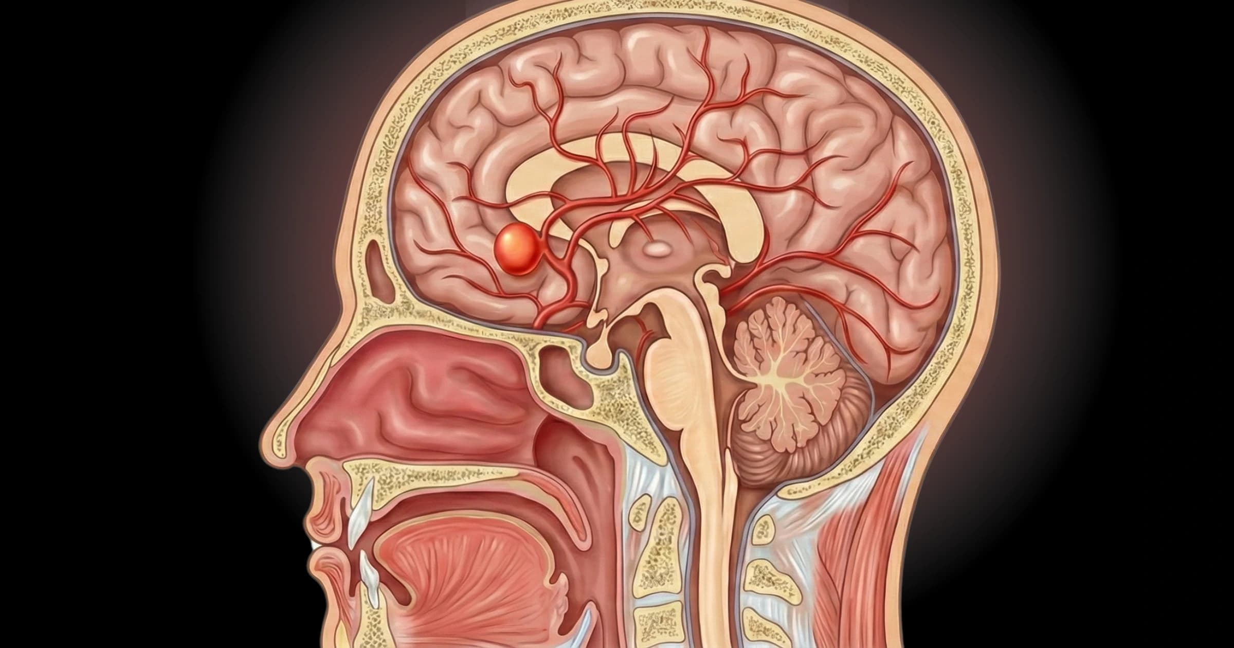
4. Cerebral (Brain) Aneurysm - Saccular at Circle of Willis
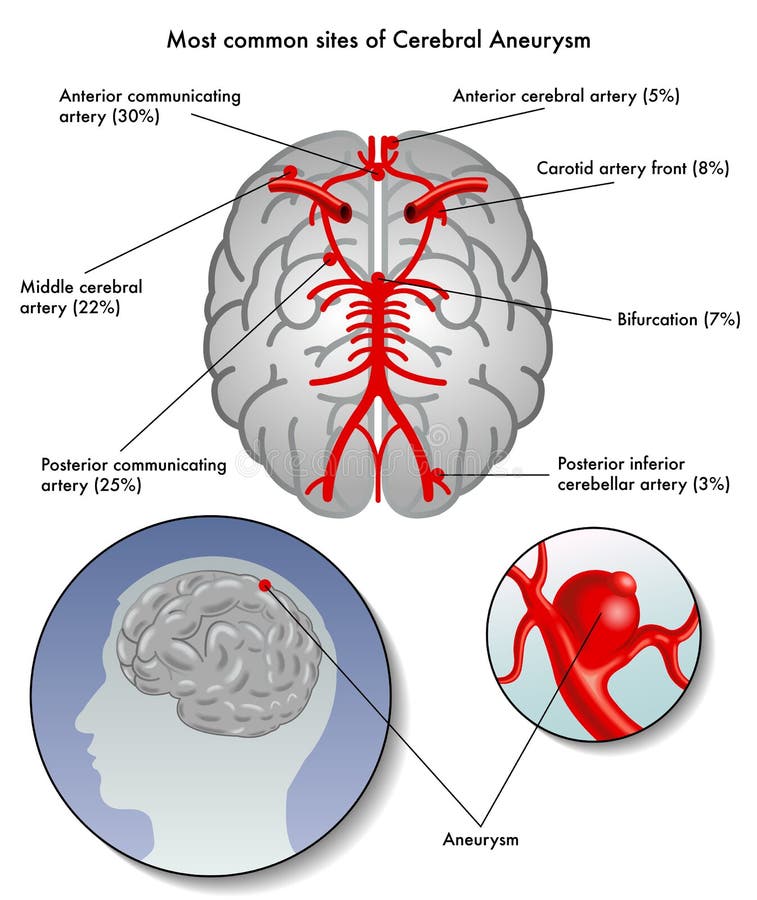
5. Most Common Sites of Cerebral Aneurysm (with % frequency)
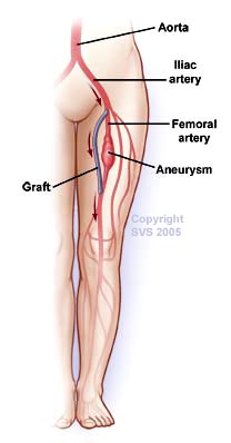
6. Peripheral Aneurysm - Femoral Artery Location
Summary Table - Aneurysm by Location
| Location | Type | Key Features |
|---|---|---|
| Abdominal Aorta | Fusiform (most common) | 95% occur infrarenal (below renal arteries); risk of rupture |
| Thoracic Aorta | Fusiform / Dissecting | Associated with hypertension, Marfan syndrome, bicuspid aortic valve |
| Cerebral (Circle of Willis) | Saccular / Berry | At arterial branch points; rupture causes subarachnoid hemorrhage |
| Popliteal Artery | Fusiform | Most common peripheral aneurysm; risk of thrombosis/embolism |
| Femoral Artery | Fusiform | 2nd most common peripheral; often bilateral |
| Splenic Artery | Saccular | Most common visceral aneurysm; risk increased in pregnancy |
| Renal Artery | Saccular | May cause hypertension; risk of dissection |
| Carotid Artery | Fusiform / Saccular | Neck mass; risk of stroke/TIA |
| Mesenteric Artery | Saccular | Visceral aneurysm; risk of bowel ischemia on rupture |
This is a shared conversation. Sign in to Orris to start your own chat.