
Endometrial hyperplasia for mbbs final year
"endometrial hyperplasia"[MeSH Terms] AND management
endometrial hyperplasia histology glands stroma microscopy
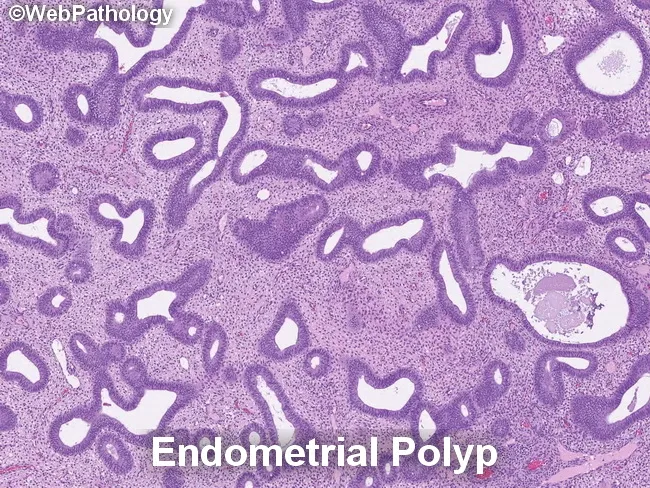
Histology – Light microscopy of paraffin-embedded endometrial tissue, stained with Hematoxylin and Eosin. The specimen demonstrates a polypoid lesion arising from the endometrium within the uterine cavity. Glandular architecture shows a spectrum from crowded, tightly packed endometrial glands to adjacent, less crowded glands embedded in a fibrous stromal background. The crowded glands are variably irregular in size and contour, with some shaping that mimics adenomatous crowding. The stromal component is relatively compact or fibrotic, containing thick-walled, dilated blood vessels. Glands generally orient parallel to the surface epithelium, a pattern characteristic of polyps, with occasional cystically dilated glands. There is no overt cytologic atypia or mitotic abnormality evident in the epithelial cells at this magnification. The background stroma lacks neoplastic infiltration and shows minimal inflammatory change. The combination of polypoid architecture, gland crowding confined to a focal lesion, and preserved noncrowded glands distinguishes endometrial polyps from diffuse endometrial hyperplasia. If crowded glands displayed nuclear enlargement, pseudostratification, or loss of polarity, the differential would broaden to atypical endometrial hyperplasia or carcinoma in situ; thus, this histology argues against EH with atypicality. Clinically, findings correlate with abnormal uterine bleeding; management includes hysteroscopic polypectomy with follow-up for recurrence or progression risk, and patient education.
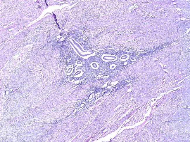
Comprehensive Description: This is a high-magnification light microscopy image of uterine tissue illustrating adenomyosis. The specimen is a myometrial section in which ectopic endometrial glands and stroma are embedded within the myometrium, below the endomyometrial junction. The pathognomonic feature is endometrial glands at least approximately 2.5 mm (100x field) deep from the junction, accompanied by relative myometrial hypertrophy and fibromuscular stroma. In the field, glands may be inactive or hormonally responsive, with basally located nuclei and stromal decidual-type changes possible depending on cyclic hormones. Occasionally, glands are scant or absent, which can complicate recognition and raise the differential with low-grade endometrial stromal sarcoma; careful assessment of architecture and invasion depth is required. The surrounding myometrium often shows hyperplasia or smooth muscle proliferation with separation of gland clusters by fibrous or edematous stroma. This histologic appearance corresponds to clinical symptoms such as dysmenorrhea, menorrhagia, and pelvic pain, and aids differentiation from fibroids or normal endometrium. The image demonstrates key features including ectopic glands within myometrium, myometrial thinning and thickening, and gland-to-stroma ratios. Clinically, adenomyosis is associated with uterine enlargement and pain; radiologic MRI findings may show thickened junctional zone, but histology confirms diagnosis. Image is educational for pathology, gynecology, and research.
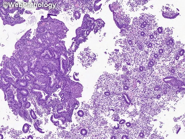
Imaging modality: Light microscopy of a hematoxylin and eosin stained endometrial tissue section, showing features used to diagnose atypical endometrial hyperplasia/endometrioid intraepithelial neoplasia (AEH/EIN). The specimen is endometrium within the uterine cavity; glandular epithelium with stromal backdrop. The histology demonstrates dense glands with back-to-back arrangement, yielding a gland:stroma ratio greater than 1, consistent with AEH/EIN criteria. Glands are cytologically and architecturally atypical relative to adjacent non-atypical glands, with nuclear enlargement, hyperchromasia, and irregular contours. The focus typically exceeds 1 mm in a single linear dimension and commonly comprises more than five to ten glands; multiple foci should not be pooled to satisfy the size criterion. Squamous morular areas are not part of the AEH/EIN assessment and are excluded from this ratio. Artifacts such as gland telescoping or artifactual displacement may create transient density increases but lack true cytologic atypia and architectural disorder. Benign mimics and invasion must be excluded; no myometrial invasion is visible at this plane. This image serves as a reference for diagnosing premalignant endometrial lesions, guiding differential diagnosis (benign hyperplasia without atypia, endometrial carcinoma, adenocarcinoma in situ), and informing clinical management decisions, including surveillance, risk stratification, and potential treatment strategies in gynecologic oncology.
Endometrial Hyperplasia - MBBS Final Year
Definition
- Robbins, Cotran & Kumar Pathologic Basis of Disease
- Berek & Novak's Gynecology
Etiopathogenesis
- Obesity - peripheral conversion of androgens to estrogens in adipose tissue
- Polycystic ovarian syndrome (PCOS) - chronic anovulation
- Functioning granulosa-theca cell tumors of the ovary (estrogen-secreting)
- Anovulatory cycles (perimenopause)
- Exogenous estrogen therapy without progestin (HRT)
- Excessive ovarian cortical function (cortical stromal hyperplasia)
- Late menopause, nulliparity
- PTEN tumor suppressor gene mutation is the key molecular alteration - found in >20% of endometrial hyperplasias (both with and without atypia) and 30-80% of endometrial carcinomas
- PTEN encodes a lipid phosphatase that negatively regulates the PI3K/AKT growth pathway; loss of PTEN leads to overactive PI3K/AKT signaling
- PTEN mutations also enhance estrogen receptor activity, creating a positive feedback loop
- Patients with Cowden syndrome (germline PTEN mutations) have a high incidence of endometrial carcinoma
Classification
WHO Classification (still widely used by pathologists)
| Type | Architecture | Cytologic Atypia | Progression to Cancer |
|---|---|---|---|
| Simple hyperplasia (cystic, without atypia) | Dilated/cystic glands, mild crowding | Absent | ~1% |
| Complex hyperplasia (adenomatous, without atypia) | Crowded, complex budding glands | Absent | ~3% |
| Simple atypical hyperplasia | Mild crowding | Present | ~8% |
| Complex atypical hyperplasia | Back-to-back crowded glands | Present | ~29% |
Modern WHO / EIN Classification (recommended since 2015 by ACOG + SGO)
| Class | Nomenclature | Treatment |
|---|---|---|
| Benign | Benign endometrial hyperplasia | Medical |
| Premalignant | EIN (Endometrial Intraepithelial Neoplasia) - replaces atypical hyperplasia | Surgical vs. medical |
| Malignant | Endometrial adenocarcinoma, endometrioid, well-differentiated | Surgical ± staging |
Key fact: 40-50% of women with atypical hyperplasia/EIN have concurrent occult carcinoma detected at hysterectomy.
Morphology (Histopathology)
Hyperplasia without Atypia
- Cardinal feature: increased gland-to-stroma ratio
- Glands vary in size and shape, may be dilated (cystic)
- Back-to-back glands may occur focally but some intervening stroma is retained
- No nuclear atypia
- Rarely progresses to adenocarcinoma (1-3%)
- May evolve into cystic atrophy when estrogen is withdrawn
Atypical Hyperplasia (EIN)
- Complex patterns of proliferating glands with nuclear atypia
- Glands are commonly back-to-back with complex branching outlines
- Individual cells are rounded with loss of normal perpendicular orientation to basement membrane
- Nuclei show: open (vesicular) chromatin, conspicuous nucleoli, irregular chromatin clumping, nuclear enlargement, increased N:C ratio, loss of polarity
- Clonal outgrowths with numerous genetic alterations
- Morphologically overlaps considerably with well-differentiated endometrioid adenocarcinoma - accurate distinction may require hysterectomy
Histology Images
Clinical Features
- Abnormal uterine bleeding (AUB) - the most common presentation
- Premenopausal: menorrhagia, intermenstrual bleeding
- Postmenopausal: any vaginal bleeding (always investigate)
- May be associated with estrogen-producing ovarian tumors
- Asymptomatic in some cases (incidental finding)
Diagnosis
- Office endometrial aspiration biopsy - accepted first step in evaluating a woman with AUB or suspected endometrial pathology
- Transvaginal ultrasound (TVUS) - endometrial thickness >4 mm postmenopausal warrants biopsy
- Hysteroscopy + directed biopsy - gold standard for visualization
- D&C (Dilation & Curettage) - when office biopsy is inconclusive
- MRI - used as surrogate staging in fertility-preservation candidates to evaluate myometrial invasion
Management
Algorithm
Endometrial hyperplasia
        |
  ------+------
  |            |
Without atypia  With atypia (EIN)
  |                    |
Progestin          Desires fertility?
therapy            /          \
(oral/LNG-IUS)   Yes          No
                  |            |
           Progestin    HYSTERECTOMY
           therapy      (definitive)
           + 3-monthly
           biopsy
Hyperplasia WITHOUT Atypia
- Progestin therapy (oral or LNG-IUS/Mirena - preferred)
- Options: Medroxyprogesterone acetate (MPA), norethisterone
- LNG-IUS (levonorgestrel intrauterine system) - most effective local delivery, higher regression rates
- Endometrial biopsy every 3-6 months to assess response
- Most regress (74%) or remain stable (18%) with treatment
Atypical Hyperplasia (EIN)
-
Hysterectomy - definitive treatment (also rules out concurrent carcinoma)
- Postmenopausal women: recommended as standard
- Women no longer desiring fertility: hysterectomy recommended
-
Fertility-sparing medical therapy (selected cases):
- Indication: young women, well-differentiated lesion, no myometrial invasion on MRI, desires pregnancy
- Agent: Megestrol acetate 40-160 mg/day (continuous progestin therapy) - most reliable for reversing complex/atypical hyperplasia
- Duration: minimum 3-6 months
- Follow-up: endometrial biopsy every 3 months (due to undiagnosed cancer risk in 25% and high recurrence after progestin)
- Overall response rate ~76%; median time to regression ~12 weeks
- Recurrence rate ~24% among responders (most within 1 year)
- After successful pregnancy or failure to regress - hysterectomy
Key Points on Medical Treatment
- LNG-IUS has emerged as superior to oral progestins for non-atypical hyperplasia (2024 network meta-analysis, PMID 39312359)
- Metformin has been investigated as adjunct therapy, especially in obese/PCOS patients, but evidence remains insufficient to recommend routine use - (Cochrane Review 2024, PMID 38695827)
- Hysteroscopic resection in atypical hyperplasia/early cancer with progestin shows promising regression and pregnancy rates (systematic review 2024, PMID 38382917)
Relationship to Endometrial Carcinoma
| Feature | Endometrial Hyperplasia | Endometrial Carcinoma |
|---|---|---|
| Type 1 (Endometrioid) | Precursor: atypical hyperplasia/EIN | Estrogen-related, perimenopausal |
| Molecular | PTEN mutation (>20%) | PTEN mutation (30-80%) |
| Prognosis | Risk of progression | Well-differentiated, favorable |
- Hyperplasia without atypia: 1-3% progress to carcinoma
- Complex atypical hyperplasia: 29% progress; 25-43% have concurrent occult carcinoma at hysterectomy
High-Yield Summary for Finals
| Feature | Without Atypia | With Atypia (EIN) |
|---|---|---|
| Gland-stroma ratio | Increased | Increased + back-to-back |
| Nuclear atypia | Absent | Present |
| PTEN mutation | ~20% | ~20% (shared with carcinoma) |
| Malignant potential | 1-3% | 8-29% |
| Concurrent carcinoma at hysterectomy | Rare | 25-43% |
| Treatment | Progestin / LNG-IUS | Hysterectomy (preferred) |
- Unopposed estrogen = root cause
- PCOS + obesity = most common clinical setting
- PTEN mutation = molecular link to endometrial carcinoma
- LNG-IUS = preferred treatment for non-atypical hyperplasia
- 40-50% with atypical hyperplasia have concurrent carcinoma - always do hysterectomy if not desiring fertility