
Short note on localized acute alveolar osteomyelitis
Reading File
Now let me search the medical library for content on localized acute alveolar osteomyelitis.
Finding Sources
Finding Sources
Finding Sources
Finding Sources
Reading File
Finding Sources
Reading File
Finding Sources
I now have good content. Let me read the alveolar osteitis sections from Rosen's and Roberts & Hedges for clinical detail, and also check Scott-Brown's.
Reading File
Reading File
Reading File
Reading File
I now have comprehensive information. Let me also quickly fetch the dry socket image from Rosen's.
Loading Image
I now have all the information needed. Here is a complete short note on localized acute alveolar osteomyelitis.
Localized Acute Alveolar Osteomyelitis (Dry Socket / Alveolar Osteitis)
Definition
Localized acute alveolar osteomyelitis - more commonly known as alveolar osteitis or "dry socket" - is a localized inflammation of the alveolar bone that occurs when the blood clot normally present in the extraction socket becomes dislodged or dissolves prematurely, leaving bone exposed. It represents the most frequent painful complication following tooth extraction.
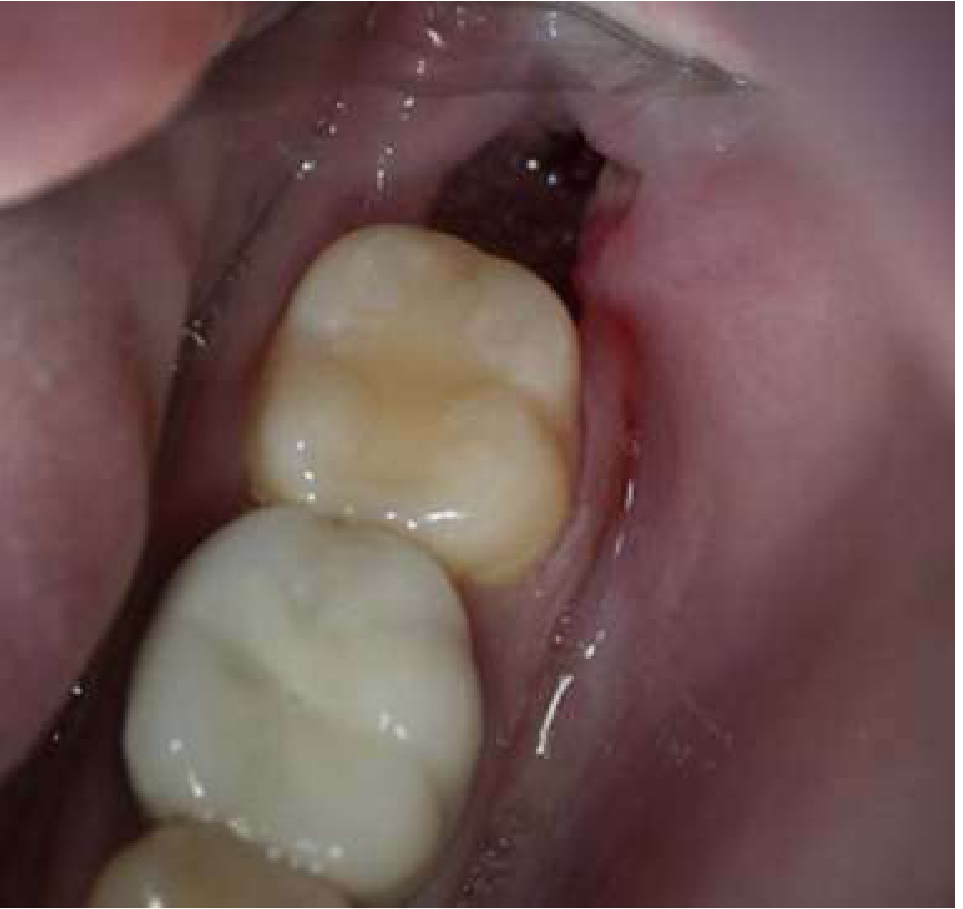
Alveolar osteitis: extraction socket devoid of clot with exposed bone - Rosen's Emergency Medicine
Pathophysiology
After a tooth extraction, a blood clot forms in the alveolar socket. This clot serves as a scaffold for healing and protects the underlying bone and nerve endings. If this clot is dislodged or fails to form, the bare alveolar bone becomes inflamed - producing intense pain from the exposed periosteum and bone marrow. It may be preceded by localized osteitis following pulpitis, which spreads through the root to the surrounding osseous tissue. If an alveolar abscess is not treated properly with antibiotics and drainage, osteomyelitis of the jaw may follow. - S. Das, Manual on Clinical Surgery, 13th Edition
Incidence
- Occurs in 2-5% of routine extractions
- Rises to 20-30% after removal of impacted mandibular third molars
- More common in the lower jaw (mandible) - the maxilla is rarely affected due to its rich anastomotic blood supply from vertical arteries - Rosen's Emergency Medicine
Predisposing Factors
- Excessive trauma during extraction
- Inadequate blood supply to the extraction site
- Pre-existing localized infection / pericoronitis
- Loss of clot from sucking, straw use, rinsing, or smoking (increased negative intraoral pressure)
- Foreign bodies remaining in the socket
- Use of oral contraceptives or corticosteroids
- Hormone replacement therapy
- Periodontal disease
- Familial tendency - Roberts & Hedges' Clinical Procedures in Emergency Medicine; Scott-Brown's Otorhinolaryngology
Clinical Features
- Onset: 2-5 days after tooth extraction (typically day 3-4)
- Pain: Moderate to severe, dull, constant, and aching - localized to the extraction site and often radiating to the ear
- Foul odor / bad taste in the mouth - in the absence of purulence or suppuration
- Duration: Can persist for 5-40 days if untreated
- Examination: Missing clot at the socket, exposed bare alveolar bone - NO significant surrounding edema, erythema, fluctuation, fever, leukocytosis, or lymphadenitis (which would suggest osteomyelitis)
Differential Diagnosis
| Feature | Alveolar Osteitis | Osteomyelitis / Infection |
|---|---|---|
| Clot | Absent | May be present |
| Swelling/erythema | Absent or minimal | Present |
| Purulence | Absent | Present |
| Fever/leukocytosis | Absent | Present |
| Lymphadenopathy | Absent | Present |
A dental nerve block provides instant relief in alveolar osteitis but not in osteomyelitis - this is a useful distinguishing feature. - Roberts & Hedges'
Treatment
- Nerve block - provides immediate pain relief; also confirms the diagnosis
- Irrigation - gently irrigate the socket with sterile saline to remove debris; do NOT curette the socket or remove residual clot (this exposes more bone and risks osteomyelitis)
- Socket packing - fill with medicated iodoform gauze impregnated with eugenol (oil of cloves) or a local anesthetic, packed to the level of the gingival cusps; change every 24-36 hours
- Dry Socket Paste (e.g., Sultan Healthcare) - a commercial alternative to eugenol-soaked gauze
- Analgesics - NSAIDs work better than narcotics for dry socket; short course of opioids (e.g., oxycodone 5 mg q4-6h) for severe pain
- Chlorhexidine rinses - shown to prevent alveolar osteitis; role in established disease is less clear
- Antibiotics - not routinely required once the socket is packed; prescribed at the discretion of the treating oral surgeon
The patient must be referred back to their dentist or oral surgeon the next day for follow-up, as socket packing must continue until healing is complete. - Roberts & Hedges'; Rosen's Emergency Medicine
Relation to Osteomyelitis of the Jaw
Alveolar osteitis represents localized alveolar bone inflammation. It can progress to true osteomyelitis if untreated. Osteomyelitis of the jaw presents in three forms:
- (i) Acute - seen in infants post-febrile illness (measles, scarlet fever)
- (ii) Subacute - the commonest form; follows dental infection or alveolar abscess; causes endarteritis, reduced blood supply, and bone necrosis; presents with pain, swelling, numbness of chin (inferior dental nerve compression)
- (iii) Chronic - follows dental infection or fractures; X-ray shows local osteitis, localized abscess (like Brodie's abscess), or sequestrum formation - S. Das, Manual on Clinical Surgery, 13th Edition
Primary herpes stomatitis with mode of transmission, clinical features with oral manifestation, histologic features, diagnosis and treatment
Finding Sources
Finding Sources
Finding Sources
Finding Sources
Excellent results. Let me now read the key sections in parallel from Robbins, Fitzpatrick's, and Tintinalli's.
Reading File
Reading File
Reading File
Reading File
Finding Sources
Reading File
Finding Sources
Finding Sources
Finding Sources
Reading File
Reading File
Reading File
I have comprehensive content. Let me now fetch the key clinical images and compile the full answer.
Loading Image
Loading Image
Excellent images obtained. I now have everything needed for a thorough, well-illustrated note. Here it is:
Primary Herpetic Stomatitis (Acute Herpetic Gingivostomatitis)
Definition
Primary herpetic stomatitis is the initial (first-time) symptomatic infection of the oral mucosa by Herpes Simplex Virus (HSV), most commonly HSV-1. It is the most common presentation of primary herpes infection in children and represents an acute, self-limited but often severe inflammatory condition affecting the gingiva and oral mucosa.
Aetiology / Causative Agent
- HSV-1 - responsible for ~90% of cases
- HSV-2 - can also cause primary orofacial infection, particularly in adolescents and young adults following genital-oral contact; clinically indistinguishable from HSV-1 disease but 120 times less likely to reactivate
- Fitzpatrick's Dermatology, 9e
Mode of Transmission
- Direct contact with infectious oral secretions - the primary route; typically transmitted from caregivers (parents, relatives) who may shed virus asymptomatically or during a cold sore
- Saliva - virus is shed in saliva even during asymptomatic periods; ~12% of oral swabs are PCR-positive for HSV in seropositive individuals
- Droplet / contact with infected mucocutaneous surfaces - kissing, shared utensils, or contact with vesicle fluid
- Self-inoculation - from oral lesions to fingers (herpetic whitlow) or eyes
- Incubation period: 2-12 days (mean 4 days)
- Important: Even asymptomatic individuals can transmit the virus; >70% of transmission occurs during asymptomatic shedding
- Tintinalli's Emergency Medicine; Fitzpatrick's Dermatology
Clinical Features
Epidemiology
- Peak age: 6 months to 5 years (most common in children 2-4 years)
- Can occur in older children and adults, though primary adult infection more commonly presents as pharyngitis
- Up to 90% of the population is infected before puberty, but only ~20% develop symptomatic disease
- Robbins Pathologic Basis of Disease; Tintinalli's
Systemic / General Features
- Abrupt onset of high fever (often the presenting complaint)
- Irritability and malaise
- Decreased oral intake and drooling - due to pain on swallowing
- Myalgias and flu-like symptoms
- Tender cervical lymphadenopathy
- Symptoms typically last up to 3 weeks but commonly resolve in less than 1 week
Oral Manifestations
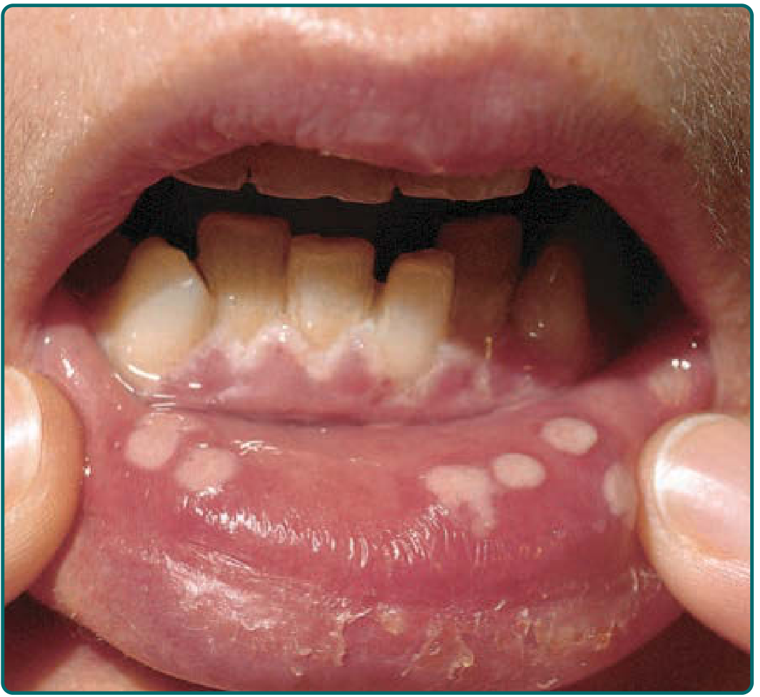
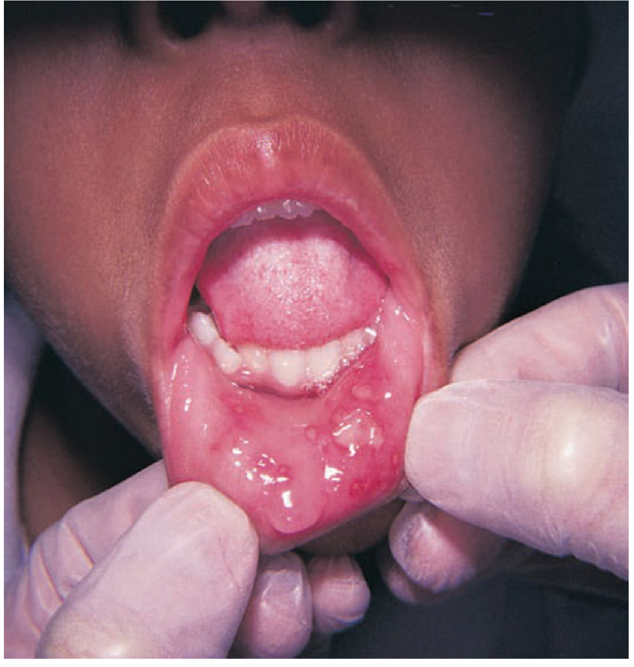
- Swollen, erythematous, friable gingiva - bright red, bleeding gingiva is often the earliest sign
- Vesicles - small fluid-filled blisters ranging from a few mm to large bullae, filled with clear serous fluid, appearing on oral mucosa
- Painful shallow ulcerations - vesicles rapidly rupture to form red-rimmed shallow ulcers with a yellowish-grey pseudomembranous base
- Distribution - hard and soft palate, tongue, buccal mucosa, gingiva, lips, and neighboring facial areas
- Halitosis due to secondary bacterial colonization of lesions
- Pain on eating/swallowing leading to dehydration in children
- Robbins Pathologic Basis of Disease; Fitzpatrick's Dermatology; Tintinalli's
Histologic Features
-
Intracellular and intercellular edema (spongiosis) within the epithelium
-
Acantholysis - dissolution of intercellular bridges creating clefts that become macroscopic vesicles
-
Ballooning degeneration of keratinocytes
-
Eosinophilic intranuclear viral inclusions (Cowdry type A bodies) - seen in individual epidermal cells at vesicle margins or free within vesicle fluid
-
Multinucleated giant cells (polykaryons) - several infected cells fuse together; these are the diagnostic hallmark seen on Tzanck smear
-
Vesicles are initially intraepidermal; roof may slough to leave ulcers
-
Underlying lamina propria shows acute inflammatory infiltrate
-
Robbins Pathologic Basis of Disease
Diagnosis
1. Clinical Diagnosis (Primary)
- Diagnosis is primarily clinical, based on the characteristic presentation of acute onset fever, irritable child with drooling, swollen red gingiva, and painful vesicles/ulcers in the oral cavity
2. Tzanck Smear
- Vesicle fluid from an unroofed lesion 24-48 hours old is smeared on a slide and stained
- Demonstrates multinucleated giant cells and eosinophilic intranuclear inclusions
- Rapid, bedside test - does not distinguish HSV-1 from HSV-2
3. Viral Culture
- The former gold standard
- Low sensitivity: only 7-25% recovery from lesions
- Swab the unroofed vesicle, place in viral transport medium
- Tintinalli's Emergency Medicine; Harriet Lane Handbook
4. PCR (Polymerase Chain Reaction)
- Most sensitive and specific - now preferred over culture
- Detects HSV DNA from vesicle fluid or lesion swab
- More accurate for lesions that have already partially healed
5. Serology
- Useful for confirming primary infection (seroconversion - rise in IgM / rising IgG titres)
- Not useful for acute diagnosis
6. Enzyme-linked immunosorbent assay (ELISA) / Direct Fluorescent Antibody (DFA)
-
Additional acceptable laboratory methods
-
Tintinalli's Emergency Medicine; Harriet Lane Handbook
Differential Diagnosis
| Condition | Distinguishing Feature |
|---|---|
| Aphthous stomatitis | No fever, no gingival involvement, no vesicles |
| Herpangina (Coxsackie A) | Lesions only in posterior oral cavity/pharynx; no gingival erythema |
| Hand, foot, and mouth disease | Concurrent skin lesions on hands and feet |
| Streptococcal pharyngitis | Exudative lesions in posterior pharynx, no anterior oral ulcers |
| Stevens-Johnson syndrome | Target lesions on skin, systemic mucosal involvement |
Treatment
Supportive Care (All Cases)
- Antipyretics - paracetamol/ibuprofen for fever
- Topical analgesics - lidocaine gel or viscous lidocaine (used cautiously in young children)
- Oral rehydration - particularly important in children who refuse oral intake
- IV fluids if severe dehydration from inadequate oral intake
- Topical acyclovir cream is ineffective for oral HSV
Antiviral Therapy
Antiviral therapy is most effective when started within 72-96 hours of symptom onset. Indicated when the child cannot drink or has significant pain.
Acyclovir (oral):
- Children: 80 mg/kg/day in 4 divided doses for 5-7 days (max 3200 mg/day) - American Academy of Pediatrics regimen
- Alternative dosing: 600 mg/m² four times daily for 10 days in children (Goodman & Gilman)
- Shortens duration of symptoms and viral shedding
Valacyclovir - generally preferred due to better bioavailability and less frequent dosing; 7-14 days
Immunocompromised patients:
- IV acyclovir 250 mg/m² every 8 h for 7 days
- Oral acyclovir 800 mg 5 times/day or valacyclovir 1000 mg twice daily for 5-10 days
Prevention
- Diligent hand washing among children and caregivers
- Avoid sharing utensils, cups, or close oral contact during active infection
- Children with active gingivostomatitis who cannot control oral secretions should be excluded from child care settings
- Tintinalli's Emergency Medicine; Goodman & Gilman's Pharmacological Basis of Therapeutics; Harriet Lane Handbook
Latency and Recurrence
After primary infection, the virus travels retrogradely along sensory nerve fibers to become latent in the trigeminal ganglion. Reactivation triggers include trauma, UV exposure, upper respiratory infection, menstruation, immunosuppression, emotional stress, and temperature extremes. Recurrent disease (herpes labialis / cold sores) is typically milder - small grouped vesicles at the vermilion border of the lips, resolving in 7-10 days. Lesions occur at or near the original site of inoculation. - Robbins Pathologic Basis of Disease
Short note on recurrent herpes labialis
Finding Sources
Finding Sources
Reading File
Reading File
Reading File
Good core content. Now let me get more clinical detail on staging, treatment, and fetch the Fitzpatrick's section on herpes labialis stages and images.
Finding Sources
Finding Sources
Reading File
Reading File
Reading File
Loading Image
Loading Image
All content and images gathered. Here is the complete short note:
Recurrent Herpes Labialis (Cold Sore / Fever Blister)
Definition
Recurrent herpes labialis is the reactivation of latent Herpes Simplex Virus type 1 (HSV-1) from the trigeminal ganglion, manifesting as recurrent clusters of vesicles - most characteristically at the vermilion border of the lips. It is the most common recurrent HSV infection in immunocompetent individuals and affects a significant proportion of the adult population.
Aetiology and Pathogenesis
Following primary herpetic gingivostomatitis, HSV-1 travels retrogradely along sensory nerve fibers and establishes lifelong latency in the trigeminal ganglion. Most adults harbor latent HSV-1. Upon triggering by various stimuli, the virus undergoes reactivation - virus particles travel anterograde along sensory axons back to the mucocutaneous site of original inoculation (or adjacent mucosa served by the same axon), where they replicate and produce new lesions.
- Robbins & Kumar Basic Pathology; Goldman-Cecil Medicine
Triggers for Reactivation
| Category | Examples |
|---|---|
| Physical | Fever, trauma, sunburn/UV irradiation, chapped lips, oral trauma |
| Hormonal | Menstruation, pregnancy |
| Psychological | Emotional stress, fatigue |
| Iatrogenic | Trigeminal nerve surgery, facial laser/chemical/abrasive cosmetic procedures, epidural morphine |
| Immune | Upper respiratory tract infections, immunosuppression |
| Miscellaneous | Season of year, allergies, temperature extremes |
- Fitzpatrick's Dermatology; Robbins & Kumar
Clinical Features
Site
- Vermilion border of the lips - two-thirds of labial lesions involve this border
- Lower lip (outer one-third) is the most commonly affected area
- Less common: nose, chin, cheek (<10% of cases), nasal orifices
- Immunocompetent patients rarely get recurrent intraoral lesions; when present, they appear on the anterior hard palate or attached gingiva (keratinized mucosa only)
Stages of Classical Lesion Progression
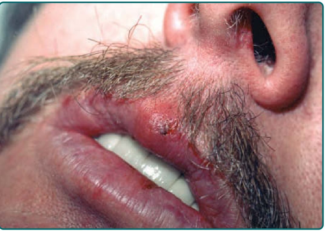
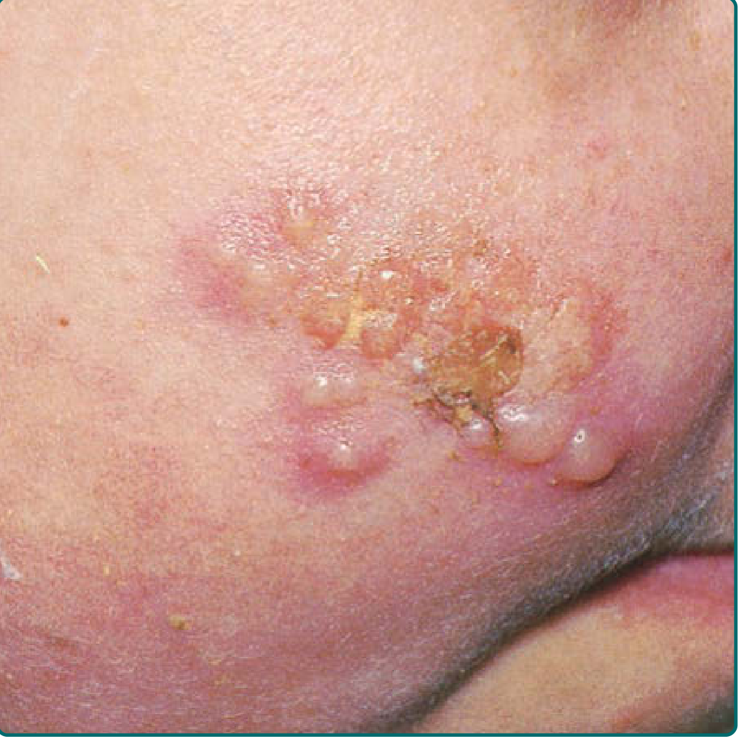
Stage 1 - Developmental Stage:
- Prodrome - pain, burning, tingling, or itching at the site of impending eruption; precedes lesions in 45-60% of episodes
- Erythema - localized redness appears at the site
- Papule - small raised firm lesion
Stage 2 - Disease Stage:
- Vesicle - small (1-3 mm) fluid-filled blisters in clusters; tense, clear, on an erythematous base
- Ulcer - vesicles rupture to form shallow, painful, red-rimmed ulcerations
- Hard crust - ulcers dry and crust over with a honey-colored scab
Stage 3 - Resolution Stage:
- Dry flaking - crust detaches
- Residual swelling - mild erythema and swelling may persist briefly
-
Lesions resolve in 5-15 days without treatment
-
Severity is highly variable - may range from prodromal symptoms alone (aborted episode) to extensive disease from severe sunburn
-
In patients with frequent recurrences, lesion location may shift slightly with each episode
-
Fitzpatrick's Dermatology
Histologic Features
-
Ballooning degeneration of keratinocytes with large eosinophilic intranuclear inclusions (Cowdry type A bodies)
-
Multinucleated polykaryons (giant cells) - adjacent infected cells fuse together
-
Intraepidermal vesicle formation from acantholysis and intercellular edema
-
Inflammatory infiltrate in the underlying dermis/lamina propria
-
Robbins & Kumar Basic Pathology
Comparison: Recurrent vs. Primary Infection
| Feature | Primary Herpetic Gingivostomatitis | Recurrent Herpes Labialis |
|---|---|---|
| First episode | Yes | No (reactivation) |
| Systemic symptoms | High fever, malaise, lymphadenopathy | Usually absent |
| Site | Diffuse oral mucosa, gingiva | Vermilion border, hard palate |
| Severity | Severe | Mild to moderate |
| Duration | Up to 3 weeks | 5-15 days |
| Intraoral involvement | Yes - keratinized and non-keratinized | Rarely (keratinized only) |
Diagnosis
- Primarily clinical - characteristic prodrome followed by grouped vesicles at the vermilion border in a known HSV-positive individual
- Tzanck smear - multinucleated giant cells from unroofed vesicle (does not distinguish HSV-1 from HSV-2)
- PCR - most sensitive; useful when diagnosis is in doubt
- Viral culture - from vesicle fluid (low sensitivity 7-25%)
- Serology - not useful for acute diagnosis in recurrent disease (IgG already positive)
Treatment
Treatment provides only modest benefit in immunocompetent hosts. It is effective only if started very early - ideally during the prodromal or early erythema stage.
Topical Agents
| Drug | Regimen | Benefit |
|---|---|---|
| Penciclovir 1% cream | Every 2 hours while awake for 4 days | FDA approved; reduces healing time (4.8 vs 5.5 days) and pain duration if started within 1 hour of symptoms |
| Docosanol 10% cream | 5 times a day at first sign of recurrence | OTC; FDA approved |
| Acyclovir 5%/10% cream | Not recommended | No benefit in recurrent herpes labialis |
Systemic (Episodic) Agents
| Drug | Regimen | Benefit |
|---|---|---|
| Valacyclovir | 2 g twice daily for 1 day (single-day therapy) | Decreases cold sore duration by ~1 day; must be started in prodrome |
| Famciclovir | 1500 mg as a single dose | Reduces healing time by ~2 days vs placebo |
| Acyclovir (oral) | 400 mg 5 times/day for 5 days | Marginal benefit only in the very earliest hours of outbreak |
| Acyclovir buccal tablet | 50 mg applied to upper gum once | Single-dose option above the incisor tooth |
Suppressive Therapy (for frequent recurrences)
- Oral acyclovir 400 mg twice daily - reduces recurrences; controversial benefit
- Valacyclovir suppressive therapy - more effective than episodic therapy for herpes labialis
- Perioperative prophylaxis (before facial laser resurfacing): famciclovir 125-250 mg twice daily or valacyclovir 500 mg twice daily starting 1-2 days before procedure
Adjunct Measures
-
Sunscreen - shown to prevent sun-triggered recurrences in some trials
-
No evidence for lysine supplementation or gammaglobulin
-
A "holiday" from suppressive therapy every year or so to reassess the continuing need
-
Fitzpatrick's Dermatology; Goodman & Gilman's Pharmacological Basis of Therapeutics
Complications
- Eczema herpeticum - widespread HSV superimposed on eczematous skin
- Herpetic whitlow - autoinoculation to fingers (especially in thumb-sucking children)
- Herpetic keratoconjunctivitis - corneal involvement; may cause dendritic ulcers and vision loss; requires ophthalmology referral and topical antivirals
- Erythema multiforme - HSV-triggered immune reaction
- Disseminated disease - in immunocompromised patients; may require IV acyclovir
Short note on Human immunodeficiency virus with a specific detail on oral lesions
Finding Sources
Finding Sources
Reading File
Finding Sources
Finding Sources
Finding Sources
Reading File
Reading File
Finding Sources
Finding Sources
Reading File
Finding Sources
Finding Sources
Finding Sources
Reading File
Reading File
Reading File
Loading Image
I now have comprehensive content from multiple authoritative sources. Here is the complete short note:
Human Immunodeficiency Virus (HIV) - with Oral Manifestations
Definition and Aetiology
Human Immunodeficiency Virus (HIV) is a lentivirus (family Retroviridae) that causes progressive destruction of cell-mediated immunity. There are two main types:
- HIV-1 - the predominant global cause; more virulent
- HIV-2 - mainly in West Africa; slower progression
HIV primarily infects and destroys CD4+ T lymphocytes, as well as macrophages and dendritic cells, leading eventually to Acquired Immune Deficiency Syndrome (AIDS). AIDS is defined by a CD4+ count below 200 cells/µL or the presence of an AIDS-defining illness.
Transmission
| Route | Detail |
|---|---|
| Sexual contact | Most common globally; unprotected vaginal, anal, or oral intercourse |
| Blood-borne | Needlestick injuries, sharing IV drug needles, contaminated blood products |
| Vertical (Mother to child) | Transplacental, during delivery (intrapartum), or breastfeeding |
| Occupational | Health care workers via needlestick (approximately 0.3% risk per exposure) |
HSV-2 co-infection significantly increases the risk of HIV acquisition and transmission. Asymptomatic viral shedding is the most common source of new HIV transmissions.
Pathogenesis
- HIV binds gp120 to the CD4 receptor and co-receptors (CCR5 or CXCR4) on T helper cells and macrophages
- Viral RNA is reverse-transcribed into DNA by reverse transcriptase
- Viral DNA integrates into the host genome as a provirus via integrase
- Progressive CD4+ T cell depletion leads to immune failure
- The virus replicates continuously throughout infection; even during the clinically asymptomatic phase, active viral replication occurs in lymphoid tissue
- When CD4 count falls below 200 cells/µL, opportunistic infections and AIDS-defining malignancies emerge
Clinical Stages
| Stage | Features | CD4 Count |
|---|---|---|
| Acute retroviral syndrome (2-4 weeks post-infection) | Flu-like illness: fever, sore throat, lymphadenopathy, rash, myalgia - often mistaken for infectious mononucleosis | Transient drop |
| Clinical latency / Chronic HIV | Asymptomatic for months to years; virus replicating in lymph nodes | Gradual decline (500-200 cells/µL) |
| Symptomatic HIV | Minor opportunistic infections, constitutional symptoms, oral manifestations begin | 200-500 cells/µL |
| AIDS | AIDS-defining illnesses - Pneumocystis pneumonia, CMV, Toxoplasma, Cryptococcus, Kaposi sarcoma, MAC | <200 cells/µL |
Oral Manifestations of HIV/AIDS
The oral cavity frequently reflects the state of HIV-related immune suppression. Since the advent of antiretroviral therapy (ART), the prevalence of oral lesions has decreased by 10-50%, but they remain clinically significant - particularly in patients with advanced or uncontrolled disease.
EC-Clearinghouse / WHO Classification of HIV-Associated Oral Lesions
Group 1 - Strongly associated with HIV:
- Oral candidiasis (pseudomembranous, erythematous, angular cheilitis)
- Oral hairy leukoplakia
- HIV-associated periodontal disease (linear gingival erythema, necrotizing ulcerative gingivitis/periodontitis)
- Kaposi sarcoma
- Non-Hodgkin lymphoma
Group 2 - Less commonly associated:
- Recurrent aphthous stomatitis
- Herpes simplex / zoster reactivation
- Oral warts (HPV)
- Salivary gland disease
Group 3 - Seen in HIV (less specific):
-
Bacterial infections (Actinomyces, Klebsiella, Cat-scratch disease)
-
Fungal infections (Cryptococcus, Histoplasma, Aspergillus)
-
CMV oral ulceration
-
Cummings Otolaryngology; K.J. Lee's Essential Otolaryngology
Detailed Oral Lesions
1. Oral Candidiasis (Most Common - ~90% of HIV/AIDS patients)
The most common oral cavity lesion in HIV/AIDS. Candida albicans is the primary pathogen. There are four types:
| Type | Appearance | Features |
|---|---|---|
| (a) Pseudomembranous (Thrush) | White curd-like / cottage cheese plaques | Can be scraped off with tongue depressor; erythematous raw base beneath; occurs on any mucosal surface |
| (b) Erythematous (Atrophic) | Red patches; loss of tongue papillations | Mild/moderately erythematous; palate, dorsal tongue, buccal mucosa |
| (c) Angular cheilitis | Cracking, fissuring, ulceration at oral commissure | Tender, erythematous; often bilateral |
| (d) Hyperplastic | Thick white plaque, cannot be scraped off | Rarest type; usually on buccal mucosa; may mimic leukoplakia |
Diagnosis: KOH preparation (pseudohyphae), Gram stain, PAS stain
Treatment:
- CD4 >200: Topical antifungals (nystatin mouthwash, clotrimazole troches)
- CD4 <200: Systemic fluconazole
- Note: Prolonged/repeated fluconazole use risks azole-resistant candidiasis
- K.J. Lee's Essential Otolaryngology; Cummings Otolaryngology
2. Oral Hairy Leukoplakia (OHL)
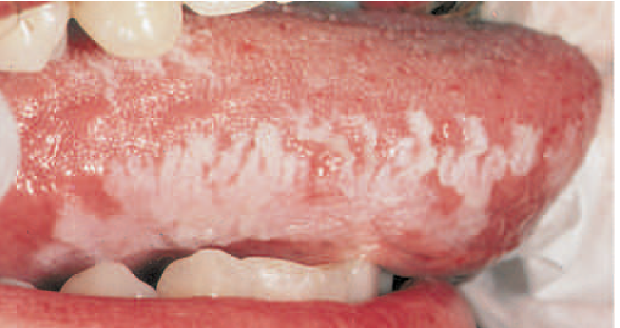
- Cause: Reactivation of Epstein-Barr virus (EBV) in HIV-infected individuals
- Appearance: White, corrugated (hair-like), hyperkeratotic plaques on the lateral aspects of the tongue (bilateral); cannot be scraped off
- Symptoms: Usually asymptomatic - an incidental finding
- Significance: Strongly associated with HIV; its presence in a previously healthy individual should prompt HIV workup; correlates with degree of immune suppression
- Differential diagnosis: Leukoplakia, lichen planus, carcinoma in situ, oral thrush
- No malignant potential
- Diagnosis: Biopsy confirming EBV presence
- Treatment: No specific treatment required if asymptomatic; resolves with ART; local options include podophyllin, gentian violet, cryotherapy
- Fitzpatrick's Dermatology; K.J. Lee's Essential Otolaryngology; Harrison's
3. HIV-Associated Periodontal Disease
Periodontal disease in HIV is staged in three categories:
(a) Linear Gingival Erythema (LGE):
- Fiery red band of marginal gingiva, out of proportion with the degree of dental plaque
- Prone to spontaneous hemorrhage
- Persists despite conventional plaque removal
- Closely associated with oral candidiasis
(b) Necrotizing Ulcerative Gingivitis / Periodontitis (NUG/NUP):
- Ulceration of the interdental papillae with gingival bleeding, pain, rapid tissue sloughing, and halitosis
- Progressive recession; may invade the alveolar bone
- Strongly associated with CD4 count below 200 cells/µL
- Its presence in an otherwise healthy individual should prompt HIV testing
(c) Necrotizing Ulcerative Stomatitis:
- Extension of necrotizing process into the mandibular or maxillary bone; the most severe form
Treatment:
- Topical chlorhexidine (0.1-0.2%) and povidone-iodine rinses
- Antifungals (co-existing candidiasis is common)
- Debridement of necrotic tissue and dental scaling
- Systemic clindamycin or metronidazole if no response
- Cummings Otolaryngology; K.J. Lee's Essential Otolaryngology
4. Kaposi Sarcoma (KS)
- Associated with HHV-8 (Human Herpesvirus 8)
- Typically affects the hard and soft palate; occasionally gingiva and buccal mucosa
- Appears as flat or raised, violaceous/purple, non-tender lesions
- May become nodular and ulcerate in advanced disease
- Diagnosis: Biopsy confirms spindle-cell vascular proliferation
- Treatment: Symptomatic; low-dose radiotherapy, intralesional vinblastine, or systemic chemotherapy when functionally problematic
- Cummings Otolaryngology; Scott-Brown's Otorhinolaryngology
5. Recurrent Aphthous Stomatitis (RAS) in HIV
- HIV patients develop unusually large, deep, and recalcitrant aphthous ulcers
- Very painful; cause severe odynophagia and substantial morbidity
- Treatment: Topical steroids, intralesional/systemic steroids, thalidomide, ART
- Biopsy confirms non-neoplastic, non-viral aetiology
- Scott-Brown's Otorhinolaryngology
6. Herpes Simplex Reactivation
- Presents as clusters of painful ulcers in the oral cavity
- In HIV/AIDS, lesions may be more extensive, persistent, and resistant to standard antiviral therapy
- Requires systemic acyclovir or valacyclovir; IV acyclovir for severe or refractory cases
7. CMV Oral Ulcers
- CMV co-infection occurs in ~90% of HIV cases
- Can cause painful oral ulcerations, often requiring biopsy to distinguish from other causes
- Also causes esophagitis and retinitis
8. Salivary Gland Disease
- Bilateral parotid swelling and xerostomia (dry mouth) can occur
- Dry mouth worsens dental caries and increases susceptibility to oral infections
Relationship Between Oral Lesions and CD4 Count
| CD4 Count (cells/µL) | Common Oral Manifestations |
|---|---|
| >500 | Oral warts, recurrent aphthous ulcers |
| 200-500 | Oral candidiasis, hairy leukoplakia, LGE |
| <200 | Severe candidiasis, necrotizing periodontitis, KS, NHL, CMV ulcers |
The presence of oral candidiasis in a patient on HAART is predictive of immune and virologic failure.
Treatment Overview
| Condition | Treatment |
|---|---|
| Oral candidiasis (mild) | Topical nystatin / clotrimazole |
| Oral candidiasis (severe / CD4 <200) | Oral/IV fluconazole |
| Hairy leukoplakia | No treatment / ART; podophyllin if desired |
| Periodontal disease | Chlorhexidine, debridement, antifungals, antibiotics |
| Kaposi sarcoma | Radiotherapy / vinblastine / chemotherapy |
| Aphthous ulcers | Topical/systemic steroids; thalidomide |
| All oral lesions | ART is the cornerstone - restores immune function and reduces oral manifestation burden |
- K.J. Lee's Essential Otolaryngology; Cummings Otolaryngology; Scott-Brown's; Fitzpatrick's Dermatology
This is a shared conversation. Sign in to Orris to start your own chat.