
Uterine factors in infertility
uterine causes of infertility hysteroscopy fibroids polyps
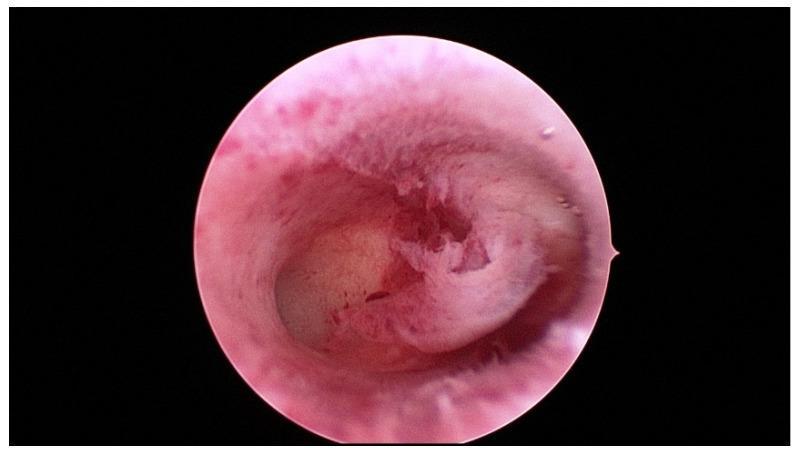
A diagnostic hysteroscopic photograph showing the interior of a human uterine cavity (cavum uteri). The image illustrates the endometrial lining, which appears pink and red with varying vascularity and thickness. In the center of the cavity, there is a prominent, raised area of endometrium, creating an irregular surface elevation against the surrounding uterine wall. The overall appearance of the cavity is described as inconspicuous, meaning it lacks obvious gross pathology like large submucosal fibroids, polyps, or significant synechiae. This visualization is characteristic of a standard diagnostic hysteroscopy used in gynecology to evaluate the uterine environment for infertility, recurrent miscarriage, or abnormal bleeding. The view captures the distal end of the uterine cavity, emphasizing the morphology and texture of the endometrial tissue under endoscopic magnification.
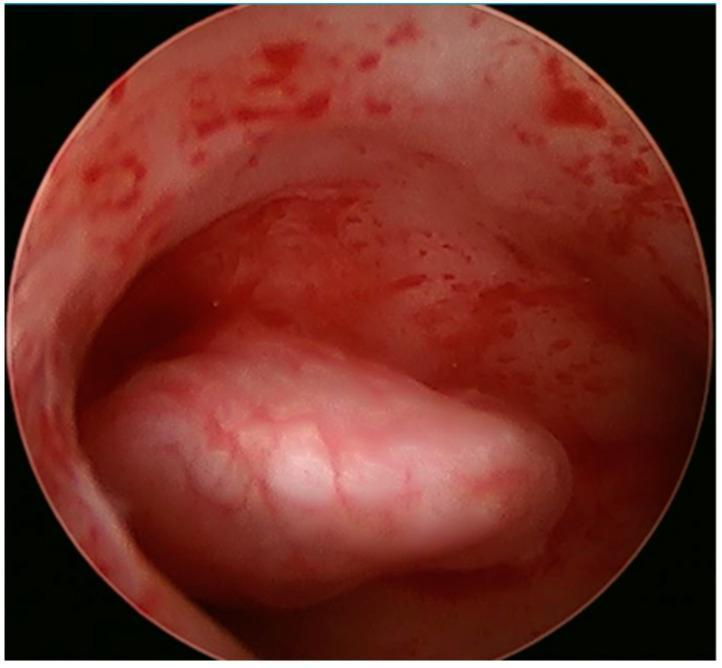
This clinical diagnostic image is a hysteroscopic view of the uterine cavity, showing an endometrial polyp. The image displays a smooth, rounded, and lobulated mass approximately 1.5 cm in size, situated on the right lateral wall of the uterus. The polyp exhibits a lighter, pinkish-white coloration compared to the surrounding pinkish-red uterine lining. Fine superficial blood vessels are visible on the surface of the polyp, which appears less vascular than the adjacent hyperemic endometrium. The surrounding uterine tissue shows evidence of mild spotting and irregular vascular patterns. This endoscopic finding is characteristic of a benign endometrial polyp, often investigated in cases of infertility or abnormal uterine bleeding. The image serves as an educational example of intrauterine pathology identified during a diagnostic hysteroscopy, illustrating the morphological differences between focal polyps and normal endometrial tissue.
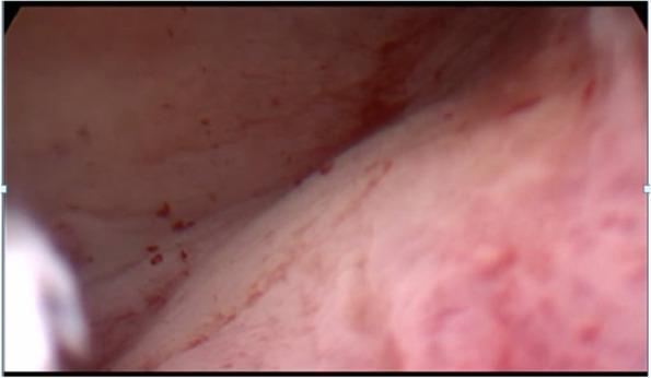
This clinical photograph shows a hysteroscopic view of a healthy-appearing endometrial cavity. The uterine lining is visualized as a pale pink to reddish mucosa with a smooth, uniform texture. Fine, branching vascular patterns are evident across the surface, along with small, scattered punctate reddish spots representing superficial capillaries or minor surface irregularities. No visible intraluminal pathologies are present; specifically, there are no polyps, submucosal fibroids, or structural distortions of the cavity. This image serves as a clinical reference for a normal uterine interior in the context of FIGO Type 3 intramural fibroids, which are known to abut the endometrium without causing visible intracavitary alterations. The anatomical focus is on the endometrial surface during a diagnostic hysteroscopy in an adult female patient, used to differentiate between cavity-distorting and non-distorting uterine myomas.
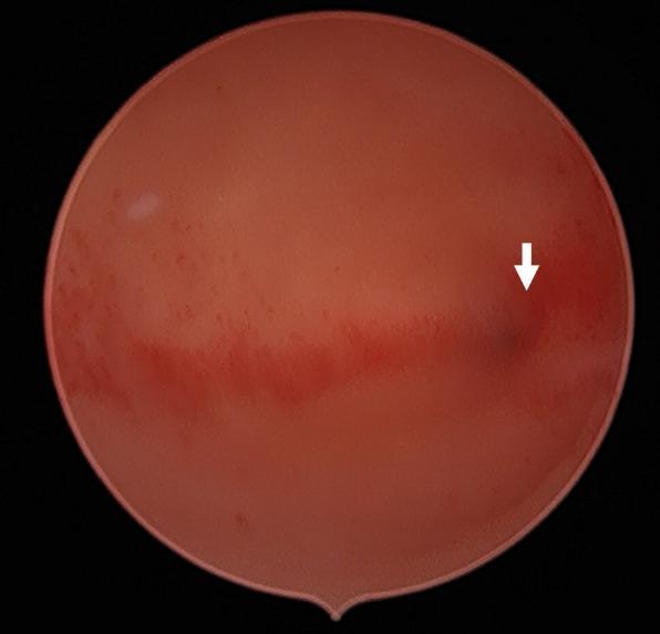
This diagnostic endoscopic image captures the internal view of a uterine cavity during a hysteroscopy. The image demonstrates the characteristic morphology of a unicornuate uterus, a Müllerian duct anomaly. The endometrial lining appears intact with a healthy reddish-pink hue and a smooth surface texture. A white arrow points toward a specific anatomical landmark, identified as the single left tubal ostium, which confirms the absence of a second communicating horn. A horizontal band of hyperemic tissue suggests variations in vascularity across the endometrium. No intracavitary lesions, adhesions, or polyps are visible, indicating a normal environment for future embryo transfer. This visual is typical of an infertility workup evaluating uterine cavity patency and endometrial health in patients with known congenital uterine anomalies.
congenital uterine anomaly Mullerian duct septate bicornuate unicornuate
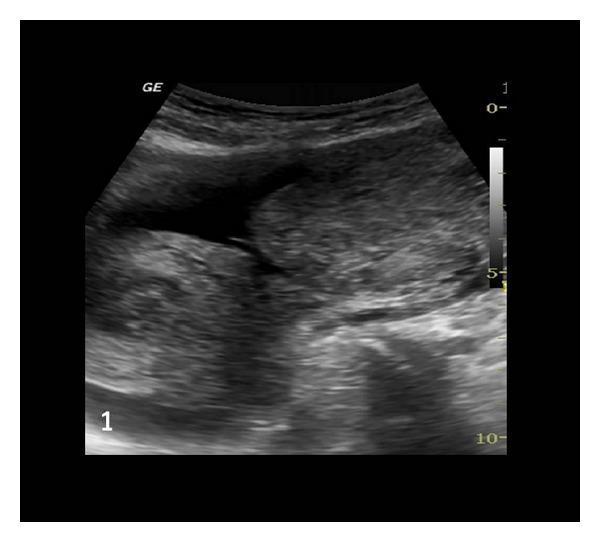
This diagnostic ultrasound image in the pelvic region demonstrates a congenital Müllerian duct anomaly. The primary finding is a unicornuate uterus, characterized by an elongated, single-sided uterine horn with a relatively uniform, intermediate echogenicity. Adjacent to the main uterine body, a distinct, noncommunicating rudimentary horn is visible. Both uterine structures appear empty, without visible gestational sacs or products of conception. Significant anechoic areas representing free fluid (hemoperitoneum) are noted surrounding the uterine structures, consistent with clinical suspicion of a ruptured rudimentary horn. The image provides a key visualization of Class II Müllerian anomalies, specifically highlighting the anatomical relationship between a unicornuate uterus and its associated rudimentary horn, which is critical for diagnostic differentiation and surgical planning in emergency gynecological presentations.
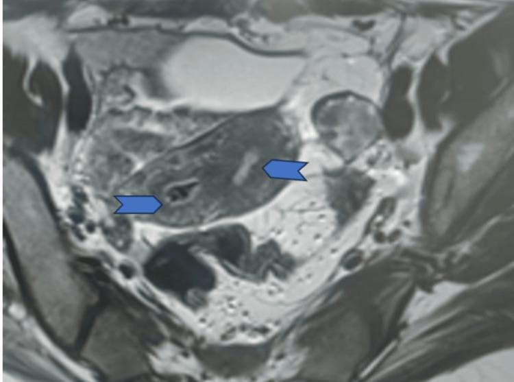
This diagnostic image is a transverse (axial) MRI section of the female pelvis, demonstrating a Müllerian duct anomaly. The scan shows a bicornuate uterus characterized by two distinct uterine horns and endometrial cavities. Blue arrowheads highlight the dual uterine cavities, which appear as two separate, low-signal intensity (hypointense) spaces within the uterine musculature. These cavities are separated by a central band of myometrial tissue of intermediate signal intensity. The external uterine contour shows a mild fundal concavity or indentation, a hallmark finding used to differentiate a bicornuate uterus from a septate uterus. Surrounding pelvic anatomy, including the iliac vessels and posterior elements, is visible but secondary to the central uterine pathology. This imaging is clinically significant for diagnosing congenital uterine malformations, which can impact reproductive outcomes and obstetric management.
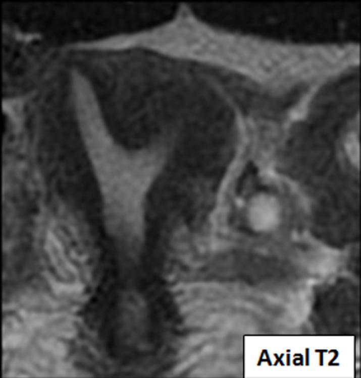
This diagnostic image is an axial T2-weighted MRI slice of the female pelvis focusing on the uterus. It demonstrates a bicornuate unicervical uterus, a congenital Mullerian duct anomaly. The image shows a single cervix and inferior uterine segment that bifurcates superiorly into two distinct, divergent uterine horns. The T2-weighted sequence provides high contrast between the hyperintense (bright) endometrial lining and the relatively hypointense (darker) myometrium. The deep fundal cleft between the two horns confirms the bicornuate morphology rather than a septate uterus. This imaging is characteristic of Class IV Mullerian duct anomalies and is clinically significant in evaluations for infertility or obstetric complications. The surrounding pelvic anatomy, including the bladder and adjacent soft tissues, is visible with varying signal intensities reflecting tissue water content.
Asherman syndrome intrauterine adhesions hysteroscopy
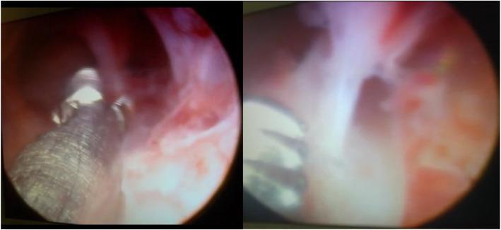
This diagnostic and interventional hysteroscopy image consists of two panels demonstrating intrauterine adhesions (Asherman syndrome) within the uterine cavity. The left panel shows the mid-uterine cavity with reddish-pink endometrial lining and whitish, fibrous tissue bands stretching across the lumen. A metallic grasping instrument with a textured surface is visible, engaging with the adhesions. The right panel provides a closer view of dense, opaque, avascular synechiae bridging the uterine walls. These adhesions partially obliterate the cavity, characteristic of moderate-to-severe intrauterine scarring. Surgical microscissors or grasping forceps are seen in the foreground, indicating the therapeutic lysis of these adhesions. The images serve to illustrate the visual appearance of fibrotic bands and the use of hysteroscopic instrumentation in the treatment of infertility related to endometrial pathology.
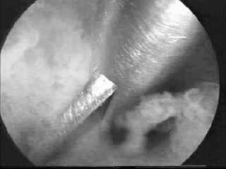
**Imaging Modality:** Hysteroscopy (intrauterine endoscopic view). **Anatomical Region:** Endometrial cavity. **Observed Pathology:** Intrauterine adhesions (synechiae). The image demonstrates thick, fibrous bands of tissue extending across the uterine cavity, characteristic of Asherman’s syndrome. **Surgical Intervention:** Hysteroscopic adhesiolysis. A semi-rigid or flexible hysteroscopic scissor instrument is visible in the foreground, positioned to perform a mechanical transection of a dense adhesive band. **Characteristic Visual Features:** * **Pathology:** Pale, avascular, or poorly vascularized fibrous bridges connecting the uterine walls, obscuring the normal glandular appearance of the endometrium. * **Instrumentation:** Metallic surgical scissors engaged with the target tissue. * **Context:** Distension of the uterine cavity is maintained, allowing for visualization of the adhesive distribution and the surgical margins. **Diagnostic Relevance:** This image illustrates the mechanical management of moderate-to-severe intrauterine synechiae. It highlights the use of "cold" (non-thermal) instruments to minimize thermal damage to the surrounding healthy endometrium during the restoration of normal uterine cavity morphology.
submucosal fibroid leiomyoma uterus
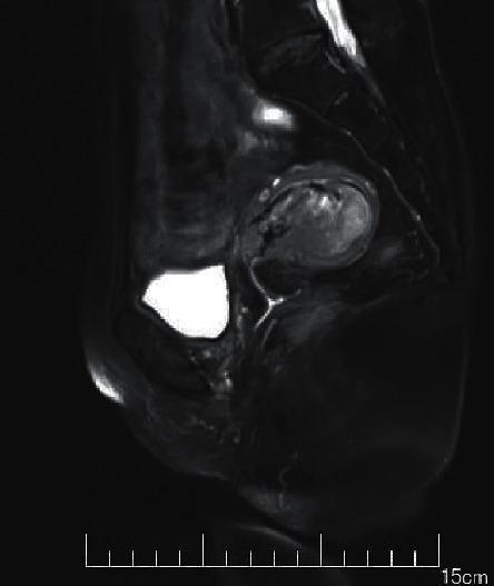
This diagnostic image is a sagittal T2-weighted MRI (T2WI) of the female pelvis, demonstrating a submucosal uterine fibroid (leiomyoma). The uterus is the central focus, containing a well-defined, nodular, and predominantly low-signal (hypointense) mass characteristic of fibrous tissue. The fibroid is situated within the endometrial cavity, consistent with a submucosal classification. Its boundaries are demarcated by a relatively hyperintense rim, although some areas appear less distinct, potentially suggesting localized hemorrhage or edema. Anterior to the uterus, the urinary bladder is highly visible, exhibiting intense high signal (hyperintensity) due to the presence of fluid (urine) on this imaging modality. The surrounding pelvic soft tissues and musculature display intermediate signal intensities. This image is an educational example used in gynecology and radiology to teach the identification of benign uterine tumors, their impact on the endometrial cavity, and the signal characteristics of leiomyomas relative to surrounding anatomical structures like the bladder.
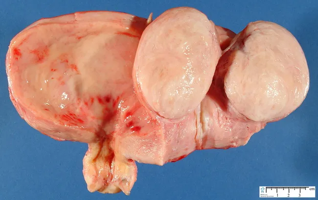
Gross pathology photograph of a uterine specimen displaying a submucosal leiomyoma, a fibroid. The image shows an external uterus surface with a prominent, rounded, pale pink‑tan mass arising beneath the endometrium and projecting toward the uterine cavity. The specimen rests on a blue background with a centimeter scale for size reference, enabling approximate measurement of the lesion. The lesion appears well circumscribed, firm, and homogeneous, with no obvious necrosis or hemorrhage on this gross view. The surface texture is smooth with mild lobulation typical of fibroid tissue composed of intersecting smooth muscle fascicles. This submucosal location explains distortion of the endometrial lining and potential abnormal uterine bleeding in affected individuals. The gross morphology supports a diagnosis of leiomyoma over other uterine neoplasms, though histopathology would confirm features such as organized smooth muscle bundles, a whorled pattern, and absence of malignant characteristics. Clinically, submucosal fibroids are important because they may cause infertility, menorrhagia, or recurrent pregnancy loss, and surgical management decisions depend on size, location, and patient factors. This image is educational for gross pathology training, surgical planning discussions, and radiologic–pathologic correlation. This representation aids medical students, residents, and clinicians in correlating gross features with imaging and histology.
Uterine Factors in Infertility
Diagnostic Modalities
| Modality | Key Use | Limitations |
|---|---|---|
| Hysteroscopy | Gold standard - direct visualization | Cannot assess myometrium/adnexa; needs anesthesia for operative cases |
| HSG | Initial screening; tubal + uterine | 50% sensitivity for polyps; false positives from bubbles |
| Transvaginal US (2D) | Good NPV for polyps (96.5%) | 0% PPV for intrauterine adhesions; poor for anomalies |
| Saline Infusion Sonography (SIS) | 100% sensitivity/specificity for polyps; better tolerated | 77.8% sensitivity for anomalies |
| Pelvic MRI | Gold standard for anomalies; best for fibroids vs. adenomyosis | Cost, availability |
I. Congenital Anomalies of the Uterus
Embryology
ASRM/AFS Classification of Mullerian Anomalies
| Class | Type | Reproductive Impact |
|---|---|---|
| I | Mullerian agenesis (Mayer-Rokitansky-Kuster-Hauser) | Infertility - needs gestational carrier; IVF possible with own oocytes |
| II | Unicornuate uterus | Preterm delivery, malpresentation; rudimentary horn must be removed |
| III | Didelphys uterus | Moderate obstetric risk |
| IV | Bicornuate uterus | Pregnancy wastage, preterm delivery |
| V | Septate uterus | Most common anomaly; highest rate of pregnancy loss |
| VI | Arcuate uterus | Mildest; live birth rates comparable to normal uteri |
| VII | DES-related (T-shaped uterus) | Declining incidence (DES banned 1971) |
- Anomalies are more strongly associated with pregnancy wastage and poor obstetric outcomes than with primary infertility - the prevalence of congenital anomalies is similar in fertile and infertile women
- Exception: Mullerian agenesis causes absolute infertility
- Because Mullerian ducts are adjacent to the urinary system, renal anomalies frequently coexist - urologic imaging is mandatory when a Mullerian anomaly is found
- Hysteroscopic metroplasty for septate uterus significantly reduces pregnancy loss rates but does not improve primary infertility rates
- Rudimentary uterine horns require surgical removal on diagnosis (risk of ectopic pregnancy)
II. Acquired Abnormalities of the Uterus
1. Intrauterine Adhesions (Asherman's Syndrome)
- Curettage after pregnancy (postpartum/post-abortion)
- Uterine surgery
- Endometritis (including tuberculous endometritis - a major cause in endemic regions)
- Partial or complete obliteration of the uterine cavity
- Destruction of the functional endometrial layer, impairing implantation
- May cause amenorrhea or hypomenorrhea (classic triad with cyclic pain)
2. Endometrial Polyps
- Focal overgrowths of endometrial glands and stroma
- Found in up to 32% of infertile women undergoing hysteroscopy
- Mechanisms of infertility: mechanical interference with implantation, altered endometrial receptivity, abnormal cytokine milieu
3. Uterine Leiomyomas (Fibroids)
| Type | Location | Impact on Fertility |
|---|---|---|
| Submucosal (FIGO Type 0, 1, 2) | Project into cavity | Most significant - clearly impair fertility and implantation |
| Intramural | Within myometrium | Some evidence of impairment, especially if large or abutting cavity |
| Subserosal | Projecting outward | Minimal to no impact on fertility |
- Altered uterine contractility impairing gamete transport
- Impaired endometrial blood supply
- Mechanical distortion of the cavity
- Endometrial dysfunction (submucosal leiomyomas have a global effect on implantation markers)
4. Adenomyosis
- Presence of endometrial glands and stroma within the myometrium
- Causes uterine enlargement, dysmenorrhea, menorrhagia
- Impairs fertility through altered uterine contractility, disrupted implantation, and impaired endometrial receptivity
- Diagnosis: MRI is preferred (junctional zone thickness >12 mm is diagnostic). Transvaginal US shows heterogeneous myometrium with cysts
- Clinically relevant in IVF patients - associated with lower implantation rates; some evidence that GnRH agonist down-regulation before IVF may improve outcomes
5. Endometrial Hyperplasia
- Abnormal proliferation of endometrial glands
- May reflect underlying anovulatory cycles (e.g., PCOS) with unopposed estrogen
- Impairs implantation
- Treatment depends on grade: progestin therapy for simple/complex hyperplasia without atypia; hysterectomy or progestin + levonorgestrel IUD for atypical hyperplasia
III. Luteal Phase Defect (LPD)
- Insufficient progesterone production by the corpus luteum
- Endometrium fails to develop secretory transformation adequate for embryo implantation
- Diagnosed by endometrial biopsy showing >2 days lag in histological dating, or subnormal luteal phase serum progesterone
- Controversial - whether true LPD is a distinct entity or a manifestation of other disorders (e.g., PCOS, hyperprolactinemia, thyroid disease) is debated
- Treatment: Progesterone supplementation (vaginal or intramuscular); luteal phase support is standard in all ART cycles. Vaginal progesterone is initiated day 0-3 post-egg retrieval and continued to 8-10 weeks of gestation
Summary Table: Uterine Factors in Infertility
| Condition | Prevalence in Infertile Women | Primary Mechanism | Gold Standard Diagnosis | Treatment |
|---|---|---|---|---|
| Intrauterine adhesions | ~5-10% | Cavity obliteration, endometrial destruction | Hysteroscopy | Hysteroscopic adhesiolysis + estrogen |
| Endometrial polyps | Up to 32% | Impaired implantation | SIS / Hysteroscopy | Hysteroscopic polypectomy |
| Submucosal fibroids | ~5-10% | Cavity distortion, endometrial dysfunction | MRI / SIS / Hysteroscopy | Hysteroscopic myomectomy |
| Intramural/subserosal fibroids | Common | Variable | MRI | Myomectomy if symptomatic |
| Congenital anomalies | 5-10% (infertile) | Implantation failure, pregnancy loss | Pelvic MRI | Metroplasty (septate); varies by type |
| Adenomyosis | ~20-30% (subfertile) | Altered contractility, receptivity | MRI | GnRH agonist; surgical excision |
| Mullerian agenesis | Rare | Absent uterus | MRI / clinical | IVF + gestational carrier |
| Luteal phase defect | 3-10% | Inadequate endometrial secretory change | Endometrial biopsy | Progesterone supplementation |