
Exophthalmus in radio active iodine vs thyroid surgery
"Graves ophthalmopathy" AND "radioactive iodine" AND "thyroidectomy"
PMID: 38686801
Graves ophthalmopathy exophthalmos proptosis
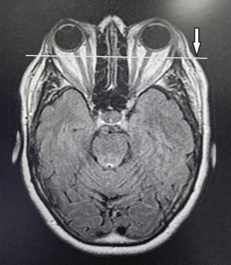
This diagnostic image is an axial view Magnetic Resonance Imaging (MRI) of the head, specifically focusing on the orbits and brain. The scan demonstrates significant bilateral proptosis (exophthalmos), characterized by the anterior displacement of the globes relative to the orbital rims. A horizontal white line and a white arrow on the left side highlight the degree to which the eyeballs protrude beyond the lateral orbital margin. Visible within the retrobulbar space is evidence of extraocular muscle enlargement and orbital fat hypertrophy, typical findings associated with Graves' ophthalmopathy (thyroid-associated orbitopathy). The optic nerves are visible and appear stretched due to the globe displacement. The brain section shows the midbrain, cerebral hemispheres with visible gyri and sulci, and the interhemispheric fissure. This image serves as an educational tool for identifying radiological signs of restrictive orbitopathy and secondary complications like proptosis.
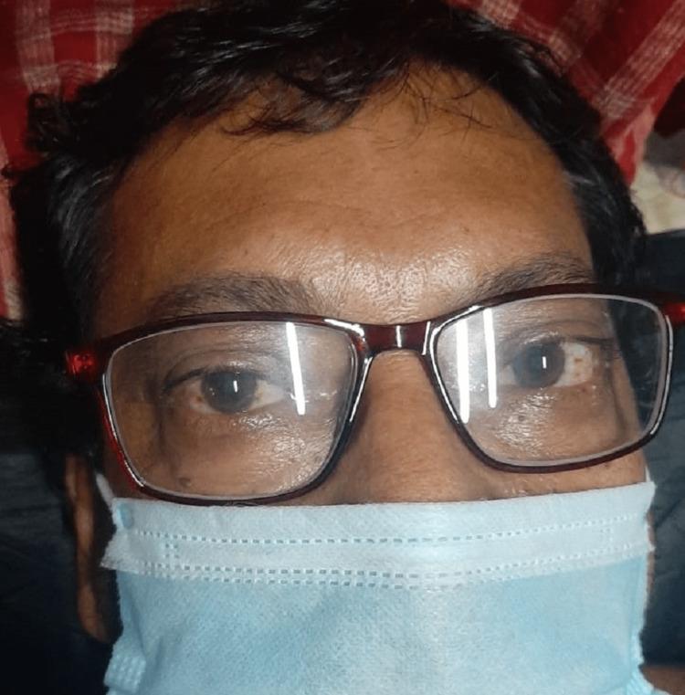
Clinical photograph of a 53-year-old male presenting with bilateral Graves' ophthalmopathy (thyroid eye disease). The image focuses on the periocular region, demonstrating bilateral exophthalmos (proptosis) characterized by the protrusion of the globes from the orbits. Significant scleral show is visible, particularly inferior to the iris, contributing to a characteristic 'staring' expression or Dalrymple's sign. The patient is wearing corrective eyeglasses and a surgical face mask, indicating a clinical setting. This visual illustrates the extrathyroidal manifestations of Graves' disease, an autoimmune condition causing hypertrophy of extraocular muscles and orbital adipose tissue. The image is an educational resource for medical professionals to recognize clinical signs of hyperthyroidism-associated orbitopathy and for calculating Clinical Activity Scores (CAS) in endocrinology and ophthalmology.
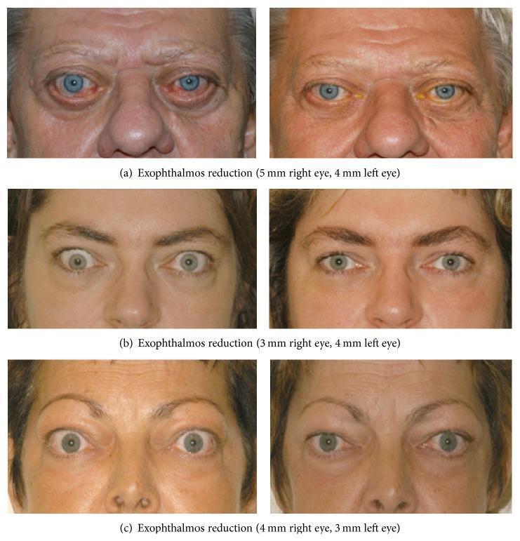
This clinical comparison chart presents three sets (a, b, c) of pre- and post-operative facial photographs documenting the results of bilateral orbital decompression in patients with Graves' ophthalmopathy (thyroid eye disease). The 'before' images on the left consistently demonstrate significant proptosis (exophthalmos), characterized by ocular prominence, upper eyelid retraction, and excessive scleral show both superiorly and inferiorly. In case (a), there is also visible conjunctival injection and chemosis. The 'after' images on the right, taken 6 months to 2 years post-surgery, show a marked reduction in globe protrusion (measured between 3–5 mm reduction per eye). Key visual improvements include a normalized eyelid position with reduced retraction, decreased scleral exposure, and a more relaxed periocular skin appearance. These images illustrate the surgical management of Graves' ophthalmopathy to reduce orbital volume, protecting the ocular surface and improving aesthetic outcomes by resolving the 'staring' expression associated with the disease.
Exophthalmos (Graves' Ophthalmopathy) - RAI vs. Thyroid Surgery
The Core Issue
Radioactive Iodine (RAI) - Effect on Exophthalmos
- RAI can worsen Graves' ophthalmopathy, especially in smokers - Cummings Otolaryngology Head & Neck Surgery explains this occurs "secondary to the development of tissue edema and worsening of the ocular symptoms" following RAI administration.
- The mechanism involves a post-ablation surge in TSH receptor antibodies (TRAb) - RAI destroys thyroid tissue, releasing thyroid antigens and transiently boosting the autoimmune response, which drives further orbital inflammation.
- Smokers are at significantly higher risk of RAI-induced ophthalmopathy worsening and should particularly avoid RAI if eye disease is present.
- RAI is not recommended in patients with moderate-to-severe Graves' ophthalmopathy, per Goldman-Cecil Medicine.
Thyroid Surgery - Effect on Exophthalmos
- Surgery is the preferred treatment when Graves' ophthalmopathy is moderate or severe. As stated in Cummings Otolaryngology, "an additional relative indication for surgery, as opposed to radioactive iodine, is moderate or severe ocular symptoms related to Graves ophthalmopathy, which may be worsened with radioactive iodine administration."
- KJ Lee's Otolaryngology lists as an indication for surgery: "concern regarding RAI and eye disease"
- Surgery does not trigger the same TSH receptor antibody surge as RAI, so it avoids the acute worsening mechanism.
- Berek & Novak's Gynecology notes: "Surgical intervention may be considered in severe Graves' ophthalmopathy. Surgery is the most rapid and consistent method of achieving a euthyroid state in Graves' disease."
- Achieving euthyroidism rapidly by surgery may actually stabilize the ophthalmopathy compared to the prolonged exposure to thyroid hormone excess while waiting for RAI to take effect (RAI typically takes 6-8 weeks to normalize thyroid hormone levels).
Summary Table
| Feature | RAI | Thyroid Surgery |
|---|---|---|
| Effect on exophthalmos | Can worsen (especially in smokers) | Preferred - does not worsen |
| Mechanism of worsening | Post-ablation TRAb surge, tissue edema | No antigen release surge |
| Mild ophthalmopathy | Usable with prophylactic steroids | Safe option |
| Moderate/severe ophthalmopathy | Contraindicated / not recommended | Treatment of choice |
| Speed of euthyroidism | 6-8 weeks | Rapid (days) |
| Risk of hypothyroidism | Up to 80% | Lower than RAI |
| Smokers with eye disease | Avoid RAI | Strongly preferred |
Recent Evidence (2024 Meta-Analysis)
- Thyroidectomy was associated with a 69% lower risk of Graves' ophthalmopathy vs. antithyroid drugs (HR 0.31, 95% CrI: 0.12-0.64) and an 82% lower risk vs. RAI (HR 0.18, 95% CrI: 0.07-0.35)
- RAI had a 70% higher risk of Graves' ophthalmopathy compared to antithyroid drugs (HR 1.70, 95% CrI: 1.02-2.99)
- This is the strongest current evidence confirming surgery as the ophthalmopathy-sparing treatment