
Pelvic supportive osteotomies
pelvic osteotomy types acetabulum hip dysplasia diagram
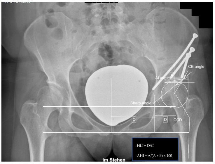
This diagnostic image is an anteroposterior (AP) pelvic radiograph demonstrating the outcome of a left-sided Periacetabular Osteotomy (PAO), a surgical procedure for hip dysplasia. The radiograph shows the pelvic girdle, proximal femurs, and sacrum. On the left hip, evidence of surgical intervention includes three large cortical screws utilized for internal fixation of the acetabular fragment into the ilium. The reconstructed acetabulum shows improved femoral head coverage. Detailed orthopedic measurements are overlaid on the image to quantify postoperative alignment, including the Center-Edge (CE) angle (38.0°), Acetabular Index (AI) angle (-0.5°), and Sharp angle (36.5°). Additional metrics include the Hip Lateralization Index (HLI = D/C, 0.56), Anterior Hip Index (AHI = A/(A+B) x 100, 91.4), and the Caput-Collum-Diaphyseal (CCD) angle (137.4°). A positive 'crossing-over sign' is noted, indicating acetabular retroversion or specific rim morphology post-correction. This clinical image serves as an educational tool for evaluating acetabular orientation and surgical success in pelvic osteotomy procedures.
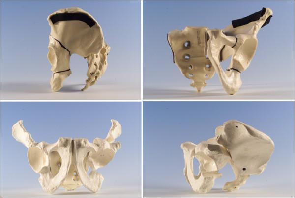
This composite of four clinical photographs displays a three-dimensional anatomical bone model of the human pelvis, specifically illustrating the technique for an Interlocking Pelvic Triple Osteotomy used in treating hip dysplasia. The images show the pelvic girdle from multiple orientations (lateral, posterior-oblique, anterior, and superior-oblique views) to highlight the three specific osteotomy sites required to mobilize the acetabulum. Key surgical cuts include: 1) a three-sided iliac osteotomy extending from the iliac crest towards the acetabulum; 2) an ischial osteotomy performed through the sub-cotyloid groove near the obturator foramen; and 3) a superior pubic ramus osteotomy. The model demonstrates how these strategic bone cuts allow for the reorientation of the acetabular fragment to improve lateral femoral head coverage and correct anteversion. The sacrum and pelvic landmarks such as the iliac crest, acetabulum, and pubic symphysis are clearly visible, providing anatomical context for the orthopedic procedure.
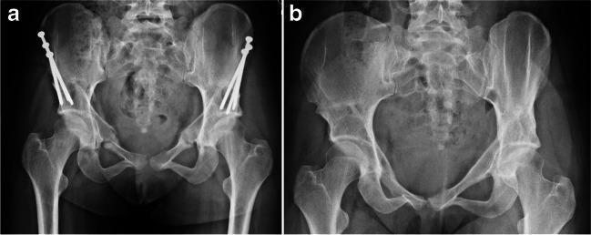
This diagnostic image displays a side-by-side comparison of two anteroposterior (AP) view radiographs of the pelvis, illustrating the progression and healing following a periacetabular osteotomy (PAO) for hip dysplasia. Radiograph (a) shows the immediate post-operative state with surgical hardware in situ. Multiple metallic cortical screws are visible, oriented from the superior ilium into the acetabular fragments to provide internal fixation across the osteotomy sites. The surgical cuts (osteotomies) around the acetabulum are visible as faint radiolucent lines. Radiograph (b) demonstrates the same pelvis at a later stage after hardware removal. The acetabular fragments have successfully united, evidenced by the absence of osteotomy lines and the presence of smooth cortical remodeling. The pelvic ring is intact, and the hip joints show improved femoral head coverage. This comparison illustrates the typical healing sequence from internal stabilization to complete bony consolidation after surgical correction of acetabular morphology.
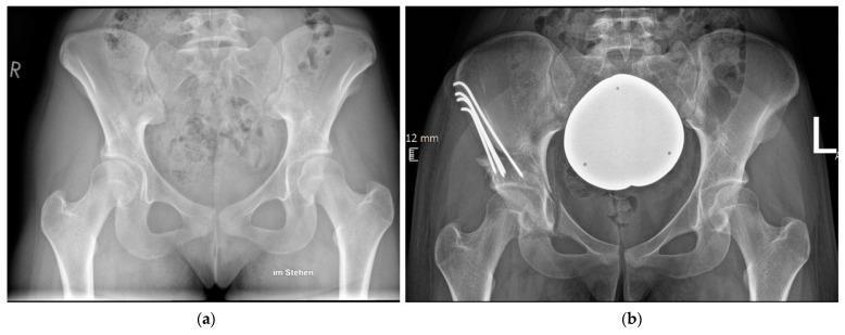
This figure presents a side-by-side comparison of anteroposterior (AP) pelvic radiographs demonstrating the surgical management of developmental dysplasia of the hip (DDH). Image (a) shows the preoperative state with bilateral acetabular dysplasia, characterized by a shallow acetabular roof and insufficient coverage of the femoral head. Image (b) displays the postoperative result following a periacetabular osteotomy (PAO) of the right hip. The acetabulum has been surgically reoriented to increase the lateral center-edge angle (LCEA) and improve superior femoral head coverage. Internal fixation is visible in the right ilium, consisting of four metallic Kirschner wires (K-wires) that are bent at their proximal ends and recessed into the iliac crest to secure the osteotomized fragment. A large, spherical radiopaque marker or artifact is visible over the pelvic midline in the postoperative view. This diagnostic comparison highlights the anatomical corrections achieved through reorientation osteotomy and the use of unthreaded K-wires as an alternative fixation method to traditional screws.
Salter innominate osteotomy hip pediatric
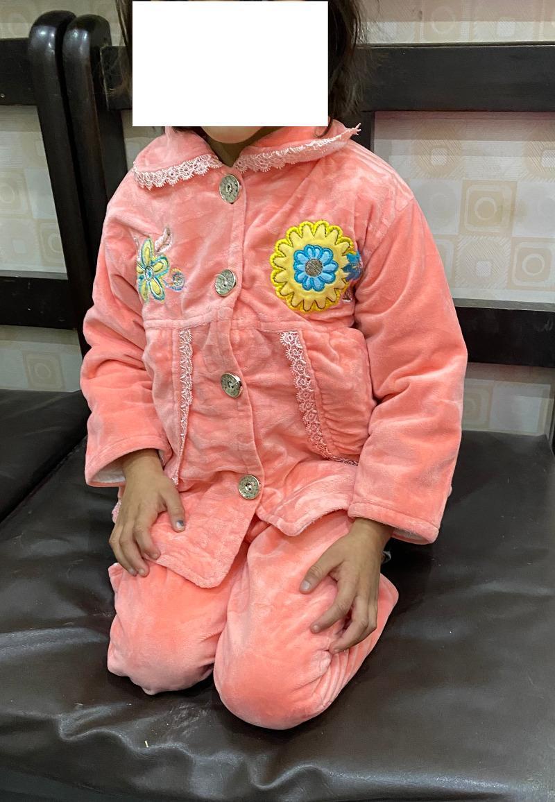
Clinical photograph of a pediatric patient demonstrating postoperative lower limb functional mobility following a Salter's innominate osteotomy (SIO) for developmental dysplasia of the hip (DDH). The subject is shown in a seated kneeling position, often used for clinical assessment in certain cultures. The image illustrates the 'tashahhud I' position, which is a component of the Bhatti Functional Scoring System (BFSS) used to evaluate clinical outcomes after pelvic osteotomies. Key visible features include the ability to achieve significant knee flexion and hip stability necessary for this specific kneeling posture. This clinical documentation is used to assess the range of motion, hip joint stability, and the success of surgical reconstruction in restoring functional activities of daily living in a pediatric orthopedic context. The patient's symmetrical posture and bilateral limb alignment are indicative of a successful surgical outcome and effective postoperative rehabilitation.
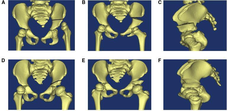
This medical visual compares the structural changes following two common pediatric pelvic osteotomies for developmental dysplasia of the hip (DDH): the Salter and Pemberton procedures. The content consists of 3D CT reconstructions of a pediatric pelvis in six panels (A–F). Panels A–C demonstrate the Salter innominate osteotomy. Panel A shows a complete horizontal osteotomy line extending from the sciatic notch to the anterior inferior iliac spine. Panel B (anteroposterior) and C (lateral) show the resulting complete separation and downward/forward rotation of the distal pelvic fragment, creating a visible gap above the acetabulum typically filled with a bone graft. Panels D–F demonstrate the Pemberton pericapsular osteotomy. Panel D shows an incomplete osteotomy line curving from the ilium toward the triradiate cartilage. Panels E (anteroposterior) and F (lateral) illustrate that the posterior column remains intact; the acetabular roof is hinged downward through the triradiate cartilage, altering the acetabular shape and volume with a less pronounced pelvic contour change compared to the Salter method. These models highlight the different biomechanical approaches to increasing acetabular coverage of the femoral head.
Pelvic Supportive Osteotomies
Classification
| Category | Operation | Mechanism |
|---|---|---|
| Redirectional | Salter innominate osteotomy | Rotates entire acetabulum anterolaterally |
| Redirectional | Pemberton acetabuloplasty | Hinges acetabular roof at triradiate cartilage |
| Redirectional | Steel triple innominate osteotomy | Completely frees acetabulum via 3 cuts |
| Redirectional | Ganz periacetabular osteotomy (PAO) | Frees acetabulum while preserving posterior column |
| Salvage/Augmentation | Shelf procedure (Staheli) | Extends acetabular roof laterally with bone graft |
| Salvage/Displacement | Chiari osteotomy | Medial displacement of pelvis, capsular interposition |
1. Salter Innominate Osteotomy
- Congruous hip reduction achievable
- Correction of acetabular index <10-15 degrees required
- Any dislocation or subluxation must be reduced (or open reduction done simultaneously)
- Patient supine; anterior iliofemoral approach
- Adductor tenotomy if contracted
- Iliac apophysis split and gluteal muscles reflected from lateral ilium down to sciatic notch
- Gigli saw passed through sciatic notch; straight-line bone cut to anterior inferior iliac spine
- Distal fragment displaced anterolaterally; iliac crest bone graft wedged into gap
- Two threaded Kirschner wires (Steinmann pins) for fixation; spica cast
2. Pemberton Acetabuloplasty (Pericapsular Osteotomy of the Ilium)
-
10-15 degrees of correction of the acetabular index required
- Small femoral head with large acetabulum (volume reduction desirable)
- Subluxation or dislocation that has been or can be reduced
- Greater degree of correction achievable (fulcrum is closer to the site of correction)
- Internal fixation not always required
- Better suited when acetabular volume reduction is desired
- Decreases acetabular volume - may create incongruity requiring remodeling
- Risk of femoroacetabular impingement (FAI) if overcorrected (though uncommon at 10-year follow-up of >150 cases)
3. Steel Triple Innominate Osteotomy
- Residual acetabular dysplasia with subluxation
- Older children/adolescents in whom acetabular remodeling can no longer be anticipated
- Symptomatic hip with a congruous joint
4. Ganz (Bernese) Periacetabular Osteotomy (PAO)
- Residual acetabular dysplasia in adolescents and adults
- Symptomatic hip with a congruous joint
- Only one surgical approach required
- Large correction achievable in all directions, including medial and lateral planes
- Blood supply to the acetabulum is preserved
- Posterior column remains intact - allows immediate crutch walking with minimal internal fixation
- Shape of the true pelvis is unaltered - normal childbirth remains possible
- Can be combined with trochanteric osteotomy if needed
- Provides similar results even in previously operated hips
5. Shelf Procedure (Staheli - Slotted Acetabular Augmentation)
- Subluxations/dislocations already reduced, where no redirectional osteotomy would establish a congruous joint
- Deficient acetabulum where redirectional osteotomy is not possible or appropriate
- Hips in which the femoral head and acetabulum are misshapen but still congruent (redirectional osteotomies would cause incongruity in these)
- Dysplastic hips with spherical congruity suitable for redirectional osteotomy
- Hips needing concurrent open reduction requiring supplementary stability
- Patients unsuited for spica cast immobilization
6. Chiari Osteotomy (Innominate Osteotomy with Medial Displacement)
- Incongruous, irreducible, subluxed, or dislocated hip
- Femoral head cannot be centered adequately in the acetabulum
- Painfully subluxated hips with early signs of osteoarthritis
- Acetabular dysplasia too severe for other pelvic osteotomies
- May improve acetabular bone stock for future total hip arthroplasty
- Corrects the lateral pathologic displacement of the femur
- Improves superolateral femoral coverage
- Deepens the deficient acetabulum by medial displacement
Summary Table (Recommended Osteotomy by Age/Indication)
| Osteotomy | Age | Indications |
|---|---|---|
| Salter innominate | 18 months - 6 years | Congruous hip; <10-15° correction needed |
| Pemberton acetabuloplasty | 18 months - 10 years | >10-15° correction needed; small head, large acetabulum |
| Steel triple innominate | Late adolescence | Residual dysplasia; open triradiate cartilage |
| Ganz PAO | Late adolescence - adult | Residual dysplasia; closed triradiate cartilage; symptomatic |
| Shelf (Staheli) | Any age (typically older) | Incongruous joint; redirectional osteotomy not possible |
| Chiari | >4 years (typically older) | Incongruous, irreducible hip; salvage situation |
General Principles
- Redirectional osteotomies (Salter, Pemberton, Steel, Ganz) are preferred when the femoral head and acetabulum are congruent and articular cartilage can be placed over the femoral head.
- Salvage/augmentation osteotomies (Shelf, Chiari) are used when the hip is incongruous, irreducible, or when redirectional procedures are technically impossible - they interpose fibrocartilage or bone, not hyaline cartilage.
- Any pelvic osteotomy can be combined with femoral osteotomy in older children to simultaneously correct both femoral and acetabular abnormalities.
- The Chiari procedure may confer a future benefit of improved acetabular bone stock for total hip arthroplasty.