
Please explain me ecg with changes and name of disease
Reading File
Now let me search the library for ECG changes and diseases simultaneously.
Finding Sources
Finding Sources
Finding Sources
Reading File
Finding Sources
Finding Sources
Reading File
Finding Sources
Reading File
Finding Sources
Now let me fetch some key ECG images from the textbook and read more on specific ECG changes:
Reading File
Finding Sources
Loading Image
Reading File
I now have comprehensive information to write a thorough, well-structured ECG guide. Let me compile everything.
ECG (Electrocardiogram): Waves, Intervals, and Disease Patterns
The ECG records the heart's electrical activity across 12 leads. Each lead "looks" at the heart from a different angle, so one disease can produce changes in specific lead groups (called territories). Below is a complete, disease-by-disease breakdown.
Normal ECG Anatomy
| Component | Normal Value | What It Represents |
|---|---|---|
| P wave | <120 ms, <2.5 mm tall | Atrial depolarization (SA node fires) |
| PR interval | 120-200 ms | AV node conduction delay |
| QRS complex | <120 ms | Ventricular depolarization |
| ST segment | Isoelectric (flat) | Early ventricular repolarization |
| T wave | Upright in I, II, V3-V6 | Ventricular repolarization |
| QT interval | <440 ms (men), <460 ms (women) | Total ventricular electrical activity |
ECG Changes by Disease
1. ST-Elevation Myocardial Infarction (STEMI)
The most time-critical ECG diagnosis.
Stages of ECG evolution:
| Time | ECG Finding |
|---|---|
| Minutes (earliest) | Hyperacute T waves - tall, broad, peaked, asymmetrical. ST segment just beginning to rise at J point |
| Hours | ST elevation - convex ("domed" or "tombstone" shape), >1 mm in 2+ contiguous limb leads; >1.5 mm in women, >2 mm in men over 40 in V2-V3 |
| Hours-days | Pathological Q waves - >40 ms wide, >25% QRS height = irreversible myocardial death |
| Days-weeks | T wave inversion - deep, symmetrical in affected leads |
| Chronic | Persistent Q waves - marker of old infarction |
Territory-based STEMI localization:
| Territory | Leads with ST elevation | Culprit Artery |
|---|---|---|
| Anterior | V1-V4 | Left anterior descending (LAD) |
| Lateral (high) | I, aVL | Left circumflex or 1st diagonal |
| Lateral (low) | V5, V6 | Left circumflex |
| Inferior | II, III, aVF | Right coronary (90%) or left circumflex |
| Posterior | Tall R in V1-V2, horizontal ST depression V1-V3 | Right coronary or circumflex |
| Right ventricle | V1 + right-sided leads V3R-V6R | Right coronary |
| Left main / multivessel | ST elevation aVR + diffuse ST depression | Left main coronary |
De Winter pattern (STEMI-equivalent): J-point depression + prominent upsloping T waves in precordial leads + ST elevation in aVR. Indicates proximal LAD occlusion.
Wellens syndrome: Deep T-wave inversions or biphasic T waves in V2-V3 in a pain-free patient. Indicates critical proximal LAD stenosis about to cause anterior STEMI.
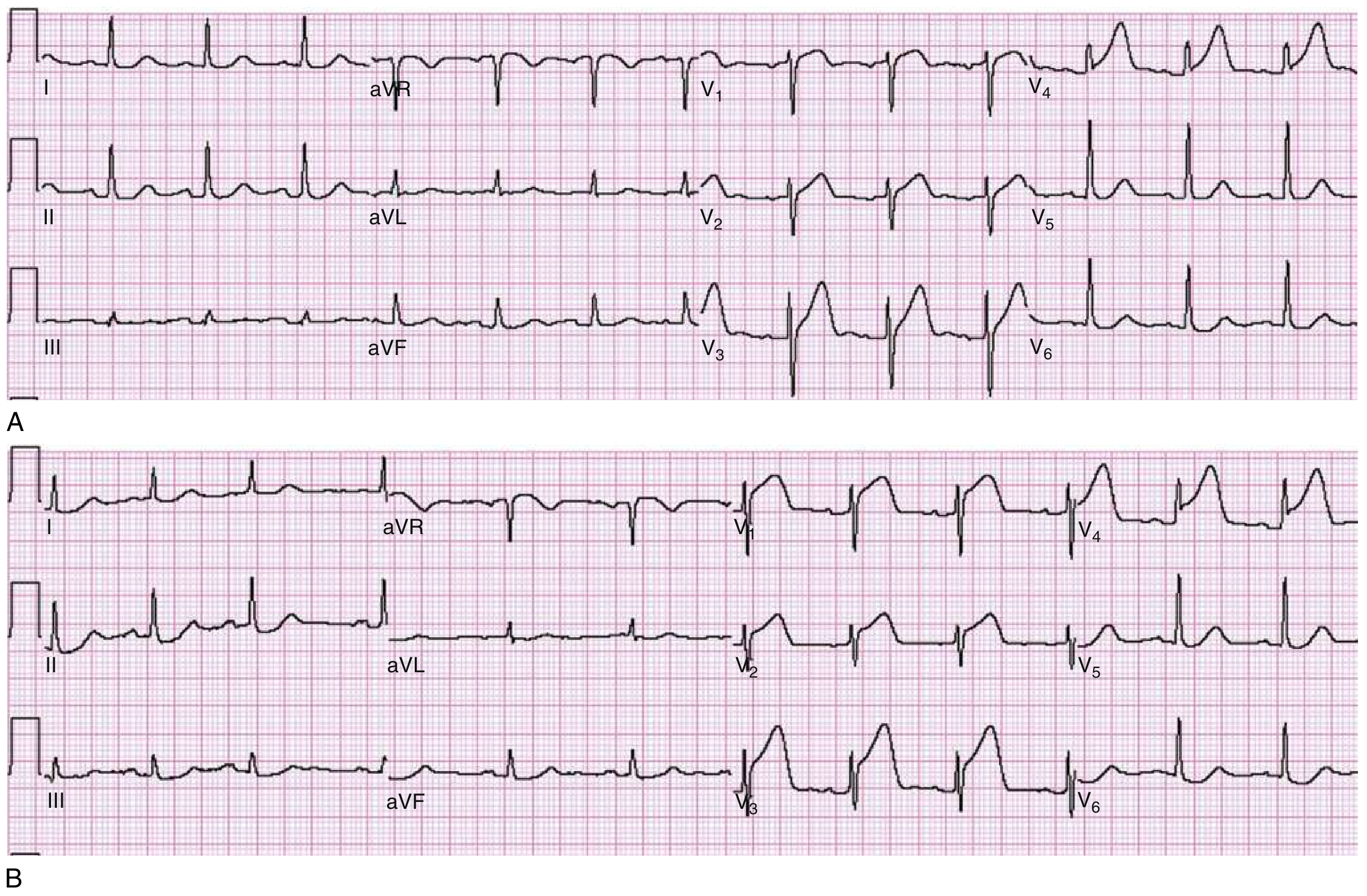
2. NSTEMI / Unstable Angina
- Horizontal or downsloping ST depression (>0.5 mm) in 2+ contiguous leads
- T wave inversion (symmetrical, deep)
- ST elevation in posterior leads (V8-V9) may indicate true posterior MI
- ECG may be normal - does NOT rule out NSTEMI
3. Atrial Fibrillation (AF)
- No visible P waves (replaced by irregular fibrillatory baseline "f" waves)
- Irregularly irregular RR intervals
- Normal narrow QRS (unless aberrant conduction)
4. Atrial Flutter
- Sawtooth flutter waves at ~300 bpm (negative in II, III, aVF)
- Regular RR intervals with 2:1, 3:1, or 4:1 block (ventricular rate ~150, 100, or 75 bpm)
- No true P waves
5. Heart Blocks
| Type | ECG Finding | Disease |
|---|---|---|
| 1st degree | PR interval >200 ms, every P conducts | Vagal tone, inferior MI, digoxin |
| 2nd degree Mobitz I (Wenckebach) | PR progressively lengthens then a QRS drops | Inferior MI, AV nodal disease |
| 2nd degree Mobitz II | Fixed PR, random dropped QRS (no warning) | Bundle of His disease, anterior MI |
| 3rd degree (complete) | P waves and QRS completely dissociated, escape rhythm | Inferior MI, Lyme disease, digoxin toxicity |
6. Bundle Branch Blocks
Left Bundle Branch Block (LBBB)
- QRS >120 ms
- Broad notched R in I, V5, V6 ("M" pattern)
- Deep S in V1 ("W" pattern)
- ST elevation in V1-V2 (discordant)
- New LBBB with chest pain = STEMI-equivalent until proven otherwise
Right Bundle Branch Block (RBBB)
- QRS >120 ms
- rSR' ("rabbit ears") in V1
- Wide S wave in I, V5, V6
- Seen in: PE, anterior MI, cor pulmonale, normal variant
7. Acute Pericarditis
- Diffuse saddle-shaped ST elevation in nearly all leads (except aVR and V1, which show ST depression)
- PR depression in II, V4-V6 (pathognomonic - due to atrial inflammation)
- PR elevation in aVR
- No reciprocal ST depression (unlike STEMI)
- No Q waves
8. Pulmonary Embolism (PE)
- Often non-specific changes
- Classic (but insensitive) S1Q3T3 pattern: deep S in I, Q wave in III, T inversion in III
- Sinus tachycardia (most common ECG finding)
- New RBBB or incomplete RBBB
- Right axis deviation
- T wave inversions in V1-V4 (right heart strain)
- Atrial fibrillation
9. Left Ventricular Hypertrophy (LVH)
- Tall R in lateral leads (I, aVL, V5, V6) + deep S in V1-V2
- Sokolow-Lyon criteria: S in V1 + R in V5 or V6 >35 mm
- ST depression + T wave inversion in lateral leads ("LVH with strain")
- May mimic STEMI (pseudo-infarction pattern)
10. Hyperkalemia
| K+ Level | ECG Change |
|---|---|
| Mild (~5.5-6.5 mEq/L) | Tall, peaked, narrow ("tented") T waves |
| Moderate (~6.5-7.5) | Widening of QRS, PR prolongation, flat P waves |
| Severe (>7.5) | Sine wave pattern - QRS merges with T wave |
| Critical | Ventricular fibrillation, asystole |
11. Hypokalemia
- Prominent U waves (after T wave, especially in V2-V3)
- Flat or inverted T waves
- ST depression
- QT prolongation (actually QU prolongation)
12. Long QT Syndrome
- QTc >440 ms (men), >460 ms (women)
- Risk of Torsades de Pointes (polymorphic VT)
- Causes: congenital (LQT1, LQT2, LQT3 syndromes), drugs (antiarrhythmics, antipsychotics, macrolides), hypokalemia, hypomagnesemia
13. Brugada Syndrome
- Right bundle branch block morphology + ST elevation in V1-V3 (Type 1: "coved" shape)
- ST elevation >2 mm with downward slope to inverted T wave
- Risk of sudden cardiac death from VF
- Sodium channelopathy (SCN5A mutation)
14. Wolff-Parkinson-White (WPW) Syndrome
- Short PR interval (<120 ms)
- Delta wave - slurred upstroke of QRS complex
- Wide QRS complex
- Pseudo-ST and T changes (secondary to abnormal depolarization)
- Risk of AF conducting rapidly down accessory pathway = life-threatening
15. Vasospastic (Prinzmetal's) Angina
- Transient ST elevation during episodes of spasm (spontaneous or provoked)
- ST elevation resolves completely with nitroglycerin or after spasm terminates
- Can trigger malignant arrhythmias
- Typically nocturnal or early morning
16. Digoxin Effect
- "Reverse tick" or "scooped" ST depression in lateral leads
- Shortened QT
- Prominent U waves
- (Note: digoxin toxicity causes AV blocks, bradycardia, PVCs in bigeminy)
17. Atrial Enlargement
| Type | ECG Finding |
|---|---|
| Left atrial enlargement | Broad notched P wave ("P mitrale") >120 ms, biphasic P in V1 |
| Right atrial enlargement | Tall peaked P wave ("P pulmonale") >2.5 mm in II, III, aVF |
18. Ventricular Tachycardia (VT)
- Wide QRS tachycardia (QRS >120 ms) at rate >100 bpm
- AV dissociation (P waves march independently)
- Fusion beats and capture beats (pathognomonic of VT)
- Concordance in precordial leads (all positive or all negative V1-V6)
Quick Reference Summary Table
| ECG Finding | Think Of |
|---|---|
| ST elevation (convex, territorial) | STEMI |
| Diffuse ST elevation (concave) + PR depression | Acute pericarditis |
| ST depression + T inversion | NSTEMI, ischemia, LVH strain |
| No P waves + irregular rhythm | Atrial fibrillation |
| Sawtooth P waves ~300/min | Atrial flutter |
| Progressive PR lengthening + dropped beat | Mobitz I (Wenckebach) |
| Fixed PR + randomly dropped QRS | Mobitz II |
| P-QRS dissociation | Complete (3rd degree) heart block |
| Tall peaked T waves | Hyperkalemia |
| Prominent U waves | Hypokalemia |
| Short PR + delta wave | WPW |
| Prolonged QTc | Long QT syndrome, drugs |
| RSR' in V1 + wide S in V1 | RBBB |
| Broad M-shaped R in I/V6 | LBBB |
| Coved ST elevation V1-V3 | Brugada syndrome |
| S1Q3T3 + sinus tach | Pulmonary embolism |
Sources:
- Rosen's Emergency Medicine: Concepts and Clinical Practice, Chapter 64 (Electrocardiographic Abnormalities in ACS, ST Segment Elevation, Differential Diagnosis)
- Fuster and Hurst's The Heart, 15th Edition (Vasospastic Angina, PE Electrocardiography)
- Guyton and Hall Textbook of Medical Physiology (Atrial Fibrillation ECG)
- Textbook of Family Medicine 9e (Arrhythmias - Long QT, Brugada)
This is a shared conversation. Sign in to Orris to start your own chat.