
Ca cervix staging
I'll look this up in the medical library for an authoritative answer on cervical cancer staging.
Reading File
Now let me search the library and PubMed in parallel for comprehensive staging information.
Finding Sources
Searching PubMed
"cervical cancer" AND "FIGO staging" AND 2021
Finding Sources
Reading Article
PMID: 35295012
Finding Sources
Finding Sources
The main staging content is in Berek & Novak's Gynecology. Let me read it in full.
Reading File
Reading File
I have all the key staging content. Let me also read the staging figure description and the 2008 table for completeness.
Reading File
I now have comprehensive staging data. Let me fetch the staging diagram too.
Loading Image
Excellent - I have the staging diagram and all the data I need. Here is the complete answer:
Carcinoma of the Cervix - FIGO Staging (2018)
The 2018 FIGO staging system replaced the 2008 version and is now the current standard. The key change is that it incorporates imaging and pathologic findings (not just clinical examination) to determine tumor size, extent, and lymph node status. Where pathological evaluation is available, it supersedes clinical and radiological findings.
Rule: When there is doubt about which stage to assign, always select the earlier stage. Once treatment begins, the stage cannot be changed.
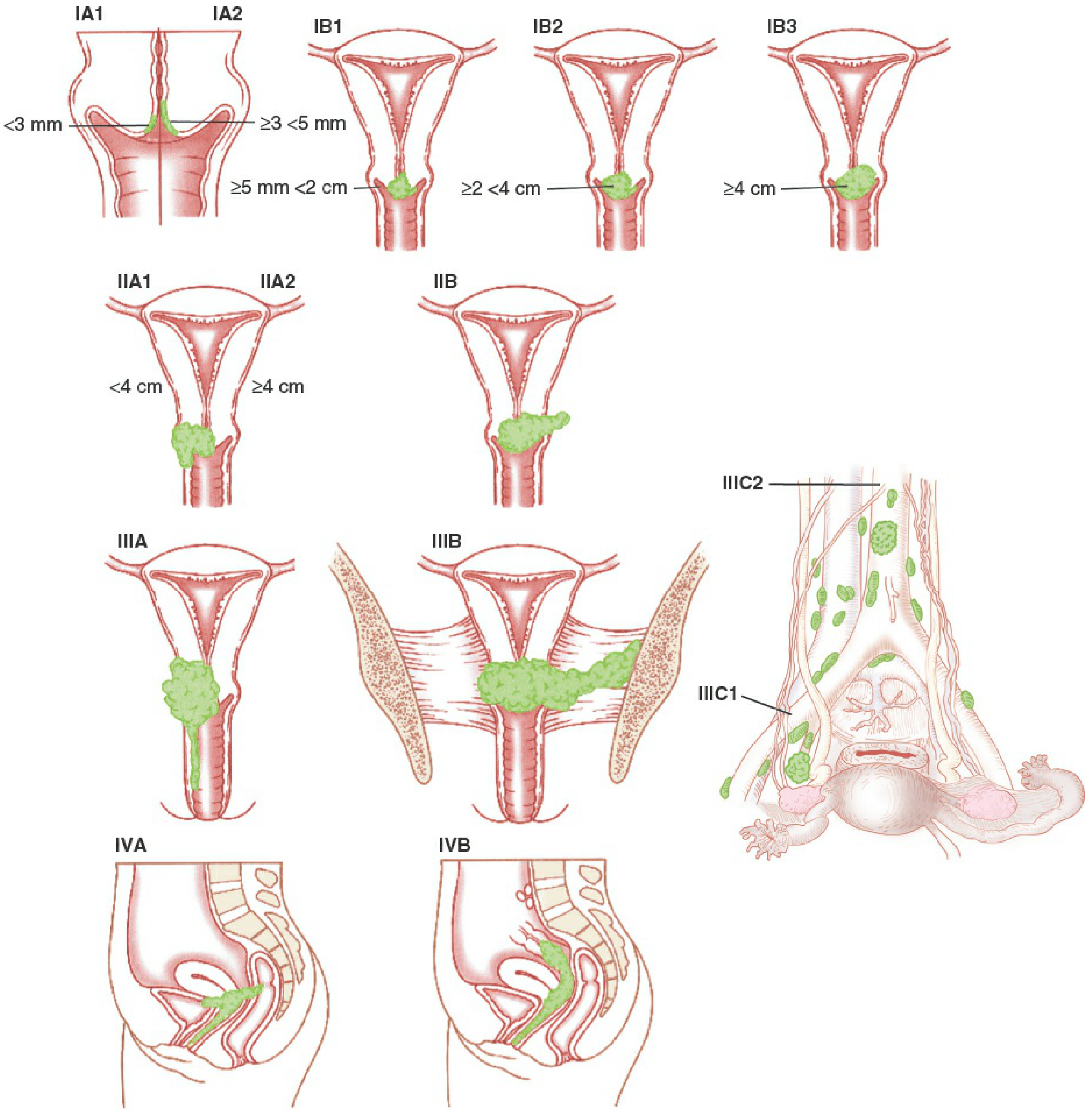
Stage I - Confined Strictly to the Cervix
(Extension to the uterine corpus is disregarded)
| Sub-stage | Definition |
|---|---|
| IA | Invasive carcinoma diagnosed only by microscopy; maximum depth of invasion <5 mm |
| IA1 | Measured stromal invasion <3 mm in depth |
| IA2 | Measured stromal invasion ≥3 mm and <5 mm in depth |
| IB | Invasive carcinoma with deepest invasion ≥5 mm, lesion limited to the cervix |
| IB1 | Invasion ≥5 mm depth, <2 cm in greatest dimension |
| IB2 | ≥2 cm and <4 cm in greatest dimension |
| IB3 | ≥4 cm in greatest dimension |
2018 Change: IB now has 3 sub-stages (was 2 in 2008). IB1 (<2 cm) was separated to reflect fertility-sparing surgical options. In 2018, horizontal spread is no longer considered in stage IA - only depth of invasion matters.
Stage II - Beyond Uterus, Not to Pelvic Wall or Lower Third of Vagina
| Sub-stage | Definition |
|---|---|
| IIA | Involvement of upper two-thirds of vagina, no parametrial involvement |
| IIA1 | <4 cm in greatest dimension |
| IIA2 | ≥4 cm in greatest dimension |
| IIB | Parametrial involvement, not reaching the pelvic wall |
Stage III - Extends to Pelvic Wall and/or Lower Vagina, or Causes Hydronephrosis, or Lymph Node Involvement
| Sub-stage | Definition |
|---|---|
| IIIA | Involves lower third of vagina, no extension to pelvic wall |
| IIIB | Extension to pelvic wall and/or hydronephrosis or non-functioning kidney (unless from another cause) |
| IIIC | Involvement of pelvic and/or para-aortic lymph nodes (regardless of tumor size/extent) |
| IIIC1 | Pelvic lymph node metastasis only |
| IIIC2 | Para-aortic lymph node metastasis |
2018 Change: Stage IIIC is entirely new - lymph node metastasis now moves a patient to at least stage III regardless of the primary tumor size. A notation is added: "r" if imaging-based (e.g., IIIC1r) or "p" if pathology-proven (e.g., IIIC1p).
Stage IV - Spread Beyond the True Pelvis or Involvement of Bladder/Rectal Mucosa
| Sub-stage | Definition |
|---|---|
| IVA | Spread to adjacent bladder or rectal mucosa (bullous oedema alone does not qualify) |
| IVB | Distant metastasis (including peritoneal spread, supraclavicular, mediastinal, or para-aortic nodes above the renal vessels) |
Key Changes: 2018 vs 2008
| Feature | 2008 | 2018 |
|---|---|---|
| Staging basis | Clinical examination only | Clinical + imaging + pathology |
| Stage IA criteria | Depth ≤5 mm AND width ≤7 mm | Depth <5 mm only (width removed) |
| Stage IB sub-stages | IB1 (<4 cm), IB2 (≥4 cm) | IB1 (<2 cm), IB2 (2-4 cm), IB3 (≥4 cm) |
| Lymph node metastasis | Not separately staged | New Stage IIIC (IIIC1 pelvic, IIIC2 para-aortic) |
| Best imaging for early disease | Not specified | MRI preferred (best soft-tissue resolution) |
Incidence by Stage at Diagnosis
- Stage I: 38%
- Stage II: 32%
- Stage III: 26%
- Stage IV: 4%
Imaging Roles
- MRI: Best for early-stage disease (soft-tissue resolution, parametrial invasion assessment)
- CT (contrast-enhanced): Lymph node metastasis, distant spread
- FDG-PET/CT: Useful in advanced disease, especially para-aortic and distant node detection
- When CT/MRI/PET shows nodal abnormality, radiographic-guided FNA can confirm metastatic disease
Sources:
- Berek & Novak's Gynecology, Table 38-1 (FIGO 2018 staging)
- Bhatla N et al., Implications of the revised cervical cancer FIGO staging system, Indian J Med Res 2021 (PMID 35295012)
- Goldman-Cecil Medicine, Chapter 184
This is a shared conversation. Sign in to Orris to start your own chat.