
Non carious cervical l3sions etiology, management and diagnosis
"non-carious cervical lesions" AND etiology
"non-carious cervical lesions" AND management
PMID: 38986732
non-carious cervical lesion abfraction abrasion erosion tooth
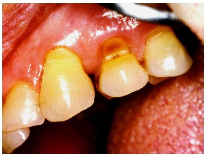
This clinical photograph is an intraoral image showing a classic example of dental abfraction on a premolar. The image highlights a non-carious cervical lesion (NCCL) characterized by a deep, wedge-shaped or V-shaped notch located at the cemento-enamel junction (CEJ). The defect exhibits a sharp internal angle and brownish discoloration within the groove, suggesting exposed dentin. The surrounding gingival tissue shows signs of recession and mild inflammation. This pathology is clinically significant in dentistry as it illustrates the loss of tooth substance due to mechanical loading and tooth flexure during occlusal stress, distinct from other forms of tooth wear such as attrition, erosion, or abrasion. The educational focus is on identifying biomechanically-induced hard tissue loss and distinguishing it from chemical or friction-related dental wear.
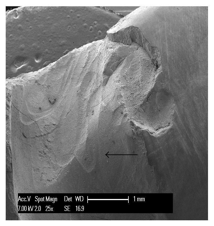
Scanning Electron Micrograph (SEM) at 25x magnification showing a non-carious cervical lesion (NCCL) in an upper human canine. The image displays a complex wedge-shaped abfraction lesion characterized by a deep, irregular morphology with clearly defined margins. Key visual features include multiple internal cavities and partially overlapping furrows. The surface texture exhibits a smear layer and distinctive mechanical wear patterns, specifically parallel striations alternating with protruding crests, indicating a superimposed process of mechanical abrasion. A black arrow points toward a prominent cavity within the lesion where these striations are visible. The imaging highlights the structural damage to dental hard tissues—enamel and dentin—and illustrates the multifactorial nature of tooth wear where tensile stress (abfraction) and physical friction (abrasion) coexist. This diagnostic image is intended for dental education regarding the classification and microscopic analysis of non-carious tooth loss.
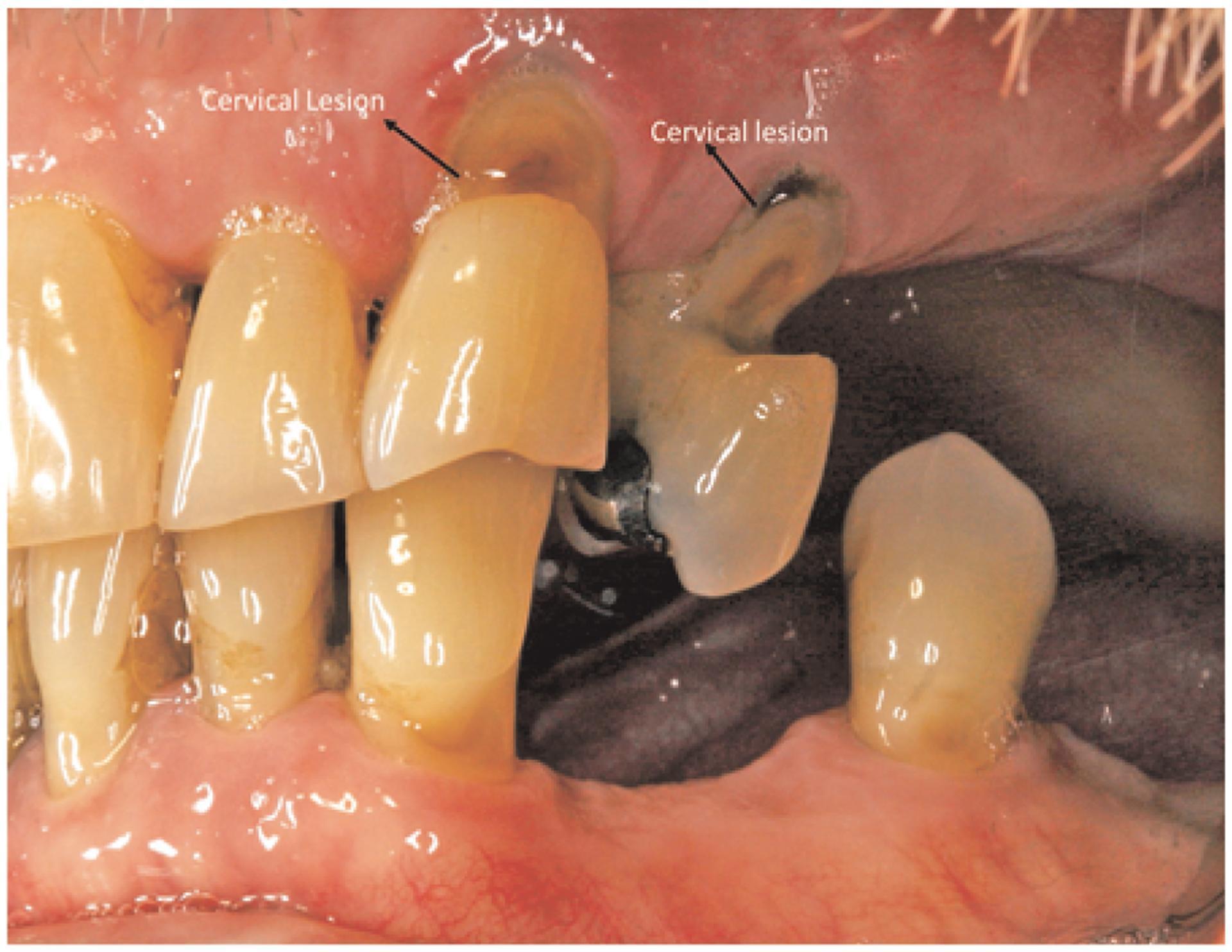
This clinical intraoral photograph demonstrates non-carious cervical lesions (NCCLs) on multiple teeth. The image highlights two primary areas of interest with arrows and text labels. One lesion, located on an upper tooth toward the center of the image, presents as an oval-shaped, yellowish-brown area of tooth loss at the cervical margin, near the gingival line. A second lesion to the right appears more advanced and darker in color, suggesting significant demineralization or staining. Both lesions occur at the cementoenamel junction, characteristic of conditions such as dental erosion, abrasion, or abfraction. The surrounding gingival tissue shows clear signs of pathology, including significant gingival recession, exposure of the anatomical roots, and generalized erythema indicating chronic inflammation. The interdental papillae appear blunted, and there is visible tooth crowding and malalignment. This image is educational for dentistry students and clinicians focusing on periodontology and restorative dentistry, illustrating the relationship between cervical tooth structure loss and gingival health.
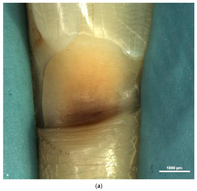
This stereomicroscopic diagnostic image (15x magnification) displays a clinical view of a human lower incisor affected by a prominent Non-Carious Cervical Lesion (NCCL). The lesion is located at the cervical margin, exhibiting a characteristic wedge-shaped morphology with a sharp internal angle. There is significant loss of hard dental tissue, with the lesion floor demonstrating exposed dentin and a dark brownish discoloration indicative of chronic mineral loss or extrinsic staining. Micromorphological features include a rough, irregular surface texture marked by visible furrows, cracks, and scratches. The adjacent enamel appears relatively intact, though fine crazing lines are visible on the tooth surface surrounding the defect. This image illustrates the macromorphological impact of mechanical or chemical wear—such as abfraction, abrasion, or erosion—on the dental hard tissues at the cementoenamel junction, serving as a primary educational example of wedge-shaped NCCLs for dental professionals and students.
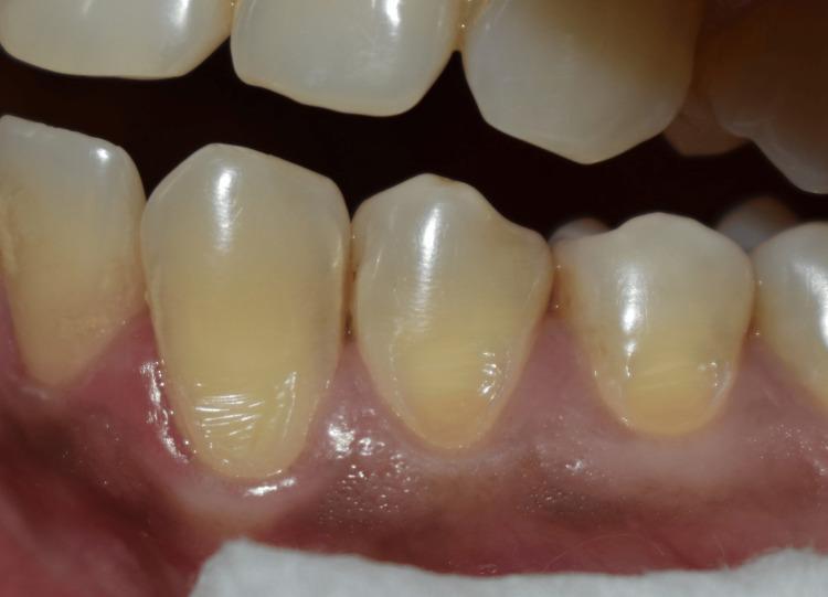
This intraoral clinical photograph provides a close-up view of the mandibular teeth, specifically highlighting a non-carious cervical lesion (NCCL) on tooth 34. The image illustrates the typical morphology of an NCCL, characterized by a smooth, wedge-shaped or saucer-shaped loss of hard tissue at the cementoenamel junction (CEJ). The affected tooth surface exhibits a glossy, yellowish-white enamel texture with distinct horizontal and vertical striations within the defect. The surrounding gingiva appears healthy, pink, and scalloped, with a slight apical recession following the contour of the lesion. To ensure a clear operative field for clinical study, soft tissue management is achieved through retraction using cotton rolls and a saliva ejector, which are visible at the inferior border of the frame. This image is an educational example for restorative dentistry and periodontology, demonstrating the clinical presentation of abfraction, erosion, or abrasion before the application of topical anesthesia or restorative composite materials.
non-carious cervical lesions etiology diagnosis management review 2023 2024
https://rsdjournal.org/rsd/article/view/41876
Non-Carious Cervical Lesions (NCCLs)
Definition
Clinical Images
Morphological Classification
| Pattern | Shape | Internal Angle | Etiology |
|---|---|---|---|
| Wedge-shaped | V-shaped notch | Acute (sharp) | Mechanical abrasion / abfraction |
| Disc/saucer-shaped | Shallow, rounded depression | Obtuse (wide) | Chemical erosion |
Epidemiology
- Prevalence increases significantly with age
- Most commonly found on buccal/vestibular surfaces
- Premolars and canines are most frequently affected (higher occlusal stress concentration)
- Can affect any permanent tooth
- Associated with gingival recession in many cases
Etiology (Multifactorial)
1. Abrasion (Mechanical)
- Loss of tooth structure from friction by external agents
- Most common cause: aggressive toothbrushing (hard bristles, horizontal scrubbing, abrasive toothpastes)
- Other causes: oral piercings, occupational habits (e.g., holding pins with teeth), nail biting
- Produces wedge-shaped lesions with sharp angles and a smooth, polished floor
- More pronounced on the side of the dominant hand (right-handed patients show more lesions on left side)
2. Erosion / Biocorrosion (Chemical)
- Loss of tooth structure from acid dissolution, without bacterial involvement
- Extrinsic sources: carbonated drinks, citric acid foods and juices, sports drinks, vinegar, wine
- Intrinsic sources: gastroesophageal reflux disease (GERD), bulimia nervosa (frequent vomiting)
- Salivary factors: decreased salivary flow (xerostomia) reduces buffering capacity and pellicle protection
- Produces smooth, shallow, saucer/cup-shaped lesions
- Softened enamel from erosion makes teeth far more susceptible to abrasion
3. Abfraction (Biomechanical)
- Proposed mechanism: eccentric occlusal loading causes tooth flexure, concentrating tensile and compressive stresses at the CEJ
- First described by Lee & Eakle (1984) and further classified by Grippo (1991)
- Enamel - brittle and weak under tension - fractures at stress concentration points
- Associated with: bruxism, parafunction, premature contacts, steep cuspal guidance, deep bite
- Position of lesion depends on direction of lateral forces; size depends on magnitude
- Still debated: some systematic reviews (Duangthip et al., 2017) find moderate evidence linking occlusal stress to NCCL formation, but direct causation is not universally proven
- Micro-cracks from abfraction increase susceptibility to erosion and abrasion
4. Attrition
- Tooth-to-tooth contact wear (less common at the cervical area; more relevant at occlusal surfaces)
- Contributes when combined with other mechanisms
Key Risk Factors Summary
| Factor | Mechanism |
|---|---|
| Hard toothbrush + horizontal technique | Abrasion |
| Acidic diet (sodas, citrus) | Erosion |
| GERD, eating disorders | Intrinsic erosion |
| Bruxism, clenching | Abfraction |
| Reduced salivary flow | Reduced buffering / pellicle |
| Thin enamel at CEJ | Increased susceptibility |
| Gingival recession | Exposes root dentin (softer) |
| Malalignment, premature contacts | Stress concentration |
Diagnosis
History Taking
- Dietary history: frequency of acidic food/drink, carbonated beverage intake, citrus consumption
- Medical history: GERD, reflux, eating disorders, xerostomia-causing medications
- Oral hygiene habits: brushing frequency, technique, type of brush/toothpaste (abrasivity - RDA value)
- Parafunctional habits: bruxism (nocturnal), clenching, nail biting
- Symptoms: dentin hypersensitivity (sharp, transient pain to thermal/tactile stimuli)
Clinical Examination
- Lesion morphology: shape (wedge vs. saucer), depth, surface texture (smooth and hard vs. rough)
- Location: buccal surface, relation to CEJ, which teeth affected
- Gingival assessment: recession (Miller's or Cairo class), attachment level
- Occlusal assessment: wear facets, fremitus, premature contacts, crossbite
- Tooth surface loss indices: BEWE (Basic Erosive Wear Examination) is commonly used for grading severity
Differential Diagnosis
| Condition | Distinguishing Features |
|---|---|
| Dental caries | Soft, discolored lesion; bacteria-induced; positive caries tests |
| Developmental defects (MIH, enamel hypoplasia) | Location not restricted to CEJ; irregular distribution; childhood history |
| Erosion alone | Smooth, rounded, shallow; multi-surface involvement; dietary/GERD history |
| Abrasion alone | Wedge-shaped; linked to oral hygiene habits; unilateral/asymmetric pattern |
| Abfraction alone | Wedge-shaped; linked to occlusal forces; consistent with stress distribution |
| Attrition | Flat, polished wear at occlusal/incisal surfaces primarily |
Diagnostic Tools
- BEWE Index - scores erosive wear on 0-3 scale per sextant; widely used in screening
- Stereomicroscopy / SEM - research tool to analyze surface microstructure and striations
- 3D morphological analysis - used in research to track lesion progression (Hayashi et al., 2022)
- Intraoral photography - documents baseline lesion size for monitoring
Management
Step 1: Etiological / Preventive Management
| Causative Factor | Intervention |
|---|---|
| Aggressive brushing | Soft toothbrush; modify to circular/Bass technique; reduce pressure |
| Abrasive toothpaste | Switch to low-RDA paste (RDA < 70 preferred) |
| Acidic diet | Reduce frequency and amount; avoid acids before brushing; use straw |
| GERD | Referral for medical management; antacids; avoid eating before sleep |
| Eating disorders | Multidisciplinary referral (psychology, nutrition) |
| Bruxism / clenching | Occlusal splint (Michigan/stabilization); manage stress |
| Dry mouth | Saliva substitutes; stimulate salivary flow (chewing gum with xylitol) |
| Premature contacts | Occlusal adjustment (selective grinding) |
Step 2: Desensitization
- In-office: fluoride varnish (5% NaF), potassium oxalate, resin sealants, dentin bonding agents, laser therapy (Nd:YAG, Er:YAG)
- At-home: desensitizing toothpastes containing potassium nitrate (5%), stannous fluoride, nano-hydroxyapatite, or arginine-calcium carbonate
- Remineralization agents: fluoride gels, CPP-ACP (casein phosphopeptide-amorphous calcium phosphate) products
- S-PRG (surface pre-reacted glass ionomer) materials show emerging evidence for remineralization and desensitization (Ferreira et al., 2026 - PMID 41309331)
Step 3: Restorative Management
- Active lesion progressing despite preventive treatment
- Dentin hypersensitivity uncontrolled by non-restorative means
- Aesthetic concern
- Risk of pulpal exposure
- Structural compromise
- Adhesive bonding to cervical area
- Best long-term retention with selective etch technique (enamel) + self-etch primer on dentin
- Cavosurface margin beveling for enamel when present
- Avoid over-preparation; often no cavity preparation needed (superficial polish only)
- Retainer for composite: cervical matrix, wedges
- Used when moisture control is difficult (sub-gingival lesions)
- Fluoride-releasing - additional caries/erosion protection
- Lower wear resistance than composite; used in non-stress-bearing areas
Step 4: Periodontal Management (when gingival recession is present)
- Connective tissue graft (CTG) + coronally advanced flap (CAF): Gold standard for root coverage; simultaneous composite restoration of the NCCL
- A 2026 updated recommendation (Santamaria et al., J Periodontol 2026 - PMID 41498281) provides evidence-based guidance on sequencing root coverage procedures with NCCL restoration
- Timing debate: some advocate restoring the NCCL first (to improve flap adaptation), others perform simultaneous grafting + restoration
Summary Table
| Aspect | Key Points |
|---|---|
| Definition | Loss of hard tissue at CEJ, non-bacterial origin |
| Most affected teeth | Premolars, canines; buccal surfaces |
| Main causes | Abrasion (brushing), erosion (acids, GERD), abfraction (occlusal stress) |
| Morphology | Wedge (mechanical) vs. saucer (erosion) |
| Diagnosis | Clinical history + examination + BEWE index |
| First-line management | Etiological control + behavior modification |
| Symptomatic relief | Desensitizing agents, fluoride, dentin sealers |
| Restorative material | Composite resin (preferred); GIC (wet/subgingival) |
| With recession | Combined CAF/CTG + composite restoration |
- Goodacre CJ et al. (2023). Noncarious cervical lesions: morphology and progression, prevalence, etiology, pathophysiology, and clinical guidelines for restoration. J Prosthodont, 32(2):e1-e18
- Grippo JO, Simring M, Coleman TA. (2012). Abfraction, abrasion, biocorrosion, and the enigma of noncarious cervical lesions: a 20-year perspective. J Esthet Restor Dent, 24(1):10-23
- Patano A et al. (2023). Conservative Treatment of Dental NCCLs: A Scoping Review. Biomedicines, 11(6):1530
- Santamaria MP et al. (2026). Updated evidence-based recommendation for treatment of gingival recession associated with NCCLs. J Periodontol - PMID 41498281
- Denucci GC, Stone S, Hara AT. (2024). Noncarious cervical lesions: A bibliometric analysis and future research trends. J Dent - PMID 38986732