explain heave
- Geology/Geotechnics - the horizontal displacement component of a fault, or upward swelling of soil/rock (e.g., frost heave, floor heave in excavations)
- Nautical/Maritime - one of the six degrees of motion of a ship (up-and-down vertical movement of the hull)
- Medicine - retching or the act of vomiting (e.g., "dry heaves")
- Structural engineering - upward movement of foundations or floors due to expansive soils, frost, or excavation stress relief
Which type of heave are you asking about?
in cardiology
cardiac heave precordium chest examination
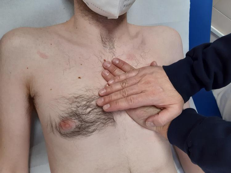
A clinical photograph depicts a procedural technique in osteopathic or physical assessment of the thoracic region. A patient is positioned supine on a medical examination table, with a clinician's hands performing palpatory listening over the precordium (heart area). The primary 'listening' hand is placed flat against the patient's upper-left chest, while the second hand is placed directly over the first to apply gentle, controlled pressure. This hand placement corresponds to the anatomical location of the heart and pericardium within the mediastinum. The patient’s chest exhibits fair skin with scattered nevi (moles) and terminal hair distribution concentrated around the nipple and upper sternum. The patient is wearing a white medical mask. The image serves as an educational resource for medical and osteopathic training, illustrating the correct hand positioning for evaluating fascial mobility, rhythmic impulses, or tissue compliance in the cardiac region.
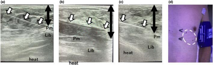
A multi-panel figure consisting of three real-time ultrasound images (a-c) and one clinical photograph (d) demonstrating the procedural steps for an implantable cardiac monitor (ICM) placement using tumescent local anesthesia (TLA). Ultrasound images a, b, and c show the cross-sectional anatomy of the left precordium, including the pectoralis major muscle (Pm), subcutaneous tissue (Lib), and the underlying heart. Image (a) depicts a 23-gauge needle (white arrows) inserted at an angle into the subcutaneous layer for TLA delivery, with a black double-headed arrow marking the target injection depth above the pectoralis major. Image (b) shows the insertion of a plunger stick (white arrows) from an ICM kit used to create a subcutaneous pocket. Image (c) visualizes the final implanted ICM (white arrows) positioned within the subcutaneous tissue. Image (d) is a clinical photograph of the patient’s chest, with a dotted white circle indicating the application area of the TLA on the left precordium. The series highlights ultrasound-guided techniques to ensure precise depth and localization of the device relative to anatomical landmarks.
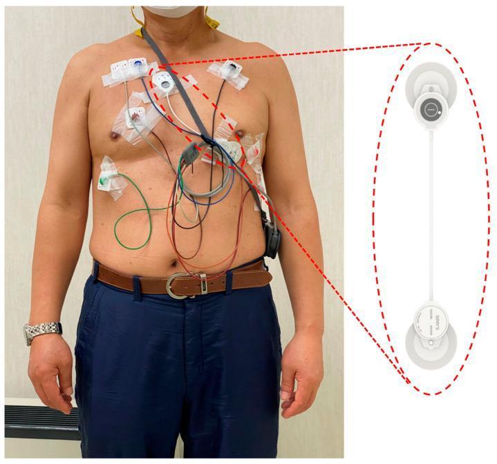
A clinical photograph and product detail illustrating a comparative setup for cardiac monitoring. The left panel shows a male participant wearing two types of Electrocardiogram (ECG) monitoring systems simultaneously on the precordium: a traditional 3-lead Holter monitor and an adhesive patch-type single-lead ECG device (mobiCARE-MC100). The Holter system consists of multiple adhesive electrodes connected via color-coded wires (red, green, blue, white, black) to a handheld recording unit worn at the waist. In contrast, the single-lead patch device is a compact, wireless system consisting of two circular adhesive electrodes connected by a thin, 120mm flexible white strip, placed diagonally across the chest to record lead II signals. The right panel provides a magnified view of the mobiCARE-MC100 device, highlighting its minimalist, patch-based design. This visual comparison demonstrates the difference in patient burden between conventional wired Holter monitoring and modern wearable adhesive patches used for long-term arrhythmia detection and heart rate variability analysis.
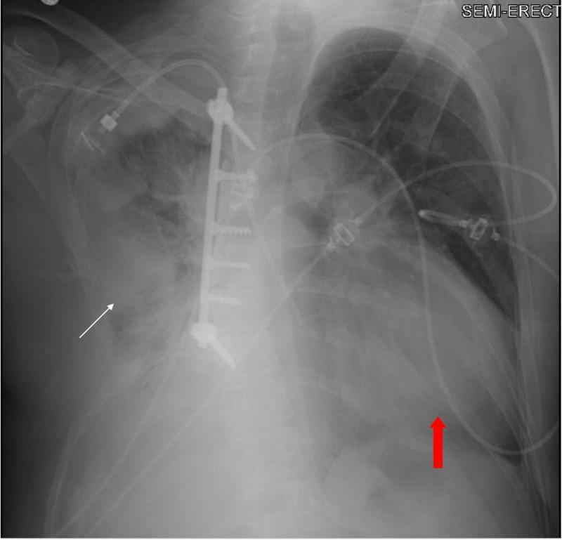
This diagnostic image is a semi-erect anteroposterior (AP) chest radiograph. The image displays a significantly enlarged cardiac silhouette (red arrow), suggesting global cardiomegaly or a large pericardial effusion, which contributes to an increased cardiothoracic ratio. In the right hemithorax, findings are consistent with status post-chest wall resection, featuring a vertical metallic fixation plate and screws along the thoracic wall. Adjacent to this surgical site, there is a dense, irregular area of increased opacity in the right mid-to-lower lung field (white arrow), representative of a remnant or recurrent neoplastic process. Multiple medical devices are visible, including ECG leads and tubing across the precordium. This radiograph illustrates complex post-surgical thoracic anatomy combined with progressive oncological and cardiovascular complications, such as metastatic spread and potential pericardial tamponade physiology.
Heave (Cardiac Heave / Parasternal Heave)
Definition
Types of Heave
1. Right Ventricular (Parasternal) Heave - Most Common
- Location: Left lower sternal border / parasternal region (3rd-5th intercostal spaces)
- Cause: Right ventricular pressure or volume overload causing RV enlargement/hypertrophy
- How to feel it: The examiner places the thenar/hypothenar surface or fingers at the left sternal border - a sustained, forceful outward lift is felt during systole
- Clinical significance: Strongly suggests pulmonary hypertension, right heart failure, pulmonary stenosis, or atrial septal defect (ASD)
2. Left Ventricular Heave (Displaced, Forceful Apex Beat)
- Location: Apex beat (normally 5th intercostal space, midclavicular line) - may be displaced laterally and inferiorly
- Cause: LV hypertrophy, LV dilation, or obstruction to LV outflow (e.g., aortic stenosis, hypertrophic cardiomyopathy)
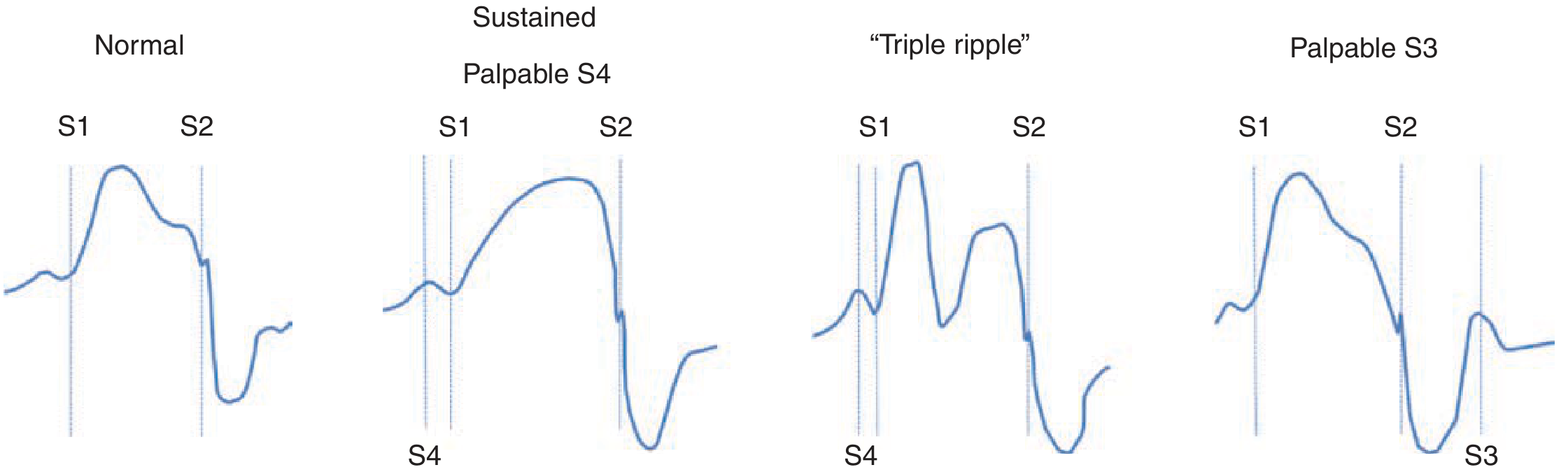
- Character: A sustained (prolonged) apex impulse that "pushes" the examiner's hand - this is the LV equivalent of a heave
- Clinical significance: Aortic stenosis, hypertension with LVH, dilated cardiomyopathy
3. Left-sided chest wall heave (hyperdynamic)
- The left anterior chest wall may heave in patients with an enlarged or hyperdynamic left or right ventricle - per Harrison's 22E
How to Detect a Heave
- Patient position: Supine at 30°; left lateral decubitus position enhances sensitivity
- Technique: Place the flat of the hand (thenar/hypothenar) or fingertips over the left sternal border
- Normal finding: Brief, gentle outward tap that lifts away quickly
- Abnormal (heave): A sustained, forceful, lifting outward impulse that moves the entire hand
Clinical Causes at a Glance
| Type | Location | Common Causes |
|---|---|---|
| RV heave (parasternal lift) | Left sternal border | Pulmonary hypertension, pulmonary stenosis, ASD, right heart failure |
| LV heave (forceful/sustained apex) | Apex (may be displaced) | Aortic stenosis, hypertension, hypertrophic cardiomyopathy, dilated cardiomyopathy |
| Biventricular heave | Diffuse precordium | Severe biventricular heart failure |
Associated Findings
- Loud or palpable P2 (pulmonary component of S2) - suggests pulmonary hypertension
- Elevated JVP with prominent cv waves - suggests tricuspid regurgitation
- Fixed wide splitting of S2 - suggests ASD
- A right ventricular S4 may also be palpable (presystolic impulse) in pressure-overloaded RV
Apical Impulse Contour (for reference)