
Hey Give me notes on Dimorphic Fungi, it should include Microbiological and Clinical Aspects along with the images.
Dimorphic Fungi - Comprehensive Notes
Definition & Core Concept
- Environmental/Mold phase (25-30°C): Filamentous hyphal form producing conidia (infectious propagules)
- Tissue/Yeast phase (37°C / body temperature): Converts to yeast forms within the host
Exception: Coccidioides spp. do not form yeasts in tissue - instead they produce spherules containing endospores. Emmonsia crescens produces diaspores.
Classification of Clinically Important Dimorphic Fungi
| Organism | Disease | Geography | Entry Route | Tissue Form |
|---|---|---|---|---|
| Histoplasma capsulatum | Histoplasmosis | Ohio/Mississippi River valleys (USA), worldwide | Inhalation | Small intracellular yeasts (2-4 µm) |
| Blastomyces dermatitidis | Blastomycosis (North American) | SE/SC USA, Canada, Africa | Inhalation | Large broad-based budding yeasts (8-15 µm) |
| Coccidioides immitis / C. posadasii | Coccidioidomycosis (Valley fever) | SW USA, Mexico, Central/South America | Inhalation | Spherules (up to 100 µm) with endospores |
| Paracoccidioides brasiliensis | Paracoccidioidomycosis (South American blastomycosis) | South America (Brazil, Colombia) | Inhalation | "Mariner's wheel" - multiple peripheral buds |
| Sporothrix schenckii | Sporotrichosis | Worldwide (warm, humid climates) | Traumatic inoculation | Cigar-shaped yeasts (2-6 µm) |
| Talaromyces marneffei (formerly Penicillium marneffei) | Talaromycosis | Southeast Asia (Thailand, China, Vietnam) | Inhalation | Small oval yeasts with transverse septa |
| Emergomyces spp. | Emergomycosis | South Africa, globally | Inhalation | Yeast |
Geographical Distribution
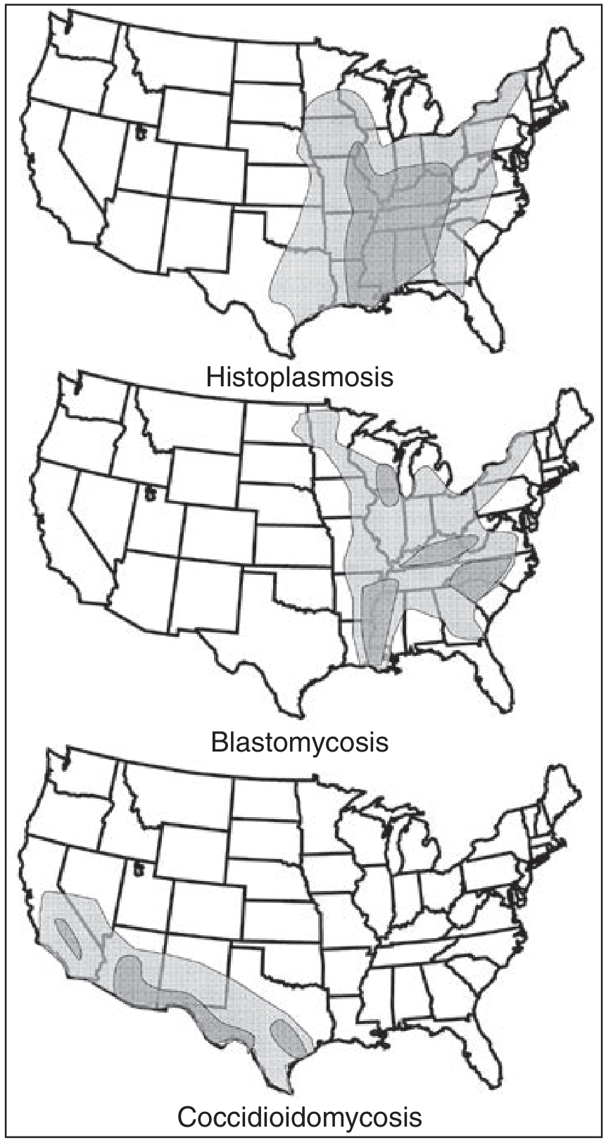
- Histoplasmosis - Ohio, Mississippi, St. Lawrence River valleys; Brazil; French Guiana; parts of Africa and Asia
- Blastomycosis - SE/SC USA, Great Lakes, Africa, rare reports from India and Saudi Arabia
- Coccidioidomycosis - SW USA (Arizona, California), northern Mexico, parts of Central/South America; expanding range now noted in Washington State
- Paracoccidioidomycosis - Exclusively South and Central America; Brazil has highest incidence
- Talaromycosis - Northern Thailand, SW China, Malaysia, Vietnam, northeast India
- Sporotrichosis - Worldwide in warm, humid climates; major urban epidemic in Rio de Janeiro via cat transmission
Shared Characteristics of Endemic Dimorphic Fungi
- Ability to cause disease in a healthy immunocompetent host
- Association with a specific ecologic niche in soil/environment
- Temperature dimorphism - mold at 25-30°C; yeast/spherules at body temperature
- Most cause latent infections that may reactivate years later, especially in immunocompromised hosts
- Clinical presentation may mimic other diseases (TB, malignancy) - travel/residence history is key
1. Histoplasmosis
Microbiology
- Causative agent: Histoplasma capsulatum (recently reclassified into multiple species: H. mississippiense, H. ohiense, H. suramericanum, etc.)
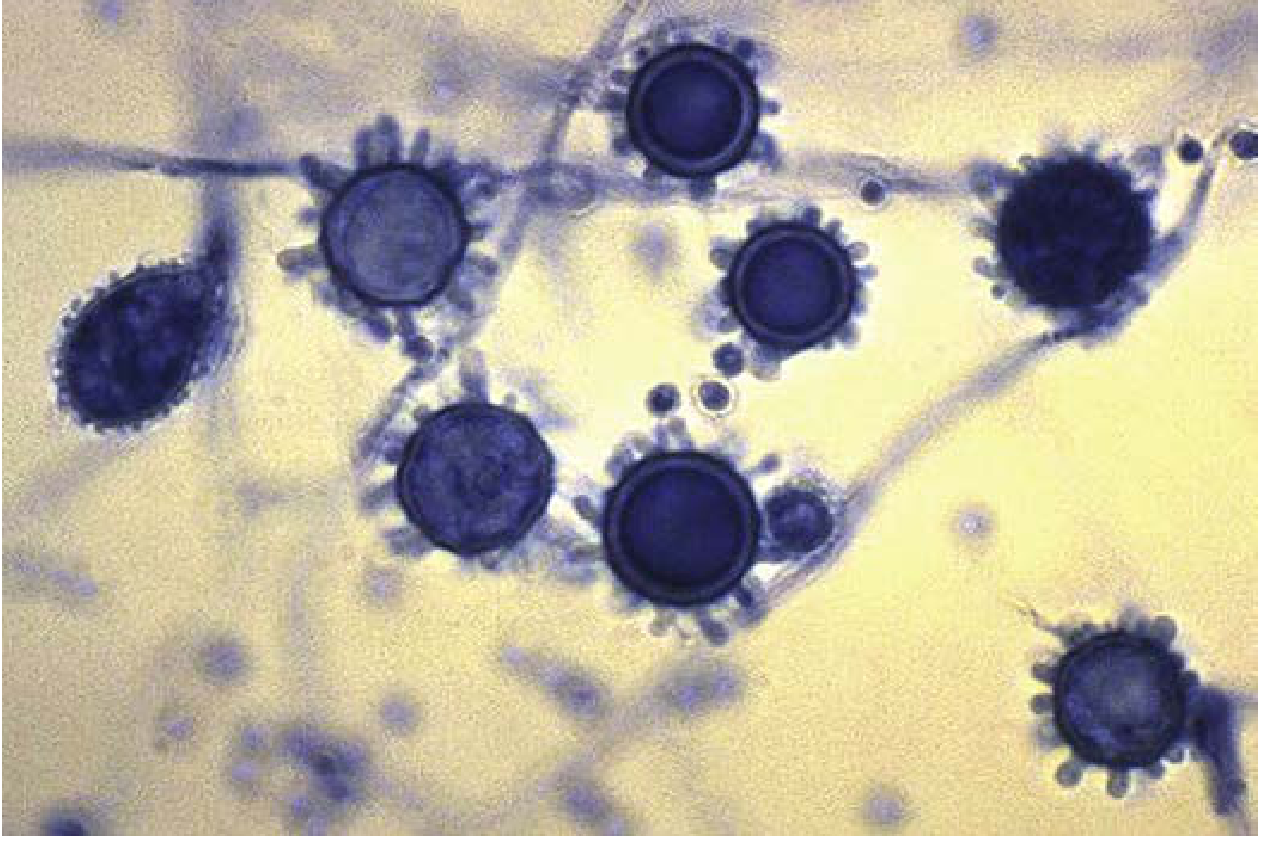
- Mold phase: Produces diagnostic tuberculate macroconidia (roughened, spiny projections) and microconidia; grows on enriched agar (brain-heart infusion) within 10-14 days; white to buff-brown fluffy colonies
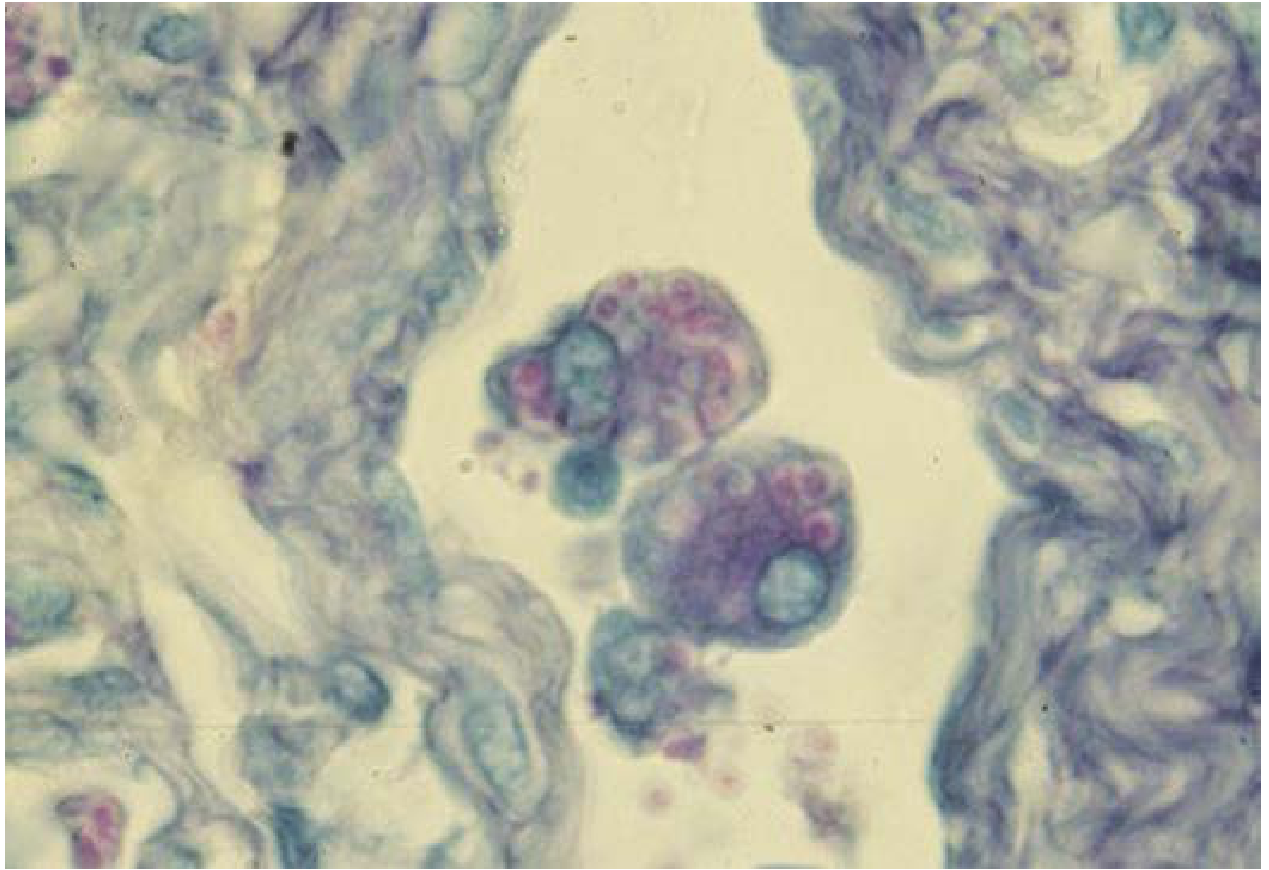
- Yeast phase: Small (2-4 µm), oval, intracellular yeasts within macrophages; multiply by narrow-based budding; NO true capsule (capsule-like artifact on staining)
- Growth stimulated by bird and bat guano in soil; birds are not infected
Clinical Aspects
- Asymptomatic - most common in normal hosts exposed to low spore counts
- Acute pulmonary - flu-like illness with fever, chills, cough, pleuritic chest pain after heavy exposure; self-limiting in immunocompetent; may leave calcified coin lesions
- Granulomatous/fibrosing mediastinitis - enlarged mediastinal lymph nodes that can obstruct airways, pulmonary vessels, or esophagus; fistula formation
- Chronic pulmonary - primarily in COPD patients; cavitation mimics TB
- Disseminated histoplasmosis - in immunosuppressed (HIV/AIDS, TNF-α blockers, transplant) and extremes of age; reticuloendothelial involvement: lymphadenopathy, hepatosplenomegaly, thrombocytopenia; AIDS-defining illness
- Culture (gold standard) - mold form from respiratory specimens; AccuProbe nucleic acid hybridization for confirmation
- Urine antigen detection: sensitivity up to 92% in disseminated disease, 75-80% in acute pulmonary
- Serology (immunodiffusion, complement fixation): delayed by 2-6 weeks; cross-reacts with Blastomyces
- Histopathology: intracellular small yeasts in macrophages (H&E, GMS, PAS)
2. Blastomycosis
Microbiology
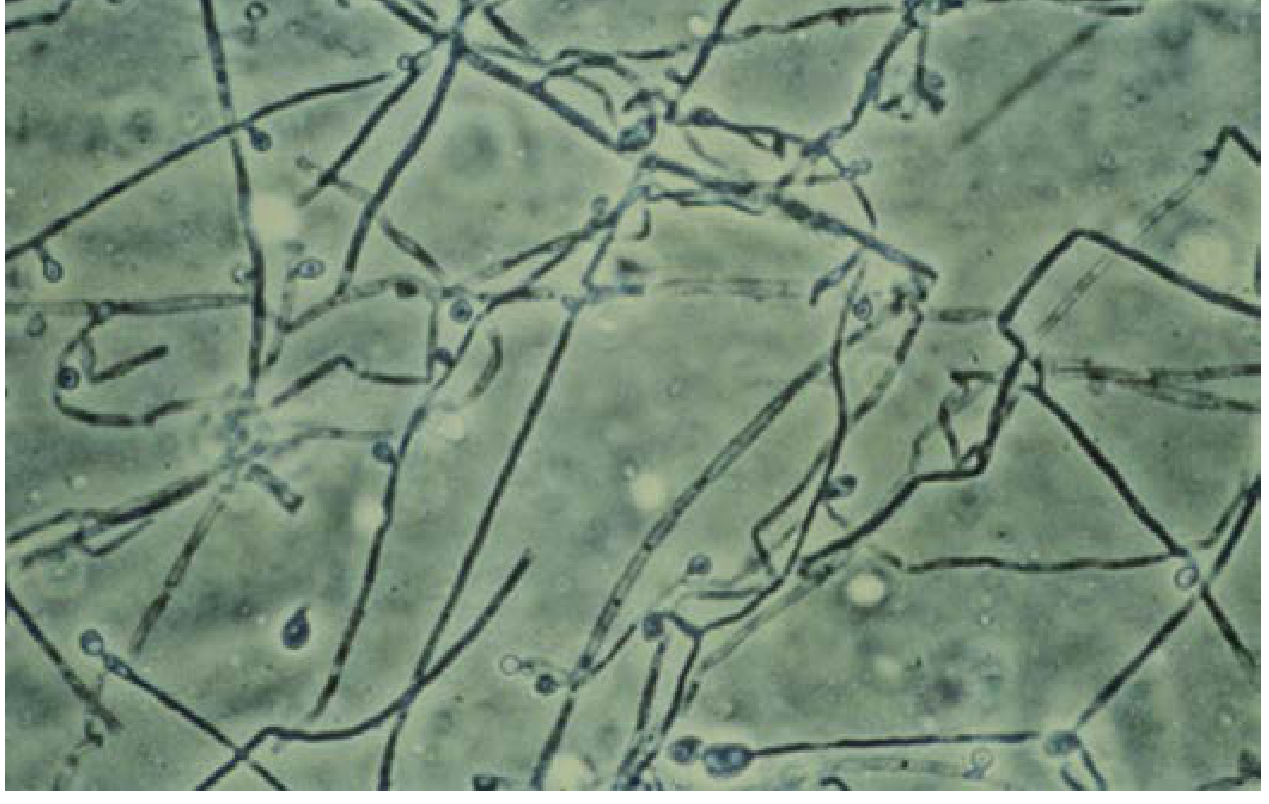
- Mold phase: Fluffy white to buff colonies; microconidia similar to Histoplasma; "lollipop" appearance of conidia on conidiophore at 25°C
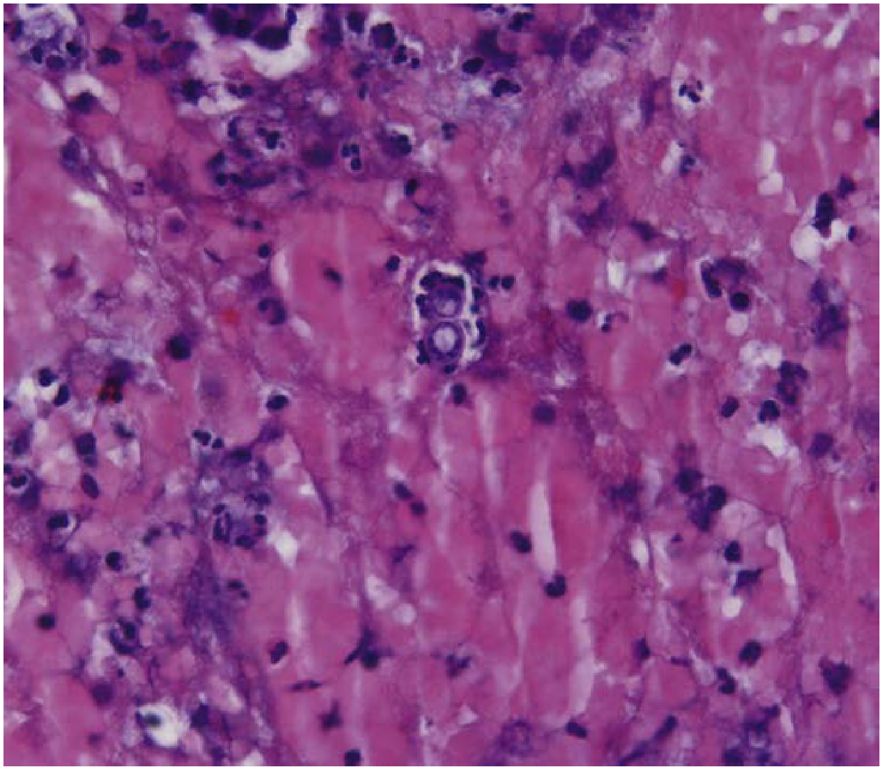
- Yeast phase: Large (8-15 µm), thick-walled yeasts with a characteristic broad-based (wide-neck) single bud - this is the key distinguishing feature
Clinical Aspects
- Called "the great masquerader" due to protean clinical manifestations
- Primarily affects men engaged in rural/outdoor activities
- Lung: Pneumonia (most common); only 5% of pulmonary cases suspected at initial presentation
- Skin: Most frequent extrapulmonary site; pseudoepitheliomatous hyperplasia; verrucous or ulcerated lesions
- Bone and joints: Osteolytic lesions, arthritis
- CNS: Rare but possible
- Histopathology: Mixed suppurative and granulomatous inflammation; microabscesses; yeast cells in giant cells; PAS or GMS staining reveals the thick-walled yeasts; diagnosis often first made by surgical pathology/cytopathology
3. Coccidioidomycosis (Valley Fever)
Microbiology
- Two species: C. immitis (California) and C. posadasii (Arizona and elsewhere) - phenotypically similar, genetically distinct
- Environmental form: Arthroconidia (highly infectious barrel-shaped segments) in soil - BIOSAFETY LEVEL 3 PATHOGEN; arthroconidia easily aerosolized; lab cultures must be handled in BSC
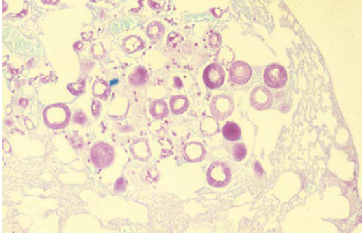
- Tissue form: Spherules (up to 100 µm diameter) containing endospores (2-5 µm); unique among dimorphic fungi - no yeast form
- Grows rapidly in culture (within 1 week); can appear on routine bacteriology media (blood agar)
Clinical Aspects
- Risk populations: Military personnel (SW US bases), construction workers, outdoor workers, immunocompromised; genetic predisposition to dissemination in Blacks, Filipinos, Asians, Native Americans
- Acute pulmonary (Valley Fever): Flu-like illness; self-limiting in most
- Skin manifestations: Erythema nodosum, erythema multiforme ("desert rheumatism")
- Disseminated disease (in immunocompromised, HIV): Skin, bone, joints, meninges
- Meningitis: Indolent/chronic; most feared complication; requires lifelong azole therapy
- Histopathology: Granulomatous with and without caseation; developing spherules in macrophages and giant cells; differential includes non-budding Blastomyces, Cryptococcus, adiaspiromycosis
- Diagnosis: Calcofluor white stain of respiratory specimens; serology (IgM for acute, IgG for complement fixation); Coccidioides-specific PCR; culture (BSC required)
4. Paracoccidioidomycosis
Microbiology
- Species: P. brasiliensis complex (P. brasiliensis, P. americana, P. restrepiensis, P. venezuelensis) and P. lutzii
- Tissue form: Yeast cells with multiple peripheral buds arranged like a "mariner's wheel" or "pilot's wheel" (4-60 µm), highly characteristic
- Female hormones (estrogens) provide protective effect - explains striking male:female ratio of 14:1 to 70:1 in chronic disease
Clinical Aspects
- Acquired by inhalation; disease typically reactivates years after initial exposure
- Acute/juvenile form (<10% of cases): Affects <30 year olds; disseminated reticuloendothelial involvement; peripheral eosinophilia; rapidly progressive
- Chronic/adult form (~90% of cases): Older rural men; progressive pulmonary disease (lower lobes, fibrosis); ulcerative and nodular mucocutaneous lesions of upper respiratory tract (must distinguish from leishmaniasis and squamous cell carcinoma); CNS, skin, adrenal gland involvement (can cause adrenal insufficiency)
- Predisposition from inborn errors affecting IL-12/IFN-γ signaling axis in severe/disseminated cases
- Diagnosis: Culture of mold form at room temperature; presumptive by seeing mariner's wheel in biopsy
5. Sporotrichosis
Microbiology
- Sporothrix schenckii complex - several species (S. brasiliensis, S. globosa, etc.) with differing geographic distributions and virulence
- Mold phase at 25-30°C: Two types of conidia: thin-walled hyaline conidia in a "rosette/daisy head" arrangement at conidiophore tip; thick-walled dark (melanized) sessile conidia directly on hyphae - gives dematiaceous (dark) appearance in culture
- Yeast phase at 37°C: Pleomorphic, small (2-6 µm), round to elongated cigar-shaped yeasts with narrow-based budding; rarely seen in tissue (unlike other dimorphic fungi)
- Splendore-Hoeppli phenomenon (asteroid bodies): Radiating eosinophilic material around yeast cells in tissue - pathognomonic but uncommon
- Culture on media with cycloheximide at 25-30°C; preferred specimen: aspirate, curetting, or biopsy of skin lesion
Clinical Aspects
- Distinct from other dimorphics: Entry by traumatic inoculation (not inhalation); disease is typically localized (not pulmonary)
- Classic presentation: Lymphocutaneous sporotrichosis - papule at entry site ulcerates, spreads via regional lymphatics producing a chain of ascending nodular lesions along the limb
- Fixed cutaneous: Localized without lymphatic spread
- Systemic dissemination: Rare; osteoarticular involvement, pulmonary (from inhalation); occurs in alcoholics, immunocompromised, AIDS
- Zoonotic transmission: Growing cat-transmitted epidemic in Rio de Janeiro (S. brasiliensis)
- Differential diagnosis: Mycobacterium marinum (swimming pool granuloma) in USA, cutaneous leishmaniasis in tropics
6. Talaromycosis (formerly Penicilliosis)
Microbiology
- Talaromyces marneffei (formerly Penicillium marneffei) - the ONLY formerly Penicillium species with true thermal dimorphism
- Natural reservoir: bamboo rats (Rhizomys sinensis)
- Mold phase at 30°C: Typical Penicillium-like reproductive structures; produces a characteristic red/wine-colored soluble diffusible pigment
- Yeast phase at 37°C: Small oval yeasts (2-5 µm) with transverse septa - reproduce by fission (not budding); key differentiating feature from Histoplasma which reproduces by budding
- Commonly isolated from blood cultures in immunocompromised patients; fungemia seen in HIV+ patients
Clinical Aspects
- Primarily opportunistic - major pathogen in HIV+ patients in SE Asia; AIDS-defining illness
- Also reported in non-HIV immunocompromised (transplant, anti-IFN-γ autoantibodies)
- Presentations: Fever, weight loss, anemia, lymphadenopathy, hepatosplenomegaly, pneumonia
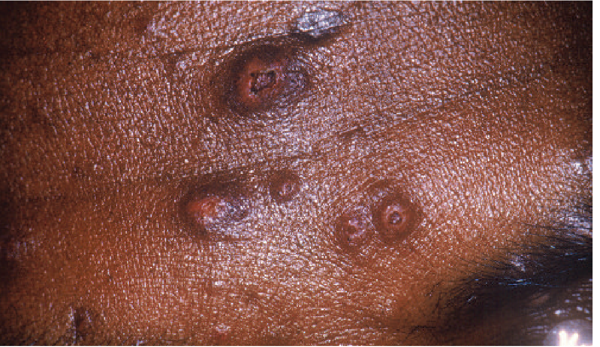
- Skin lesions: Characteristic cutaneous papules with central necrosis/umbilication resembling molluscum contagiosum - a clinical clue
7. Other Dimorphic Fungi
African Histoplasmosis (H. capsulatum var. duboisii)
- Limited to equatorial Africa
- Larger yeast cells (8-15 µm) with thicker walls resembling Blastomyces but WITHOUT broad-based buds
- Emerging in HIV-infected African patients
Emergomycosis (Emergomyces spp.)
- Newly described genus (5 species: E. africanus, E. canadensis, E. orientalis, E. europaeus, E. pasteurianus)
- E. africanus: Most common endemic mycosis in South Africa; primarily in AIDS patients
- Frequently misidentified as Blastomyces; sequencing necessary for differentiation
- Symptoms: Fever, weight loss, elevated liver enzymes, TB-like chest findings, skin disease
- Treatment: Amphotericin B initially, then itraconazole/voriconazole/posaconazole
Laboratory Diagnosis Summary
| Method | Utility | Notes |
|---|---|---|
| Culture | Gold standard | Slow (weeks for most); AccuProbe nucleic acid probes for confirmation; BSL-3 precautions for Coccidioides |
| Direct microscopy | Rapid presumptive diagnosis | KOH/Calcofluor white; GMS; PAS; H&E - each organism has characteristic tissue morphology |
| Antigen detection | Best for disseminated disease | Urine/serum Histoplasma antigen (92% sensitivity in disseminated); cross-reactivity between fungi |
| Serology | Useful but limited | 2-6 week delay; cross-reactions; poor sensitivity in immunocompromised |
| Nucleic acid probe (AccuProbe) | From culture isolates | Confirms Histoplasma, Blastomyces, Coccidioides |
| PCR/Molecular | Emerging | Direct detection from specimens; better sensitivity |
| Histopathology | Often diagnostic | Tissue biopsy with GMS or PAS staining |
- H&E - routine; may miss small organisms (Histoplasma)
- GMS (Gomori's methenamine silver) - preferred for fungi; highlights cell walls black
- PAS - alternative silver stain
- Calcofluor white - fluorescent; enhances detection; used on respiratory specimens
Treatment Overview
| Organism | Mild-Moderate Disease | Severe/Disseminated Disease |
|---|---|---|
| Histoplasma | Itraconazole | Amphotericin B → Itraconazole |
| Blastomyces | Itraconazole | Amphotericin B → Itraconazole |
| Coccidioides | Fluconazole or Itraconazole | Amphotericin B; lifelong azole for meningitis |
| Paracoccidioides | Itraconazole | Amphotericin B → Itraconazole/TMP-SMX |
| Sporothrix | Itraconazole | Amphotericin B; then Itraconazole |
| Talaromyces | Itraconazole | Amphotericin B → Itraconazole |
- Itraconazole is the azole of choice for most dimorphic fungi
- Posaconazole has activity against dimorphic fungi and mucorales
- Disseminated Coccidioides, Histoplasma, and Talaromyces are AIDS-defining infections in HIV
Key Clinical Pearls
- Travel/residence history is essential - most dimorphic fungi have restricted geographic distributions
- Dimorphic fungi can mimic TB, sarcoidosis, and malignancy - always consider in endemic area patients
- Most infections are asymptomatic in immunocompetent hosts - disease results from heavy exposure or immunosuppression
- Latent infections can reactivate years later - question patients on past residence in endemic regions
- Sporothrix is the odd one out: inoculation (not inhalation), lymphocutaneous pattern, cigar-shaped yeasts rarely seen in tissue
- Coccidioides is the most infectious in the lab (BSL-3) - arthroconidia easily aerosolized from cultures
- Paracoccidioides has a striking male predominance due to estrogen's protective effect (estrogens inhibit mold-to-yeast conversion)
- T. marneffei yeast cells divide by fission (with transverse septa), not budding - distinguishes from Histoplasma
Make a PowerPoint presentation on dimorphic fungi clinical cases
mkdir -p /home/daytona/workspace/dimorphic-fungi-cases && node /home/daytona/skills/shared/scripts/fetch_images.js \ "https://cdn.orris.care/cdss_images/90b69c075e29cf2a4d7f0d288b7d6ab0e40f6a3bac6945d87dfd2497c26ab32b.png" \ "https://cdn.orris.care/cdss_images/2b2ad3f37f4e5004226900eb74ea4c14ad798bd09e95ba7a552e22c80a22323f.png" \ "https://cdn.orris.care/cdss_images/760308f7920ea158187f6ee6134f4e0dc06ef8cfb8358ea15772930f2945ceef.png" \ "https://cdn.orris.care/cdss_images/913fc46a67c0c2d95cd7fd96b001b870f6dca2d7ed3a4ec7cbdf5e4003b10f2b.png" \ "https://cdn.orris.care/cdss_images/94539d50435c02242aee234496daa8758d4b80abd443c1cbf4abab88a437f6ad.png" \ "https://cdn.orris.care/cdss_images/aab07ebed44bf0ef5cceb6a4f4102596ae66f04d32cd83e2a5dd0472d68be6ff.png" \ "https://cdn.orris.care/cdss_images/850276cde41e057b58156a99168a1d47cea97167e305879172f33806c2a74cda.png" \ 2>/dev/null | node -e "const d=require('fs');let s='';process.stdin.on('data',c=>s+=c);process.stdin.on('end',()=>{const r=JSON.parse(s);r.forEach((x,i)=>{if(!x.error&&x.base64){d.writeFileSync('/home/daytona/workspace/dimorphic-fungi-cases/img'+i+'.txt',x.base64);}});console.log(r.map((x,i)=>i+': '+(x.error||'OK '+x.base64.slice(0,30))).join('\n'));})"
~/dimorphic-fungi-cases/make_pptx.js
const pptxgen = require("pptxgenjs");
const fs = require("fs");
const pres = new pptxgen();
pres.layout = "LAYOUT_16x9";
pres.title = "Dimorphic Fungi – Clinical Cases";
pres.author = "Medical Mycology";
// Load images
const imgs = [];
for (let i = 0; i <= 6; i++) {
try { imgs[i] = fs.readFileSync(`/home/daytona/workspace/dimorphic-fungi-cases/img${i}.txt`, "utf8"); }
catch(e) { imgs[i] = null; }
}
// ── COLORS ──────────────────────────────────────────────────────────────────
const C = {
dark: "0D1B2A", // deep navy
mid: "1B3A5C", // medium navy
accent: "2A9D8F", // teal-green
gold: "E9C46A", // warm amber
alert: "E76F51", // coral/orange
light: "F4F1EC", // off-white
white: "FFFFFF",
gray: "7A8FA6",
};
// ── HELPERS ──────────────────────────────────────────────────────────────────
function addDarkBg(slide, hex) {
slide.addShape(pres.shapes.RECTANGLE, { x:0, y:0, w:10, h:5.625, fill:{ color: hex || C.dark }, line:{ type:"none" } });
}
function addLightBg(slide) {
slide.addShape(pres.shapes.RECTANGLE, { x:0, y:0, w:10, h:5.625, fill:{ color: C.light }, line:{ type:"none" } });
}
function sectionTag(slide, label, color) {
slide.addShape(pres.shapes.RECTANGLE, { x:0, y:0, w:10, h:0.42, fill:{ color: color || C.accent }, line:{ type:"none" } });
slide.addText(label, { x:0.2, y:0, w:9.6, h:0.42, fontSize:9, bold:true, color:C.white, valign:"middle", margin:0 });
}
function caseLabel(slide, num, title, accent) {
slide.addShape(pres.shapes.RECTANGLE, { x:0, y:0.42, w:10, h:1.1, fill:{ color: C.mid }, line:{ type:"none" } });
slide.addText(`CASE ${num}`, { x:0.35, y:0.46, w:1.5, h:0.38, fontSize:9, bold:true, color:accent, margin:0 });
slide.addText(title, { x:0.35, y:0.78, w:9.3, h:0.6, fontSize:22, bold:true, color:C.white, valign:"middle", margin:0 });
}
function keyBadge(slide, label, value, x, y, accent) {
slide.addShape(pres.shapes.RECTANGLE, { x, y, w:2.9, h:0.72,
fill:{ color: C.dark }, line:{ color: accent, pt:1.5 },
shadow:{ type:"outer", color:"000000", blur:5, offset:2, angle:135, opacity:0.2 }
});
slide.addText([
{ text: label + "\n", options:{ bold:true, color:accent, fontSize:7.5 } },
{ text: value, options:{ color:C.white, fontSize:9.5 } }
], { x, y, w:2.9, h:0.72, align:"center", valign:"middle", margin:4 });
}
function diagBox(slide, items, x, y, w, h, accent) {
slide.addShape(pres.shapes.RECTANGLE, { x, y, w, h, fill:{ color:"132940" }, line:{ color: accent, pt:1 } });
slide.addText(items, { x:x+0.1, y, w:w-0.2, h, fontSize:10, color:C.white, valign:"middle", margin:6 });
}
function addSectionDivider(slide, title, subText, accent) {
addDarkBg(slide, C.dark);
slide.addShape(pres.shapes.RECTANGLE, { x:0, y:2.2, w:10, h:1.25, fill:{ color: accent }, line:{ type:"none" } });
slide.addText(title, { x:0.5, y:2.22, w:9, h:0.75, fontSize:32, bold:true, color:C.white, align:"center", valign:"middle", margin:0 });
slide.addText(subText, { x:0.5, y:3.0, w:9, h:0.4, fontSize:13, color:C.light, align:"center", valign:"middle", margin:0 });
}
// ══════════════════════════════════════════════════════════════════════════
// SLIDE 1 – TITLE
// ══════════════════════════════════════════════════════════════════════════
{
const s = pres.addSlide();
addDarkBg(s, C.dark);
// Teal bar left accent
s.addShape(pres.shapes.RECTANGLE, { x:0, y:0, w:0.18, h:5.625, fill:{ color:C.accent }, line:{ type:"none" } });
// Gold bottom stripe
s.addShape(pres.shapes.RECTANGLE, { x:0, y:5.1, w:10, h:0.525, fill:{ color:C.gold }, line:{ type:"none" } });
s.addText("DIMORPHIC FUNGI", { x:0.4, y:0.9, w:9.2, h:0.7, fontSize:13, bold:true, color:C.accent, charSpacing:6, margin:0 });
s.addText("Clinical Cases", { x:0.4, y:1.5, w:9.2, h:1.4, fontSize:44, bold:true, color:C.white, margin:0 });
s.addText("Microbiological & Clinical Perspectives", { x:0.4, y:3.0, w:9.2, h:0.5, fontSize:16, color:C.gold, margin:0 });
s.addText("Histoplasmosis · Blastomycosis · Coccidioidomycosis · Sporotrichosis · Talaromycosis", {
x:0.4, y:3.6, w:9.2, h:0.38, fontSize:10, color:C.gray, margin:0 });
s.addText("Case-based Learning Series", { x:0.4, y:5.1, w:9.2, h:0.45, fontSize:11, color:C.dark, align:"center", valign:"middle", bold:true, margin:0 });
}
// ══════════════════════════════════════════════════════════════════════════
// SLIDE 2 – OVERVIEW: What are Dimorphic Fungi?
// ══════════════════════════════════════════════════════════════════════════
{
const s = pres.addSlide();
addLightBg(s);
sectionTag(s, "OVERVIEW");
s.addShape(pres.shapes.RECTANGLE, { x:0, y:0.42, w:10, h:0.95, fill:{ color:C.mid }, line:{ type:"none" } });
s.addText("What Are Dimorphic Fungi?", { x:0.35, y:0.42, w:9.3, h:0.95, fontSize:24, bold:true, color:C.white, valign:"middle", margin:0 });
// Central concept box
s.addShape(pres.shapes.RECTANGLE, { x:0.3, y:1.55, w:4.1, h:1.5, fill:{ color:C.accent }, line:{ type:"none" },
shadow:{ type:"outer", color:"000000", blur:8, offset:3, angle:135, opacity:0.2 } });
s.addText([
{ text:"MOLD PHASE (25–30°C)\n", options:{ bold:true, fontSize:10 } },
{ text:"Filamentous, produces infectious conidia\nFound in soil/environment", options:{ fontSize:9.5 } }
], { x:0.3, y:1.55, w:4.1, h:1.5, color:C.white, align:"center", valign:"middle", margin:8 });
// Arrow
s.addShape(pres.shapes.RIGHT_ARROW, { x:4.6, y:2.05, w:0.9, h:0.5, fill:{ color:C.gold }, line:{ type:"none" } });
s.addText("37°C\n(body)", { x:4.55, y:2.6, w:1.0, h:0.45, fontSize:8, color:C.dark, align:"center", margin:0 });
s.addShape(pres.shapes.RECTANGLE, { x:5.6, y:1.55, w:4.1, h:1.5, fill:{ color:C.mid }, line:{ type:"none" },
shadow:{ type:"outer", color:"000000", blur:8, offset:3, angle:135, opacity:0.2 } });
s.addText([
{ text:"YEAST PHASE (37°C)\n", options:{ bold:true, fontSize:10 } },
{ text:"Single cells / spherules in tissue\nReplication & dissemination", options:{ fontSize:9.5 } }
], { x:5.6, y:1.55, w:4.1, h:1.5, color:C.white, align:"center", valign:"middle", margin:8 });
// Bullets
s.addText([
{ text: "Key shared features: ", options:{ bold:true, color:C.dark, fontSize:10.5 } },
{ text: "Infect immunocompetent hosts · Geographically restricted · Latent reactivation · Mimic TB/malignancy", options:{ color:"333333", fontSize:10 } }
], { x:0.3, y:3.25, w:9.4, h:0.45, margin:0 });
// Table of organisms
const rows = [
[{ text:"Organism", options:{ bold:true, color:C.white } }, { text:"Disease", options:{ bold:true, color:C.white } }, { text:"Geography", options:{ bold:true, color:C.white } }, { text:"Entry", options:{ bold:true, color:C.white } }],
["Histoplasma capsulatum","Histoplasmosis","Ohio/Mississippi valleys, Worldwide","Inhalation"],
["Blastomyces dermatitidis","Blastomycosis","SE/SC USA, Africa","Inhalation"],
["Coccidioides spp.","Coccidioidomycosis (Valley Fever)","SW USA, Mexico","Inhalation"],
["Sporothrix schenckii","Sporotrichosis","Worldwide (warm, humid)","Traumatic inoculation"],
["Talaromyces marneffei","Talaromycosis","SE Asia","Inhalation"],
];
s.addTable(rows, {
x:0.3, y:3.78, w:9.4, h:1.7,
fontSize:9, color:"222222", valign:"middle",
rowH:0.28,
border:{ type:"solid", color:"CCCCCC", pt:0.5 },
fill:"F4F1EC",
colW:[2.6, 2.3, 2.7, 1.8],
});
// Override header row fill
// pptxgenjs doesn't support per-row fill in addTable without per-cell opts
}
// ══════════════════════════════════════════════════════════════════════════
// SLIDE 3 – GEOGRAPHIC DISTRIBUTION (IMAGE)
// ══════════════════════════════════════════════════════════════════════════
{
const s = pres.addSlide();
addDarkBg(s, C.mid);
sectionTag(s, "EPIDEMIOLOGY", C.accent);
s.addText("Geographic Distribution of Endemic Dimorphic Fungi", {
x:0.3, y:0.5, w:9.4, h:0.65, fontSize:20, bold:true, color:C.white, margin:0 });
if (imgs[0]) s.addImage({ data:imgs[0], x:0.2, y:1.25, w:4.0, h:4.0 });
// Right side bullets
const pts = [
["Histoplasmosis", "Ohio & Mississippi River valleys; extends into Caribbean, South America, Asia"],
["Blastomycosis", "SE/SC USA, Great Lakes, Canada; also sub-Saharan Africa"],
["Coccidioidomycosis", "SW USA (Arizona, California), Mexico, Central/South America"],
["Paracoccidioidomycosis", "Exclusively South & Central America; Brazil highest burden"],
["Talaromycosis", "SE Asia: Thailand, S China, Vietnam, NE India"],
["Sporotrichosis", "Worldwide; Brazil epidemic via cat transmission"],
];
let yy = 1.28;
pts.forEach(([org, desc]) => {
s.addShape(pres.shapes.RECTANGLE, { x:4.5, y:yy, w:5.25, h:0.62, fill:{ color:"132940" }, line:{ color:C.accent, pt:0.8 } });
s.addText([
{ text:org + " ", options:{ bold:true, color:C.accent, fontSize:9 } },
{ text:desc, options:{ color:C.light, fontSize:8.5 } }
], { x:4.6, y:yy, w:5.05, h:0.62, valign:"middle", margin:4 });
yy += 0.68;
});
}
// ══════════════════════════════════════════════════════════════════════════
// SLIDE 4 – CASE 1: HISTOPLASMOSIS (Presentation)
// ══════════════════════════════════════════════════════════════════════════
{
const s = pres.addSlide();
addDarkBg(s, C.dark);
sectionTag(s, "CASE 1 – HISTOPLASMOSIS", C.accent);
caseLabel(s, "01", "Histoplasmosis – The Cave Explorer", C.accent);
// Left panel: case scenario
s.addShape(pres.shapes.RECTANGLE, { x:0.25, y:1.62, w:5.5, h:3.8, fill:{ color:"0F2535" }, line:{ color:C.accent, pt:1 } });
s.addText("CLINICAL PRESENTATION", { x:0.35, y:1.68, w:5.3, h:0.32, fontSize:8.5, bold:true, color:C.accent, margin:0 });
s.addText([
{ text:"• ", options:{ color:C.gold } }, { text:"32-year-old male, spelunker (caver) from Ohio\n", options:{ color:C.white, fontSize:10 } },
{ text:"• ", options:{ color:C.gold } }, { text:"3-week history of fever, dry cough, fatigue, night sweats\n", options:{ color:C.white, fontSize:10 } },
{ text:"• ", options:{ color:C.gold } }, { text:"Severe headache and weight loss (4 kg)\n", options:{ color:C.white, fontSize:10 } },
{ text:"• ", options:{ color:C.gold } }, { text:"Explored bat caves 3–4 weeks ago\n\n", options:{ color:C.white, fontSize:10 } },
{ text:"EXAMINATION\n", options:{ bold:true, color:C.gold, fontSize:9 } },
{ text:"• ", options:{ color:C.gold } }, { text:"Temp 38.8°C, HR 102, SpO₂ 94% on room air\n", options:{ color:C.white, fontSize:10 } },
{ text:"• ", options:{ color:C.gold } }, { text:"Hepatosplenomegaly, cervical lymphadenopathy\n", options:{ color:C.white, fontSize:10 } },
{ text:"• ", options:{ color:C.gold } }, { text:"CXR: bilateral diffuse interstitial infiltrates\n\n", options:{ color:C.white, fontSize:10 } },
{ text:"LABS\n", options:{ bold:true, color:C.gold, fontSize:9 } },
{ text:"• WBC 3.2 × 10⁹/L • LDH elevated • Ferritin 2400 ng/mL\n", options:{ color:C.white, fontSize:10 } },
{ text:"• HIV negative • No immunosuppressive medications", options:{ color:C.white, fontSize:10 } },
], { x:0.35, y:2.05, w:5.3, h:3.25, valign:"top", margin:4 });
// Right panel: key clues
s.addText("KEY DIAGNOSTIC CLUES", { x:6.0, y:1.68, w:3.7, h:0.32, fontSize:8.5, bold:true, color:C.gold, margin:0 });
const clues = [
["🦇", "Bat Cave Exposure", "Primary epidemiologic risk"],
["🌡️", "Flu-like Syndrome", "Fever, cough, fatigue"],
["🫁", "Bilateral Infiltrates", "CXR / CT finding"],
["🔬", "Intracellular Yeasts", "In macrophages (2–4 µm)"],
["📍", "Ohio River Valley", "Classic endemic area"],
];
let cy = 2.05;
clues.forEach(([icon, hd, sub]) => {
s.addShape(pres.shapes.RECTANGLE, { x:6.0, y:cy, w:3.72, h:0.64, fill:{ color:"0F2535" }, line:{ color:C.gold, pt:0.6 } });
s.addText([
{ text:icon + " ", options:{ fontSize:13 } },
{ text:hd + "\n", options:{ bold:true, color:C.gold, fontSize:9.5 } },
{ text:" " + sub, options:{ color:C.gray, fontSize:8.5 } }
], { x:6.05, y:cy, w:3.62, h:0.64, valign:"middle", margin:4 });
cy += 0.7;
});
}
// ══════════════════════════════════════════════════════════════════════════
// SLIDE 5 – CASE 1: HISTOPLASMOSIS (Micro + Diagnosis + Treatment)
// ══════════════════════════════════════════════════════════════════════════
{
const s = pres.addSlide();
addLightBg(s);
sectionTag(s, "CASE 1 – HISTOPLASMOSIS: MICROBIOLOGY & MANAGEMENT", C.accent);
s.addShape(pres.shapes.RECTANGLE, { x:0, y:0.42, w:10, h:0.75, fill:{ color:C.mid }, line:{ type:"none" } });
s.addText("Histoplasma capsulatum – Microbiological Features & Workup", {
x:0.3, y:0.42, w:9.4, h:0.75, fontSize:17, bold:true, color:C.white, valign:"middle", margin:0 });
// Image: macroconidia
if (imgs[2]) s.addImage({ data:imgs[2], x:0.25, y:1.28, w:3.2, h:2.4 });
s.addShape(pres.shapes.RECTANGLE, { x:0.25, y:3.68, w:3.2, h:0.35, fill:{ color:C.accent }, line:{ type:"none" } });
s.addText("Tuberculate macroconidia — mold phase (LPCB 400×)", { x:0.25, y:3.68, w:3.2, h:0.35, fontSize:7.5, color:C.white, align:"center", valign:"middle", margin:0 });
// Image: yeasts in macrophage
if (imgs[1]) s.addImage({ data:imgs[1], x:3.65, y:1.28, w:3.2, h:2.4 });
s.addShape(pres.shapes.RECTANGLE, { x:3.65, y:3.68, w:3.2, h:0.35, fill:{ color:C.accent }, line:{ type:"none" } });
s.addText("H. capsulatum yeasts inside macrophages (H&E 1000×)", { x:3.65, y:3.68, w:3.2, h:0.35, fontSize:7.5, color:C.white, align:"center", valign:"middle", margin:0 });
// Right panel: diagnosis + treatment
s.addShape(pres.shapes.RECTANGLE, { x:7.1, y:1.28, w:2.65, h:4.1, fill:{ color:C.dark }, line:{ color:C.accent, pt:1 } });
s.addText([
{ text:"DIAGNOSIS\n", options:{ bold:true, color:C.accent, fontSize:9.5, breakLine:true } },
{ text:"✓ Urine antigen\n (92% sensitivity, disseminated)\n", options:{ color:C.white, fontSize:8.5, breakLine:true } },
{ text:"✓ Bronchoalveolar lavage\n + GMS stain\n", options:{ color:C.white, fontSize:8.5, breakLine:true } },
{ text:"✓ Culture: BHI agar 25°C\n (10–28 days); AccuProbe\n", options:{ color:C.white, fontSize:8.5, breakLine:true } },
{ text:"✓ Serology: complement\n fixation / immunodiffusion\n\n", options:{ color:C.white, fontSize:8.5, breakLine:true } },
{ text:"TREATMENT\n", options:{ bold:true, color:C.gold, fontSize:9.5, breakLine:true } },
{ text:"Mild-moderate:\n Itraconazole 200 mg TID\n", options:{ color:C.white, fontSize:8.5, breakLine:true } },
{ text:"Severe/disseminated:\n Amphotericin B → Itraconazole\n", options:{ color:C.white, fontSize:8.5, breakLine:true } },
{ text:"Duration: 12 weeks\n (disseminated: 12 months)", options:{ color:C.white, fontSize:8.5 } },
], { x:7.15, y:1.33, w:2.55, h:4.0, valign:"top", margin:6 });
s.addText("Answer: Disseminated Histoplasmosis | Confirmed by urine antigen + bone marrow biopsy",
{ x:0.25, y:4.12, w:6.7, h:0.38, fontSize:8.5, bold:true, color:C.dark,
fill:{ color:C.gold }, align:"center", valign:"middle", margin:4 });
}
// ══════════════════════════════════════════════════════════════════════════
// SLIDE 6 – CASE 2: BLASTOMYCOSIS (Presentation)
// ══════════════════════════════════════════════════════════════════════════
{
const s = pres.addSlide();
addDarkBg(s, C.dark);
sectionTag(s, "CASE 2 – BLASTOMYCOSIS", C.gold);
caseLabel(s, "02", "Blastomycosis – The Great Masquerader", C.gold);
s.addShape(pres.shapes.RECTANGLE, { x:0.25, y:1.62, w:5.5, h:3.8, fill:{ color:"0F2535" }, line:{ color:C.gold, pt:1 } });
s.addText("CLINICAL PRESENTATION", { x:0.35, y:1.68, w:5.3, h:0.32, fontSize:8.5, bold:true, color:C.gold, margin:0 });
s.addText([
{ text:"• ", options:{ color:C.accent } }, { text:"45-year-old male logger from Wisconsin\n", options:{ color:C.white, fontSize:10 } },
{ text:"• ", options:{ color:C.accent } }, { text:"6-week history of productive cough, fever, pleuritic chest pain\n", options:{ color:C.white, fontSize:10 } },
{ text:"• ", options:{ color:C.accent } }, { text:"3 courses of antibiotics — no improvement\n", options:{ color:C.white, fontSize:10 } },
{ text:"• ", options:{ color:C.accent } }, { text:"New raised, verrucous skin lesions on face and arms\n\n", options:{ color:C.white, fontSize:10 } },
{ text:"EXAMINATION\n", options:{ bold:true, color:C.accent, fontSize:9 } },
{ text:"• ", options:{ color:C.accent } }, { text:"Temp 38.2°C, decreased breath sounds at right base\n", options:{ color:C.white, fontSize:10 } },
{ text:"• ", options:{ color:C.accent } }, { text:"Verrucous crusted plaques with irregular borders\n", options:{ color:C.white, fontSize:10 } },
{ text:"• ", options:{ color:C.accent } }, { text:"CXR: right lower lobe mass-like consolidation\n\n", options:{ color:C.white, fontSize:10 } },
{ text:"LABS\n", options:{ bold:true, color:C.accent, fontSize:9 } },
{ text:"• WBC 14 × 10⁹/L • CRP 85 mg/L • HIV negative\n", options:{ color:C.white, fontSize:10 } },
{ text:"• CT chest: consolidation + cavitation (mimics lung cancer)", options:{ color:C.white, fontSize:10 } },
], { x:0.35, y:2.05, w:5.3, h:3.25, valign:"top", margin:4 });
// Right clues
s.addText("THE GREAT MASQUERADER", { x:6.0, y:1.68, w:3.7, h:0.32, fontSize:8.5, bold:true, color:C.gold, margin:0 });
const clues = [
["🌲", "Rural/Outdoor Worker", "Logging, farming — soil exposure"],
["🩹", "Antibiotic Failure", "Key red flag for fungal etiology"],
["🩺", "Skin Lesions", "Verrucous, ulcerated plaques"],
["🔬", "Broad-based Buds", "8–15 µm thick-walled yeasts"],
["📍", "Ohio/Mississippi", "Great Lakes endemic region"],
];
let cy = 2.05;
clues.forEach(([icon, hd, sub]) => {
s.addShape(pres.shapes.RECTANGLE, { x:6.0, y:cy, w:3.72, h:0.64, fill:{ color:"0F2535" }, line:{ color:C.gold, pt:0.6 } });
s.addText([
{ text:icon + " ", options:{ fontSize:13 } },
{ text:hd + "\n", options:{ bold:true, color:C.gold, fontSize:9.5 } },
{ text:" " + sub, options:{ color:C.gray, fontSize:8.5 } }
], { x:6.05, y:cy, w:3.62, h:0.64, valign:"middle", margin:4 });
cy += 0.7;
});
}
// ══════════════════════════════════════════════════════════════════════════
// SLIDE 7 – CASE 2: BLASTOMYCOSIS (Micro + Diagnosis)
// ══════════════════════════════════════════════════════════════════════════
{
const s = pres.addSlide();
addLightBg(s);
sectionTag(s, "CASE 2 – BLASTOMYCOSIS: MICROBIOLOGY & MANAGEMENT", C.gold);
s.addShape(pres.shapes.RECTANGLE, { x:0, y:0.42, w:10, h:0.75, fill:{ color:C.mid }, line:{ type:"none" } });
s.addText("Blastomyces dermatitidis – Microbiological Features & Workup", {
x:0.3, y:0.42, w:9.4, h:0.75, fontSize:17, bold:true, color:C.white, valign:"middle", margin:0 });
// Image: Blastomyces yeast in tissue
if (imgs[3]) s.addImage({ data:imgs[3], x:0.25, y:1.28, w:3.2, h:2.4 });
s.addShape(pres.shapes.RECTANGLE, { x:0.25, y:3.68, w:3.2, h:0.4, fill:{ color:"B8860B" }, line:{ type:"none" } });
s.addText("B. dermatitidis — broad-based budding yeasts in tissue (H&E 400×)", { x:0.25, y:3.68, w:3.2, h:0.4, fontSize:7.5, color:C.white, align:"center", valign:"middle", margin:0 });
// Image: mold form
if (imgs[6]) s.addImage({ data:imgs[6], x:3.65, y:1.28, w:3.2, h:2.4 });
s.addShape(pres.shapes.RECTANGLE, { x:3.65, y:3.68, w:3.2, h:0.4, fill:{ color:"B8860B" }, line:{ type:"none" } });
s.addText("Mold form — \"lollipop\" conidia on conidiophore (LPCB 400×)", { x:3.65, y:3.68, w:3.2, h:0.4, fontSize:7.5, color:C.white, align:"center", valign:"middle", margin:0 });
// Right: diagnosis + treatment
s.addShape(pres.shapes.RECTANGLE, { x:7.1, y:1.28, w:2.65, h:4.1, fill:{ color:C.dark }, line:{ color:C.gold, pt:1 } });
s.addText([
{ text:"DIAGNOSIS\n", options:{ bold:true, color:C.gold, fontSize:9.5, breakLine:true } },
{ text:"✓ Wet prep / KOH of sputum\n or skin scraping\n", options:{ color:C.white, fontSize:8.5, breakLine:true } },
{ text:"✓ Culture: BHI agar\n White fluffy colonies\n AccuProbe confirmation\n", options:{ color:C.white, fontSize:8.5, breakLine:true } },
{ text:"✓ Urine antigen\n (cross-reacts w/ Histoplasma)\n", options:{ color:C.white, fontSize:8.5, breakLine:true } },
{ text:"✓ Biopsy: GMS/PAS stain\n\n", options:{ color:C.white, fontSize:8.5, breakLine:true } },
{ text:"TREATMENT\n", options:{ bold:true, color:C.accent, fontSize:9.5, breakLine:true } },
{ text:"Mild-moderate:\n Itraconazole 200 mg BID\n × 6 months\n", options:{ color:C.white, fontSize:8.5, breakLine:true } },
{ text:"Severe/CNS:\n Amphotericin B\n → Itraconazole\n", options:{ color:C.white, fontSize:8.5, breakLine:true } },
], { x:7.15, y:1.33, w:2.55, h:4.0, valign:"top", margin:6 });
s.addText("Answer: Pulmonary + Cutaneous Blastomycosis | Confirmed by culture + broad-based budding yeasts",
{ x:0.25, y:4.12, w:6.7, h:0.38, fontSize:8.5, bold:true, color:C.dark,
fill:{ color:C.gold }, align:"center", valign:"middle", margin:4 });
}
// ══════════════════════════════════════════════════════════════════════════
// SLIDE 8 – CASE 3: COCCIDIOIDOMYCOSIS (Presentation)
// ══════════════════════════════════════════════════════════════════════════
{
const s = pres.addSlide();
addDarkBg(s, C.dark);
sectionTag(s, "CASE 3 – COCCIDIOIDOMYCOSIS (VALLEY FEVER)", C.alert);
caseLabel(s, "03", "Coccidioidomycosis – Valley Fever", C.alert);
s.addShape(pres.shapes.RECTANGLE, { x:0.25, y:1.62, w:5.5, h:3.8, fill:{ color:"0F2535" }, line:{ color:C.alert, pt:1 } });
s.addText("CLINICAL PRESENTATION", { x:0.35, y:1.68, w:5.3, h:0.32, fontSize:8.5, bold:true, color:C.alert, margin:0 });
s.addText([
{ text:"• ", options:{ color:C.gold } }, { text:"28-year-old Filipino male, military recruit training in Arizona\n", options:{ color:C.white, fontSize:10 } },
{ text:"• ", options:{ color:C.gold } }, { text:"2-week fever, dry cough, chest tightness\n", options:{ color:C.white, fontSize:10 } },
{ text:"• ", options:{ color:C.gold } }, { text:"Painful red nodules on shins (erythema nodosum)\n", options:{ color:C.white, fontSize:10 } },
{ text:"• ", options:{ color:C.gold } }, { text:"Joint aches, myalgia — called \"desert rheumatism\"\n\n", options:{ color:C.white, fontSize:10 } },
{ text:"EXAMINATION\n", options:{ bold:true, color:C.accent, fontSize:9 } },
{ text:"• ", options:{ color:C.accent } }, { text:"Temp 38.5°C, tender anterior tibial nodules\n", options:{ color:C.white, fontSize:10 } },
{ text:"• ", options:{ color:C.accent } }, { text:"Reduced air entry at right apex\n", options:{ color:C.white, fontSize:10 } },
{ text:"• ", options:{ color:C.accent } }, { text:"CXR: right upper lobe infiltrate with hilar adenopathy\n\n", options:{ color:C.white, fontSize:10 } },
{ text:"LABS\n", options:{ bold:true, color:C.accent, fontSize:9 } },
{ text:"• WBC 11.2 × 10⁹/L • Eosinophilia (8%)\n", options:{ color:C.white, fontSize:10 } },
{ text:"• HIV negative • IgM coccidioides serology positive", options:{ color:C.white, fontSize:10 } },
], { x:0.35, y:2.05, w:5.3, h:3.25, valign:"top", margin:4 });
s.addText("KEY DIAGNOSTIC CLUES", { x:6.0, y:1.68, w:3.7, h:0.32, fontSize:8.5, bold:true, color:C.alert, margin:0 });
const clues = [
["🏜️", "Desert Exposure", "SW USA military base/construction"],
["🦶", "Erythema Nodosum", "\"Desert rheumatism\" triad"],
["⚠️", "Filipino Ethnicity", "Genetic risk for dissemination"],
["🔬", "Spherules", "Up to 100 µm, filled with endospores"],
["⚗️", "BSL-3 Pathogen", "Most infectious dimorphic fungus"],
];
let cy = 2.05;
clues.forEach(([icon, hd, sub]) => {
s.addShape(pres.shapes.RECTANGLE, { x:6.0, y:cy, w:3.72, h:0.64, fill:{ color:"0F2535" }, line:{ color:C.alert, pt:0.6 } });
s.addText([
{ text:icon + " ", options:{ fontSize:13 } },
{ text:hd + "\n", options:{ bold:true, color:C.alert, fontSize:9.5 } },
{ text:" " + sub, options:{ color:C.gray, fontSize:8.5 } }
], { x:6.05, y:cy, w:3.62, h:0.64, valign:"middle", margin:4 });
cy += 0.7;
});
}
// ══════════════════════════════════════════════════════════════════════════
// SLIDE 9 – CASE 3: COCCIDIOIDOMYCOSIS (Micro + Diagnosis)
// ══════════════════════════════════════════════════════════════════════════
{
const s = pres.addSlide();
addLightBg(s);
sectionTag(s, "CASE 3 – COCCIDIOIDOMYCOSIS: MICROBIOLOGY & MANAGEMENT", C.alert);
s.addShape(pres.shapes.RECTANGLE, { x:0, y:0.42, w:10, h:0.75, fill:{ color:C.mid }, line:{ type:"none" } });
s.addText("Coccidioides spp. – Unique Tissue Form & Workup", {
x:0.3, y:0.42, w:9.4, h:0.75, fontSize:17, bold:true, color:C.white, valign:"middle", margin:0 });
// Coccidioides spherules image
if (imgs[4]) s.addImage({ data:imgs[4], x:0.25, y:1.28, w:4.5, h:2.8 });
s.addShape(pres.shapes.RECTANGLE, { x:0.25, y:4.08, w:4.5, h:0.4, fill:{ color:C.alert }, line:{ type:"none" } });
s.addText("Coccidioides spherules of varying size in tissue (H&E 100×)", { x:0.25, y:4.08, w:4.5, h:0.4, fontSize:7.5, color:C.white, align:"center", valign:"middle", margin:0 });
// Right panel: micro + treatment
s.addShape(pres.shapes.RECTANGLE, { x:5.0, y:1.28, w:4.75, h:3.2, fill:{ color:C.dark }, line:{ color:C.alert, pt:1 } });
s.addText([
{ text:"UNIQUE MICROBIOLOGICAL FEATURES\n", options:{ bold:true, color:C.alert, fontSize:9.5, breakLine:true } },
{ text:"• Spherules (up to 100 µm) — NOT yeasts\n", options:{ color:C.white, fontSize:9 } },
{ text:"• Endospores (2–5 µm) — disseminate on rupture\n", options:{ color:C.white, fontSize:9 } },
{ text:"• BSL-3 biohazard — arthroconidia infectious in lab\n", options:{ color:C.white, fontSize:9 } },
{ text:"• C. immitis (CA) vs. C. posadasii (AZ/elsewhere)\n\n", options:{ color:C.white, fontSize:9 } },
{ text:"DIAGNOSIS\n", options:{ bold:true, color:C.gold, fontSize:9.5, breakLine:true } },
{ text:"• Serology: IgM (acute), IgG/complement fixation\n", options:{ color:C.white, fontSize:9 } },
{ text:"• Culture in BSC — rapid growth (1 week)\n", options:{ color:C.white, fontSize:9 } },
{ text:"• Calcofluor white stain on BAL/sputum\n", options:{ color:C.white, fontSize:9 } },
{ text:"• Urine/serum antigen (less sensitive)\n\n", options:{ color:C.white, fontSize:9 } },
{ text:"TREATMENT\n", options:{ bold:true, color:C.accent, fontSize:9.5, breakLine:true } },
{ text:"Mild: Fluconazole 400 mg/day or Itraconazole\n", options:{ color:C.white, fontSize:9 } },
{ text:"Severe: Amphotericin B then azole maintenance\n", options:{ color:C.white, fontSize:9 } },
{ text:"Meningitis: Fluconazole LIFELONG", options:{ color:C.alert, fontSize:9, bold:true } },
], { x:5.05, y:1.33, w:4.65, h:3.1, valign:"top", margin:6 });
s.addText("Answer: Acute Pulmonary Coccidioidomycosis (Valley Fever) | Monitor for dissemination (Filipino ethnicity = high risk)",
{ x:0.25, y:4.58, w:9.5, h:0.42, fontSize:8.5, bold:true, color:C.dark,
fill:{ color:C.gold }, align:"center", valign:"middle", margin:4 });
}
// ══════════════════════════════════════════════════════════════════════════
// SLIDE 10 – CASE 4: SPOROTRICHOSIS (Presentation)
// ══════════════════════════════════════════════════════════════════════════
{
const s = pres.addSlide();
addDarkBg(s, C.dark);
sectionTag(s, "CASE 4 – SPOROTRICHOSIS", C.accent);
caseLabel(s, "04", "Sporotrichosis – The Rose Thorn Infection", C.accent);
s.addShape(pres.shapes.RECTANGLE, { x:0.25, y:1.62, w:5.5, h:3.8, fill:{ color:"0F2535" }, line:{ color:C.accent, pt:1 } });
s.addText("CLINICAL PRESENTATION", { x:0.35, y:1.68, w:5.3, h:0.32, fontSize:8.5, bold:true, color:C.accent, margin:0 });
s.addText([
{ text:"• ", options:{ color:C.gold } }, { text:"38-year-old female florist from Brazil\n", options:{ color:C.white, fontSize:10 } },
{ text:"• ", options:{ color:C.gold } }, { text:"Initially: small painless papule on right index finger\n", options:{ color:C.white, fontSize:10 } },
{ text:"• ", options:{ color:C.gold } }, { text:"Over 4 weeks: ulcerated, then new nodules appeared\n extending up the forearm\n", options:{ color:C.white, fontSize:10 } },
{ text:"• ", options:{ color:C.gold } }, { text:"Works with roses and potted plants daily\n\n", options:{ color:C.white, fontSize:10 } },
{ text:"EXAMINATION\n", options:{ bold:true, color:C.accent, fontSize:9 } },
{ text:"• ", options:{ color:C.accent } }, { text:"Afebrile, no systemic symptoms\n", options:{ color:C.white, fontSize:10 } },
{ text:"• ", options:{ color:C.accent } }, { text:"Chain of 5 nodular lesions tracking along lymphatics\n from finger to elbow — classical sporotrichoid spread\n", options:{ color:C.white, fontSize:10 } },
{ text:"• ", options:{ color:C.accent } }, { text:"No hepatosplenomegaly, normal CXR\n\n", options:{ color:C.white, fontSize:10 } },
{ text:"LABS\n", options:{ bold:true, color:C.accent, fontSize:9 } },
{ text:"• Normal CBC, ESR mildly elevated\n", options:{ color:C.white, fontSize:10 } },
{ text:"• HIV negative • Culture: moist colony, turns dark", options:{ color:C.white, fontSize:10 } },
], { x:0.35, y:2.05, w:5.3, h:3.25, valign:"top", margin:4 });
s.addText("KEY DIAGNOSTIC CLUES", { x:6.0, y:1.68, w:3.7, h:0.32, fontSize:8.5, bold:true, color:C.accent, margin:0 });
const clues = [
["🌹", "Rose / Plant Thorn", "Traumatic inoculation — NOT inhalation"],
["📈", "Lymphocutaneous", "Ascending chain of nodules"],
["🔬", "Cigar-shaped Yeasts", "Rarely seen in tissue (2–6 µm)"],
["⭐", "Asteroid Bodies", "Splendore-Hoeppli phenomenon"],
["🌍", "Worldwide", "Warm, humid environments"],
];
let cy = 2.05;
clues.forEach(([icon, hd, sub]) => {
s.addShape(pres.shapes.RECTANGLE, { x:6.0, y:cy, w:3.72, h:0.64, fill:{ color:"0F2535" }, line:{ color:C.accent, pt:0.6 } });
s.addText([
{ text:icon + " ", options:{ fontSize:13 } },
{ text:hd + "\n", options:{ bold:true, color:C.accent, fontSize:9.5 } },
{ text:" " + sub, options:{ color:C.gray, fontSize:8.5 } }
], { x:6.05, y:cy, w:3.62, h:0.64, valign:"middle", margin:4 });
cy += 0.7;
});
}
// ══════════════════════════════════════════════════════════════════════════
// SLIDE 11 – CASE 4: SPOROTRICHOSIS (Micro + Treatment)
// ══════════════════════════════════════════════════════════════════════════
{
const s = pres.addSlide();
addLightBg(s);
sectionTag(s, "CASE 4 – SPOROTRICHOSIS: MICROBIOLOGY & MANAGEMENT", C.accent);
s.addShape(pres.shapes.RECTANGLE, { x:0, y:0.42, w:10, h:0.75, fill:{ color:C.mid }, line:{ type:"none" } });
s.addText("Sporothrix schenckii – Features & Treatment", {
x:0.3, y:0.42, w:9.4, h:0.75, fontSize:17, bold:true, color:C.white, valign:"middle", margin:0 });
// Content boxes
s.addShape(pres.shapes.RECTANGLE, { x:0.25, y:1.3, w:4.7, h:2.2, fill:{ color:C.dark }, line:{ color:C.accent, pt:1 } });
s.addText([
{ text:"MOLD PHASE (25–30°C)\n", options:{ bold:true, color:C.accent, fontSize:10, breakLine:true } },
{ text:"• Two conidial types:\n", options:{ color:C.white, fontSize:9.5 } },
{ text:" - Thin-walled hyaline: rosette/daisy-head arrangement\n", options:{ color:C.white, fontSize:9 } },
{ text:" - Thick-walled dark (melanized) sessile on hyphae\n", options:{ color:C.white, fontSize:9 } },
{ text:"• Colonies: initially cream, turn dark brown/black\n", options:{ color:C.white, fontSize:9 } },
{ text:"• Dematiaceous (melanized) appearance in culture\n", options:{ color:C.white, fontSize:9 } },
{ text:"• Cycloheximide: grows well (selective advantage)", options:{ color:C.white, fontSize:9 } },
], { x:0.35, y:1.35, w:4.5, h:2.1, valign:"top", margin:4 });
s.addShape(pres.shapes.RECTANGLE, { x:0.25, y:3.58, w:4.7, h:1.75, fill:{ color:C.dark }, line:{ color:C.gold, pt:1 } });
s.addText([
{ text:"YEAST PHASE (37°C in tissue)\n", options:{ bold:true, color:C.gold, fontSize:10, breakLine:true } },
{ text:"• Cigar-shaped or pleomorphic (2–6 µm)\n", options:{ color:C.white, fontSize:9 } },
{ text:"• Narrow-based budding — rarely seen in tissue\n", options:{ color:C.white, fontSize:9 } },
{ text:"• Asteroid bodies (Splendore-Hoeppli): eosinophilic\n radiating material around yeast — pathognomonic\n", options:{ color:C.white, fontSize:9 } },
{ text:"• Differential: M. marinum, cutaneous leishmaniasis", options:{ color:C.white, fontSize:9 } },
], { x:0.35, y:3.63, w:4.5, h:1.65, valign:"top", margin:4 });
// Right: diagnosis + treatment
s.addShape(pres.shapes.RECTANGLE, { x:5.2, y:1.3, w:4.55, h:4.03, fill:{ color:C.dark }, line:{ color:C.accent, pt:1 } });
s.addText([
{ text:"DIAGNOSIS\n", options:{ bold:true, color:C.accent, fontSize:9.5, breakLine:true } },
{ text:"✓ Culture (preferred)\n Aspirate / biopsy of skin lesion\n BHI or Sabouraud's agar at 25–30°C\n", options:{ color:C.white, fontSize:9, breakLine:true } },
{ text:"✓ Histopathology\n PAS/GMS stain (yeasts rarely visible)\n", options:{ color:C.white, fontSize:9, breakLine:true } },
{ text:"✓ Sporotrichin skin test\n (epidemiologic surveys only)\n\n", options:{ color:C.white, fontSize:9, breakLine:true } },
{ text:"TREATMENT\n", options:{ bold:true, color:C.gold, fontSize:9.5, breakLine:true } },
{ text:"Lymphocutaneous/Fixed cutaneous:\n Itraconazole 200 mg/day × 3–6 months\n OR Saturated KI (SSKI) drops\n", options:{ color:C.white, fontSize:9, breakLine:true } },
{ text:"Disseminated/Pulmonary:\n Amphotericin B → Itraconazole\n", options:{ color:C.white, fontSize:9, breakLine:true } },
{ text:"Heat therapy: local heat application\n (yeast sensitive to temperature)\n\n", options:{ color:C.white, fontSize:9, breakLine:true } },
{ text:"⚠️ Terbinafine may be used for\n cutaneous disease (S. brasiliensis)", options:{ color:C.alert, fontSize:9 } },
], { x:5.3, y:1.35, w:4.35, h:3.93, valign:"top", margin:6 });
s.addText("Answer: Lymphocutaneous Sporotrichosis | Confirmed by culture of skin aspirate",
{ x:0.25, y:5.1, w:9.5, h:0.38, fontSize:8.5, bold:true, color:C.dark,
fill:{ color:C.gold }, align:"center", valign:"middle", margin:4 });
}
// ══════════════════════════════════════════════════════════════════════════
// SLIDE 12 – CASE 5: TALAROMYCOSIS (Presentation)
// ══════════════════════════════════════════════════════════════════════════
{
const s = pres.addSlide();
addDarkBg(s, C.dark);
sectionTag(s, "CASE 5 – TALAROMYCOSIS (formerly Penicilliosis)", C.alert);
caseLabel(s, "05", "Talaromycosis – The AIDS-Defining Mycosis", C.alert);
s.addShape(pres.shapes.RECTANGLE, { x:0.25, y:1.62, w:5.5, h:3.8, fill:{ color:"0F2535" }, line:{ color:C.alert, pt:1 } });
s.addText("CLINICAL PRESENTATION", { x:0.35, y:1.68, w:5.3, h:0.32, fontSize:8.5, bold:true, color:C.alert, margin:0 });
s.addText([
{ text:"• ", options:{ color:C.gold } }, { text:"35-year-old HIV-positive male, CD4 count 45 cells/µL\n", options:{ color:C.white, fontSize:10 } },
{ text:"• ", options:{ color:C.gold } }, { text:"Resident of Chiang Mai, northern Thailand\n", options:{ color:C.white, fontSize:10 } },
{ text:"• ", options:{ color:C.gold } }, { text:"3 weeks of fever, productive cough, weight loss (6 kg)\n", options:{ color:C.white, fontSize:10 } },
{ text:"• ", options:{ color:C.gold } }, { text:"Multiple skin lesions on face resembling molluscum\n\n", options:{ color:C.white, fontSize:10 } },
{ text:"EXAMINATION\n", options:{ bold:true, color:C.accent, fontSize:9 } },
{ text:"• ", options:{ color:C.accent } }, { text:"Temp 39.4°C, tachycardic, pallor\n", options:{ color:C.white, fontSize:10 } },
{ text:"• ", options:{ color:C.accent } }, { text:"Papules with central umbilication on face/trunk\n", options:{ color:C.white, fontSize:10 } },
{ text:"• ", options:{ color:C.accent } }, { text:"Massive hepatosplenomegaly, lymphadenopathy\n", options:{ color:C.white, fontSize:10 } },
{ text:"• ", options:{ color:C.accent } }, { text:"Bilateral crackles, diffuse infiltrates on CXR\n\n", options:{ color:C.white, fontSize:10 } },
{ text:"LABS\n", options:{ bold:true, color:C.accent, fontSize:9 } },
{ text:"• Hb 7.2 g/dL • WBC 2.1 × 10⁹/L • Platelets 58 × 10⁹/L\n", options:{ color:C.white, fontSize:10 } },
{ text:"• LDH markedly elevated • Blood cultures × 2 POSITIVE", options:{ color:C.alert, fontSize:10, bold:true } },
], { x:0.35, y:2.05, w:5.3, h:3.25, valign:"top", margin:4 });
s.addText("KEY DIAGNOSTIC CLUES", { x:6.0, y:1.68, w:3.7, h:0.32, fontSize:8.5, bold:true, color:C.alert, margin:0 });
const clues = [
["🌏", "SE Asia + HIV", "CD4 <100 — opportunistic pathogen"],
["🐀", "Bamboo Rat", "Environmental reservoir"],
["🩺", "Skin Lesions", "Umbilicated papules = molluscum-like"],
["🔬", "Transverse Septa", "Fission (NOT budding) — unique feature"],
["🎨", "Red Pigment", "Soluble pigment in culture = diagnostic"],
];
let cy = 2.05;
clues.forEach(([icon, hd, sub]) => {
s.addShape(pres.shapes.RECTANGLE, { x:6.0, y:cy, w:3.72, h:0.64, fill:{ color:"0F2535" }, line:{ color:C.alert, pt:0.6 } });
s.addText([
{ text:icon + " ", options:{ fontSize:13 } },
{ text:hd + "\n", options:{ bold:true, color:C.alert, fontSize:9.5 } },
{ text:" " + sub, options:{ color:C.gray, fontSize:8.5 } }
], { x:6.05, y:cy, w:3.62, h:0.64, valign:"middle", margin:4 });
cy += 0.7;
});
}
// ══════════════════════════════════════════════════════════════════════════
// SLIDE 13 – CASE 5: TALAROMYCOSIS (Micro + Diagnosis + Skin image)
// ══════════════════════════════════════════════════════════════════════════
{
const s = pres.addSlide();
addLightBg(s);
sectionTag(s, "CASE 5 – TALAROMYCOSIS: MICROBIOLOGY & MANAGEMENT", C.alert);
s.addShape(pres.shapes.RECTANGLE, { x:0, y:0.42, w:10, h:0.75, fill:{ color:C.mid }, line:{ type:"none" } });
s.addText("Talaromyces marneffei – Unique Features & Workup", {
x:0.3, y:0.42, w:9.4, h:0.75, fontSize:17, bold:true, color:C.white, valign:"middle", margin:0 });
// Skin image
if (imgs[5]) s.addImage({ data:imgs[5], x:0.25, y:1.28, w:4.0, h:2.8 });
s.addShape(pres.shapes.RECTANGLE, { x:0.25, y:4.08, w:4.0, h:0.4, fill:{ color:C.alert }, line:{ type:"none" } });
s.addText("Disseminated talaromycosis — umbilicated papules (Tietz Textbook)", { x:0.25, y:4.08, w:4.0, h:0.4, fontSize:7.5, color:C.white, align:"center", valign:"middle", margin:0 });
// Right panels
s.addShape(pres.shapes.RECTANGLE, { x:4.5, y:1.28, w:5.25, h:1.6, fill:{ color:C.dark }, line:{ color:C.alert, pt:1 } });
s.addText([
{ text:"UNIQUE MICROBIOLOGICAL FEATURES\n", options:{ bold:true, color:C.alert, fontSize:9.5, breakLine:true } },
{ text:"• Only dimorphic member of former Penicillium genus\n", options:{ color:C.white, fontSize:9 } },
{ text:"• Yeast divides by FISSION (transverse septa) — NOT budding\n", options:{ color:C.white, fontSize:9 } },
{ text:"• Soluble red/wine pigment diffuses into agar\n", options:{ color:C.white, fontSize:9 } },
{ text:"• Mold: Penicillium-like structures (biseriate phialides)\n", options:{ color:C.white, fontSize:9 } },
{ text:"• Natural host: Rhizomys sinensis (bamboo rat)\n", options:{ color:C.white, fontSize:9 } },
], { x:4.6, y:1.33, w:5.05, h:1.5, valign:"top", margin:5 });
s.addShape(pres.shapes.RECTANGLE, { x:4.5, y:3.0, w:5.25, h:1.4, fill:{ color:C.dark }, line:{ color:C.gold, pt:1 } });
s.addText([
{ text:"DIAGNOSIS\n", options:{ bold:true, color:C.gold, fontSize:9.5, breakLine:true } },
{ text:"✓ Blood / bone marrow culture (fungemia in ~50%)\n", options:{ color:C.white, fontSize:9 } },
{ text:"✓ Skin scraping / biopsy: Wright/Giemsa stain\n", options:{ color:C.white, fontSize:9 } },
{ text:"✓ Serology and antigen: not widely available\n", options:{ color:C.white, fontSize:9 } },
{ text:"✓ Molecular: ITS sequencing for confirmation", options:{ color:C.white, fontSize:9 } },
], { x:4.6, y:3.05, w:5.05, h:1.3, valign:"top", margin:5 });
s.addShape(pres.shapes.RECTANGLE, { x:4.5, y:4.5, w:5.25, h:0.85, fill:{ color:C.dark }, line:{ color:C.accent, pt:1 } });
s.addText([
{ text:"TREATMENT\n", options:{ bold:true, color:C.accent, fontSize:9.5, breakLine:true } },
{ text:"Induction: Amphotericin B × 2 weeks → Itraconazole × 10 weeks\n", options:{ color:C.white, fontSize:9 } },
{ text:"Secondary prophylaxis: Itraconazole until CD4 >100 cells/µL", options:{ color:C.white, fontSize:9 } },
], { x:4.6, y:4.55, w:5.05, h:0.75, valign:"top", margin:5 });
s.addText("Answer: Disseminated Talaromycosis (AIDS-defining illness) | Confirmed by blood cultures + skin biopsy",
{ x:0.25, y:5.1, w:9.5, h:0.38, fontSize:8.5, bold:true, color:C.dark,
fill:{ color:C.gold }, align:"center", valign:"middle", margin:4 });
}
// ══════════════════════════════════════════════════════════════════════════
// SLIDE 14 – COMPARISON TABLE
// ══════════════════════════════════════════════════════════════════════════
{
const s = pres.addSlide();
addLightBg(s);
sectionTag(s, "SUMMARY COMPARISON", C.mid);
s.addShape(pres.shapes.RECTANGLE, { x:0, y:0.42, w:10, h:0.75, fill:{ color:C.mid }, line:{ type:"none" } });
s.addText("Dimorphic Fungi at a Glance", { x:0.3, y:0.42, w:9.4, h:0.75, fontSize:22, bold:true, color:C.white, valign:"middle", margin:0 });
const rows = [
[
{ text:"Organism", options:{ bold:true, color:C.white, fill:"1B3A5C" } },
{ text:"Mold Features", options:{ bold:true, color:C.white, fill:"1B3A5C" } },
{ text:"Tissue Form", options:{ bold:true, color:C.white, fill:"1B3A5C" } },
{ text:"Classic Presentation", options:{ bold:true, color:C.white, fill:"1B3A5C" } },
{ text:"Key Test", options:{ bold:true, color:C.white, fill:"1B3A5C" } },
{ text:"1st-Line Rx", options:{ bold:true, color:C.white, fill:"1B3A5C" } },
],
["H. capsulatum","Tuberculate macroconidia","Small yeasts 2–4 µm intracellular","Flu + hepatosplenomegaly (bat cave)","Urine antigen","Itraconazole"],
["B. dermatitidis","Lollipop conidia","Broad-based budding yeasts 8–15 µm","Pneumonia + verrucous skin lesions","KOH / culture","Itraconazole"],
["Coccidioides spp.","Barrel arthroconidia (BSL-3)","Spherules up to 100 µm","Valley fever; erythema nodosum","IgM serology","Fluconazole / Itraz"],
["Sporothrix spp.","Rosette & melanized conidia","Cigar yeasts 2–6 µm (rare in tissue)","Lymphocutaneous nodules (thorn)","Culture of aspirate","Itraconazole"],
["T. marneffei","Penicillium-like; red pigment","Small yeasts w/ transverse septa","Disseminated in HIV; molluscum-like","Blood culture","AmB → Itraconazole"],
];
s.addTable(rows, {
x:0.2, y:1.28, w:9.6, h:4.1,
fontSize:8.5, color:"1A1A1A", valign:"middle",
rowH:0.68,
border:{ type:"solid", color:"CCCCCC", pt:0.5 },
fill:"FFFFFF",
colW:[1.9, 1.95, 1.95, 1.95, 1.5, 1.35],
});
}
// ══════════════════════════════════════════════════════════════════════════
// SLIDE 15 – KEY PEARLS / TAKE-HOME MESSAGES
// ══════════════════════════════════════════════════════════════════════════
{
const s = pres.addSlide();
addDarkBg(s, C.dark);
s.addShape(pres.shapes.RECTANGLE, { x:0, y:0, w:10, h:0.42, fill:{ color:C.accent }, line:{ type:"none" } });
s.addText("CLINICAL PEARLS", { x:0.2, y:0, w:9.6, h:0.42, fontSize:9, bold:true, color:C.white, valign:"middle", margin:0 });
s.addText("Take-Home Messages", { x:0.3, y:0.48, w:9.4, h:0.65, fontSize:24, bold:true, color:C.white, margin:0 });
const pearls = [
["1", C.accent, "Travel History is Diagnostic", "Always ask about residence or recent travel to endemic regions — most dimorphic fungi are geographically restricted."],
["2", C.gold, "Antibiotic Failure = Think Fungi", "Blastomycosis, histoplasmosis, and coccidioidomycosis all mimic bacterial pneumonia. Failure of antibiotics is a key red flag."],
["3", C.alert, "Immunocompromised = High Risk", "HIV (CD4 <100), TNF-α inhibitors, transplant recipients are at risk for severe/disseminated disease."],
["4", C.accent, "Lab Safety for Coccidioides", "BSL-3 pathogen — arthroconidia are highly infectious. All cultures must be handled in a biosafety cabinet."],
["5", C.gold, "Sporothrix is the Odd One Out", "Traumatic inoculation (not inhalation), lymphocutaneous disease, worldwide distribution, yeast rarely seen in tissue."],
["6", C.alert, "Talaromyces Divides by Fission", "Unlike all other dimorphic fungi that bud, T. marneffei divides by fission — transverse septa on yeast cells."],
];
let xPos = 0.25, yPos = 1.25;
pearls.forEach(([ num, col, title, body ], i) => {
if (i === 3) { xPos = 5.15; yPos = 1.25; }
s.addShape(pres.shapes.RECTANGLE, { x:xPos, y:yPos, w:4.65, h:1.25,
fill:{ color:"0F2535" }, line:{ color:col, pt:1 },
shadow:{ type:"outer", color:"000000", blur:6, offset:2, angle:135, opacity:0.25 }
});
s.addShape(pres.shapes.RECTANGLE, { x:xPos, y:yPos, w:0.35, h:1.25, fill:{ color:col }, line:{ type:"none" } });
s.addText(num, { x:xPos, y:yPos, w:0.35, h:1.25, fontSize:14, bold:true, color:C.white, align:"center", valign:"middle", margin:0 });
s.addText([
{ text:title + "\n", options:{ bold:true, color:col, fontSize:9.5 } },
{ text:body, options:{ color:C.light, fontSize:8.5 } }
], { x:xPos+0.42, y:yPos+0.08, w:4.15, h:1.1, valign:"top", margin:4 });
yPos += 1.38;
});
}
// ══════════════════════════════════════════════════════════════════════════
// SLIDE 16 – CLOSING
// ══════════════════════════════════════════════════════════════════════════
{
const s = pres.addSlide();
addDarkBg(s, C.dark);
s.addShape(pres.shapes.RECTANGLE, { x:0, y:0, w:10, h:0.18, fill:{ color:C.accent }, line:{ type:"none" } });
s.addShape(pres.shapes.RECTANGLE, { x:0, y:5.42, w:10, h:0.2, fill:{ color:C.gold }, line:{ type:"none" } });
s.addText("DIMORPHIC FUNGI", { x:0.5, y:1.3, w:9, h:0.5, fontSize:11, bold:true, color:C.accent, align:"center", charSpacing:8, margin:0 });
s.addText("Clinical Cases", { x:0.5, y:1.85, w:9, h:1.0, fontSize:38, bold:true, color:C.white, align:"center", margin:0 });
s.addText("Thank You", { x:0.5, y:2.85, w:9, h:0.55, fontSize:22, color:C.gold, align:"center", margin:0 });
s.addText("Histoplasmosis · Blastomycosis · Coccidioidomycosis · Sporotrichosis · Talaromycosis", {
x:0.5, y:3.6, w:9, h:0.38, fontSize:9.5, color:C.gray, align:"center", margin:0 });
s.addText("Sources: Henry's Clinical Diagnosis & Management | Tietz Textbook of Laboratory Medicine 7e | Harrison's Internal Medicine 22e",
{ x:0.5, y:4.9, w:9, h:0.38, fontSize:7.5, color:C.gray, align:"center", margin:0 });
}
// ── WRITE FILE ───────────────────────────────────────────────────────────────
pres.writeFile({ fileName: "/home/daytona/workspace/dimorphic-fungi-cases/Dimorphic_Fungi_Clinical_Cases.pptx" })
.then(() => console.log("✅ Saved: Dimorphic_Fungi_Clinical_Cases.pptx"))
.catch(e => { console.error("ERROR:", e); process.exit(1); });
cd /home/daytona/workspace/dimorphic-fungi-cases && node make_pptx.js
ls -lh /home/daytona/workspace/dimorphic-fungi-cases/Dimorphic_Fungi_Clinical_Cases.pptx
Dimorphic Fungi Clinical Cases
Presentation · PPTX
| Slide | Content |
|---|---|
| 1 | Title slide |
| 2 | Overview — What are dimorphic fungi? (mold vs. yeast phase, comparison table) |
| 3 | Geographic distribution map (with real textbook image) |
| 4-5 | Case 1: Histoplasmosis — The Cave Explorer (32M, Ohio, bat cave exposure) — clinical + microbiology (tuberculate macroconidia + yeast-in-macrophage images) |
| 6-7 | Case 2: Blastomycosis — The Great Masquerader (45M, Wisconsin logger, antibiotic failure + verrucous skin) — broad-based buds + lollipop mold images |
| 8-9 | Case 3: Coccidioidomycosis — Valley Fever (28M, Filipino military recruit in Arizona, erythema nodosum) — spherule images, BSL-3 warning |
| 10-11 | Case 4: Sporotrichosis — The Rose Thorn (38F, Brazilian florist, lymphocutaneous nodular chain) |
| 12-13 | Case 5: Talaromycosis — AIDS-defining mycosis (35M, HIV+, CD4 45, Thailand) — skin papule image, fission vs. budding |
| 14 | Summary comparison table (all 5 fungi side-by-side) |
| 15 | 6 clinical pearls / take-home messages |
| 16 | Closing slide |