
Upper respiratory tract lower respiratory tract muscle of respiration in detail
upper respiratory tract anatomy nose pharynx larynx diagram
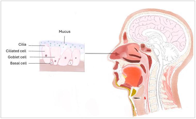
This medical illustration presents a sagittal anatomical diagram of the human head and neck, coupled with a histological inset of the nasal mucosa. The main diagram depicts the upper respiratory tract including the nasal cavity, hard and soft palate, tongue, pharynx, larynx, and trachea, alongside the brain and cervical spine. An arrow points from the nasal conchae to a detailed histological cross-section of the respiratory epithelium. This inset illustrates a pseudostratified ciliated columnar epithelium, highlighting its cellular composition: basal cells positioned along the basement membrane; secretory goblet cells; and ciliated cells featuring hair-like cilia at the apical surface. A superficial layer of mucus is shown above the cilia, representing the mucociliary clearance mechanism. This educational visual is designed to demonstrate the relationship between gross anatomy and microscopic tissue structure in the context of respiratory physiology and physical defense barriers against pathogens.
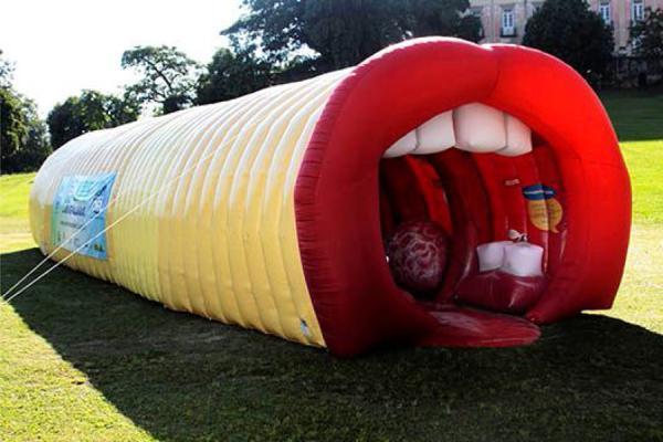
This photograph depicts a large-scale, walk-through educational tool known as a 'giant larynx' model, used in public health campaigns to teach human anatomy and voice production. The structure is an elongated inflatable tunnel with a segmented yellow exterior. One end is designed as a large open mouth featuring oversized red lips and white anatomical representations of upper teeth. The red interior visible through the mouth opening represents the pharynx and larynx, including structural interpretations of the tongue and vocal cords. This interactive anatomical diagram serves as a public education device for the specialty of otolaryngology (ENT), focusing on the upper respiratory tract and phonation. It is designed to simplify complex internal structures for a general audience, demonstrating the pathway from the mouth through the throat.
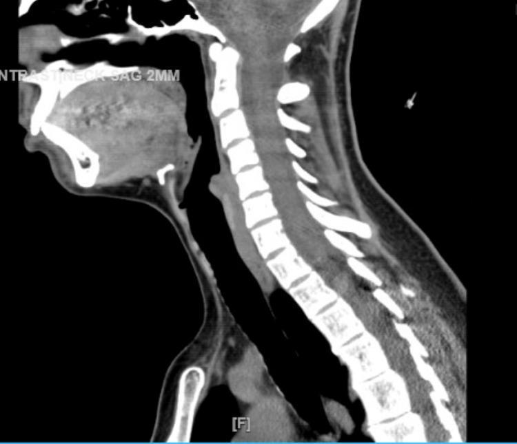
This diagnostic image is a non-contrast Computed Tomography (CT) scan of the neck in a sagittal view. The image illustrates the upper respiratory and digestive tract anatomy, specifically focusing on the soft tissues of the pharynx and larynx. Following resolution of a previous obstruction, this scan demonstrates a patent airway with visible restoration of the air column in the hypopharynx and laryngeal regions. Key anatomical landmarks include the cervical vertebrae (C1-C7), the base of the tongue, the epiglottis, and the hyoid bone. The soft tissues surrounding the airway show normal thickness and density, indicating a significant reduction in the edema previously noted in the clinical history. The image serves as a comparison to illustrate the resolution of drug-induced angioedema or inflammatory swelling that had compromised the airway at the level of the hyoid cartilage. It is a critical teaching tool for understanding airway patency and the radiological monitoring of soft tissue emergencies in otolaryngology and emergency medicine.
muscles of respiration diaphragm intercostal accessory muscles diagram
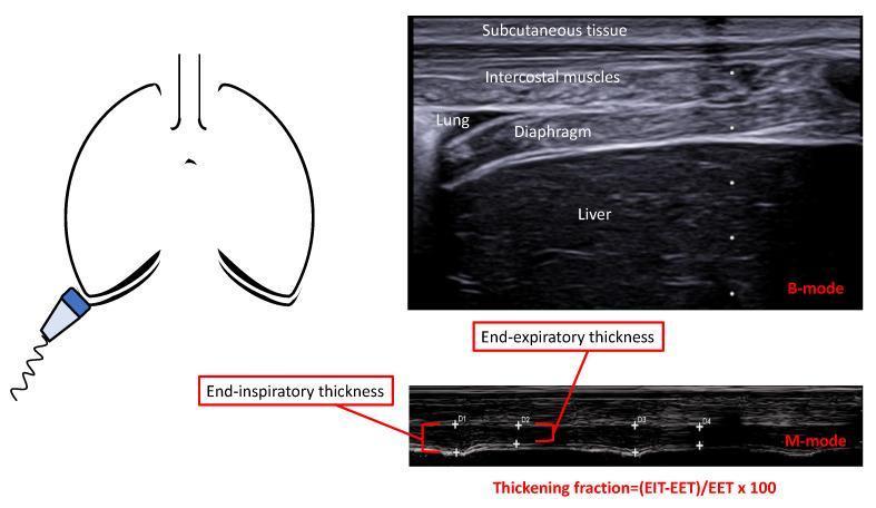
This composite educational graphic illustrates the ultrasonographic assessment of the diaphragm in the zone of opposition. It includes an anatomical diagram showing a linear probe placed in the lower intercostal space, a B-mode ultrasound image, and an M-mode ultrasound image. The B-mode image provides a cross-sectional anatomical view, labeling structures from superficial to deep: subcutaneous tissue, intercostal muscles, the lung, the hypoechogenic diaphragm, and the liver. The M-mode image displays the diaphragm's movement and thickness changes over the respiratory cycle. Key educational concepts shown include the measurement of end-inspiratory thickness (EIT) and end-expiratory thickness (EET). A mathematical formula for calculating the Thickening Fraction (TF) is provided: (EIT - EET) / EET x 100. This methodology is used in clinical settings to evaluate diaphragmatic function, muscle atrophy, and readiness for ventilator weaning by quantifying the muscle's contractile capacity and thickness.
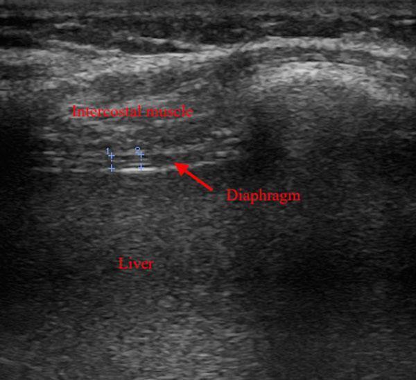
This diagnostic ultrasound image demonstrates the anatomical assessment of the diaphragm using a high-frequency linear probe. The imaging captures the zone of apposition, where the diaphragm is visible as a thin, relatively hypoechoic muscular layer sandwiched between two echogenic lines (the pleural and peritoneal membranes). Superficially, the intercostal muscle exhibits a heterogeneous echotexture, while deep to the diaphragm, the liver parenchyma presents with a more homogeneous, mid-level echogenicity. A red arrow identifies the diaphragm, and two vertical blue electronic calipers (labeled 1 and 2) demonstrate the clinical technique for measuring diaphragm thickness. This imaging modality is used in critical care settings to monitor for diaphragmatic atrophy or dysfunction in patients undergoing mechanical ventilation. Key landmarks included are the intercostal muscles, diaphragm, and liver, highlighting the use of point-of-care ultrasound (POCUS) for respiratory muscle monitoring.
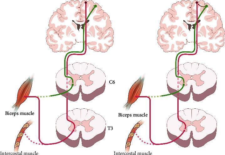
This pathophysiology diagram illustrates neuroplasticity and cortical reorganization following an intercostal-to-musculocutaneous nerve transfer for brachial plexus injury. The figure consists of two side-by-side panels showing a coronal brain section, spinal cord segments (C6 and T3), and the target muscles (biceps and intercostals). In the early phase (left panel), motor control for the biceps originates from the medial primary motor cortex (intercostal area). The descending pathway (red) bypasses the injured C6 segment to synapse at the T3 thoracic segment, where the donor intercostal nerve provides the motor signal to the reinnervated biceps brachii. This reflects synergistic movement where biceps contraction is tied to respiration. In the late phase (right panel), the 'cortical shifting phenomenon' is shown. A new functional connection (curved arrow) has formed between the lateral arm representation area and the medial intercostal area. While the physical nerve route via T3 remains (red line), the green pathway signifies that the lateral motor cortex now exerts control over elbow flexion. This illustrates the central nervous system's ability to adapt, allowing the patient to perform independent voluntary biceps contraction separate from respiratory activity.
Respiratory System: Upper Tract, Lower Tract, and Muscles of Respiration
PART 1: UPPER RESPIRATORY TRACT
1. Nose (External and Internal)
- The external nose is the entry portal; air enters via the nares (nostrils).
- Internally, the nasal cavity is divided by the nasal septum into right and left passages.
- The lateral walls bear three turbinates (conchae): superior, middle, and inferior. These create turbulent airflow, dramatically increasing surface area for:
- Warming inspired air to body temperature
- Humidifying inspired air to near 100% relative humidity
- Filtering particles >10 µm via nasal hairs and mucus
- The olfactory epithelium occupies the superior nasal cavity for smell.
- The nasal mucosa is lined by pseudostratified ciliated columnar epithelium with goblet cells that secrete mucus; cilia beat toward the pharynx to clear particles (mucociliary clearance).
- The nasal cavity opens into the nasopharynx posteriorly via the choanae.
2. Paranasal Sinuses
- Air-filled cavities in the skull bones that drain into the nasal cavity: maxillary, frontal, ethmoid, and sphenoid sinuses.
- They lighten the skull, add resonance to the voice, and produce mucus.
- Obstruction of their ostia leads to sinusitis.
3. Pharynx
| Region | Location | Key Features |
|---|---|---|
| Nasopharynx | Behind choanae, above soft palate | Contains adenoids (pharyngeal tonsil); Eustachian tubes open here; purely respiratory |
| Oropharynx | Soft palate to epiglottis | Contains palatine tonsils; shared food-and-air passage; bounded by soft palate superiorly, epiglottis inferiorly |
| Laryngopharynx (Hypopharynx) | Epiglottis to cricoid cartilage | Leads to esophagus posteriorly and larynx anteriorly |
4. Larynx
- Thyroid cartilage - largest; forms the "Adam's apple"; shields the vocal cords
- Cricoid cartilage - ring-shaped; the only complete cartilaginous ring in the airway; lies below the thyroid cartilage; the narrowest part of the adult airway (avg diameter 17 mm in men, 13 mm in women - Morgan and Mikhail's, p. 911)
- Epiglottis - leaf-shaped; folds over the glottis during swallowing to prevent aspiration
- Arytenoid cartilages (paired) - articulate with the cricoid; the vocal cords attach to them
- Corniculate and cuneiform cartilages - small accessory cartilages in the aryepiglottic folds
- True vocal cords - vibrate to produce sound; form the glottis
- False vocal cords (vestibular folds) - above the true cords; no phonation function but assist in airway protection
PART 2: LOWER RESPIRATORY TRACT
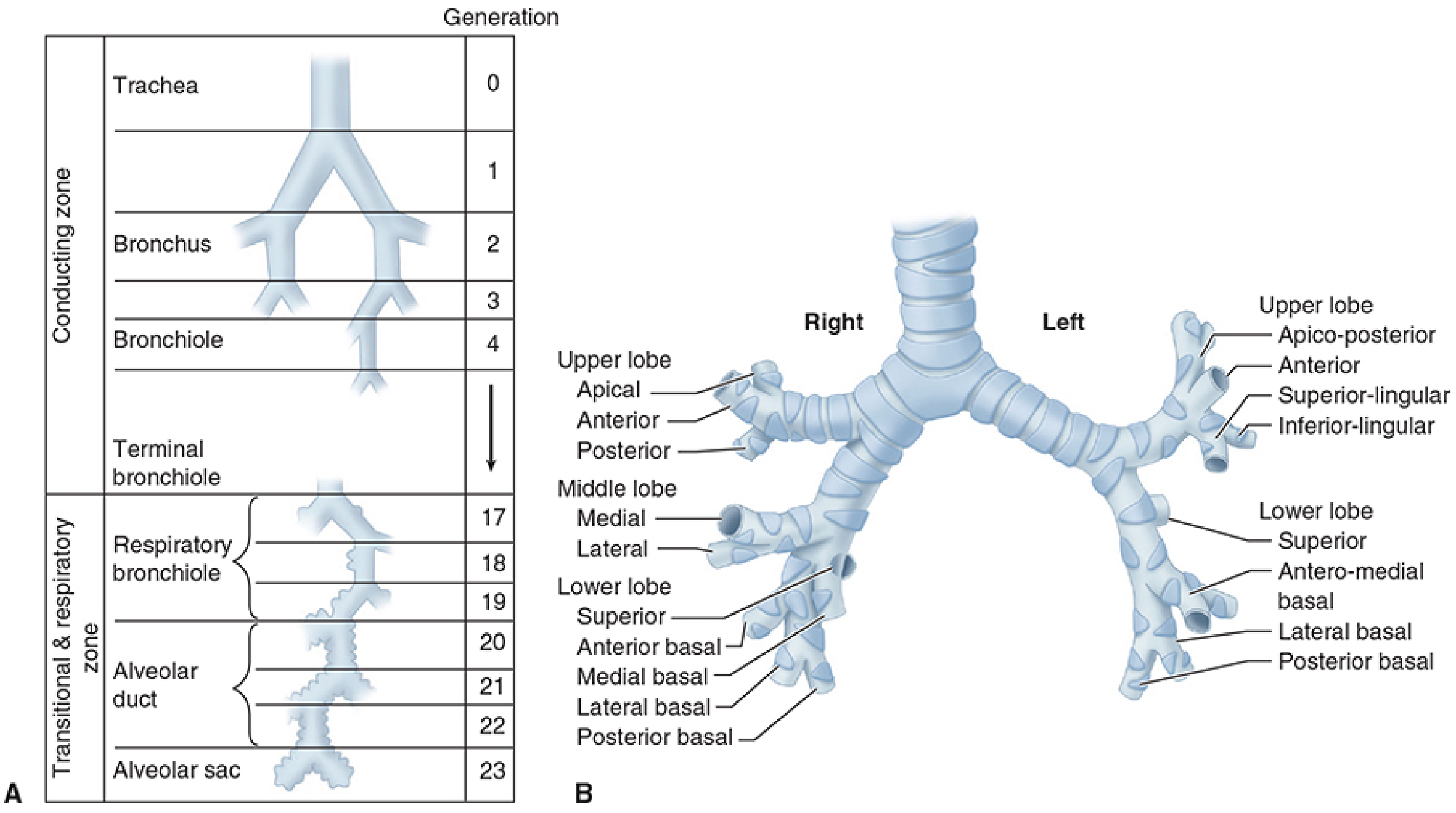
1. Trachea
- Begins at the lower border of the cricoid cartilage and extends to the carina.
- Average length: 10-13 cm in adults.
- Composed of 16-20 C-shaped cartilaginous rings, open posteriorly and connected by the membranous (posterior) wall containing the trachealis muscle.
- The carina marks the bifurcation into the right and left main bronchi, located at the level of the sternal angle (angle of Louis), approximately at T4-T5.
- Lined by pseudostratified ciliated columnar epithelium with goblet cells. - Morgan and Mikhail's Clinical Anesthesiology, 7e, p. 910-911
2. Mainstem Bronchi
| Feature | Right Main Bronchus | Left Main Bronchus |
|---|---|---|
| Angle from trachea | More vertical (~25°) | More horizontal (~45°) |
| Length | Shorter (~2 cm to RUL takeoff) | Longer (~4.5-5.0 cm) |
| Clinical significance | Foreign bodies more likely to lodge here | Longer → more time to act if intubation endobronchial |
3. Lobar and Segmental Bronchi
- Each lobe receives a lobar bronchus which then divides into segmental bronchi supplying bronchopulmonary segments:
- Right lung: 3 lobes, 10 segments (3 upper, 2 middle, 5 lower)
- Left lung: 2 lobes, 8-10 segments (4-5 upper, 4-5 lower)
- Bronchopulmonary segments are the smallest surgically resectable units.
4. Bronchioles
- Airways <1 mm in diameter with no cartilage; maintained open by elastic radial traction from surrounding lung tissue.
- Terminal bronchioles - last purely conducting generation (generation 16); no gas exchange occurs here.
- Respiratory bronchioles (generations 17-19) - first airways where alveoli begin to appear in the walls; partial gas exchange begins.
5. Alveolar Ducts and Alveoli
- Alveolar ducts (generations 20-22) lead to alveolar sacs (generation 23), each containing approximately 17 alveoli.
- An estimated 300-500 million alveoli provide a surface area of 50-100 m² for gas exchange in the adult. - Morgan and Mikhail's Clinical Anesthesiology, 7e, p. 912
- Alveolar cells:
- Type I pneumocytes (~95% of alveolar surface): flat, thin cells across which gas exchange occurs; cannot regenerate
- Type II pneumocytes (~5%): cuboidal cells that produce surfactant (reduces surface tension) and can regenerate Type I cells after injury
- Alveolar macrophages: mobile phagocytes that clear debris and pathogens
6. Conducting vs. Respiratory Zone
| Zone | Generations | Function |
|---|---|---|
| Conducting zone | 0 (trachea) to 16 (terminal bronchiole) | Air conduction only; forms the "anatomical dead space" (~150 mL) |
| Transitional zone | 17-19 (respiratory bronchioles) | Mixed conduction and gas exchange |
| Respiratory zone | 20-23 (alveolar ducts and sacs) | Gas exchange |
PART 3: MUSCLES OF RESPIRATION
Overview of the Ventilatory Pump
- Respiratory control centers (cortex, brainstem)
- Spinal cord tracts (corticospinal for voluntary; reticulospinal for automatic)
- Lower motor neurons (phrenic nerve, intercostal nerves)
- The respiratory muscles themselves
- Feedback receptors (chemoreceptors, mechanoreceptors) - Murray & Nadel's Textbook of Respiratory Medicine, p. 3039
A. Inspiratory Muscles
1. Diaphragm (Primary Muscle of Inspiration)
- The most important muscle of breathing, accounting for approximately 70-75% of inhaled tidal volume during quiet breathing.
- A dome-shaped musculotendinous partition between the thoracic and abdominal cavities.
- Contraction causes the dome to descend 1.5-7 cm, pulling the lungs downward and expanding thoracic volume; simultaneously, increased abdominal pressure pushes the lower ribs outward (bucket-handle motion via the zone of apposition).
- Innervation: Phrenic nerve (C3, C4, C5) - "C3, 4, 5 keeps the diaphragm alive."
- Bilateral phrenic nerve injury causes respiratory failure; unilateral injury causes ipsilateral hemidiaphragm paralysis with paradoxical movement. - Murray & Nadel's, p. 3040; Morgan and Mikhail's, p. 910
2. External Intercostal Muscles
- 11 pairs of thin muscle sheets running between adjacent ribs; fibers run downward and forward.
- Function: Elevate the ribs during inspiration → increases anteroposterior and transverse diameter of the chest ("bucket handle" and "pump handle" motions).
- Innervated by intercostal nerves (T1-T11).
- Contribute about 25% of resting tidal volume (the rest from the diaphragm).
3. Parasternal Intercartilaginous Muscles
- The most medial fibers of the internal intercostals that run between costal cartilages; they paradoxically act as inspiratory muscles (despite being "internal").
B. Expiratory Muscles
1. Abdominal Muscles (most important for active expiration)
| Muscle | Origin/Insertion | Action |
|---|---|---|
| Rectus abdominis | Pubic crest → xiphoid/costal cartilages | Compresses abdomen, depresses lower ribs |
| External oblique | Lower 8 ribs → iliac crest, linea alba | Compresses abdomen |
| Internal oblique | Iliac crest → lower 3 ribs, linea alba | Compresses abdomen |
| Transversus abdominis | Iliac crest, lumbar fascia → xiphoid, linea alba | Compresses abdomen; most important for cough/forced expiration |
- Abdominal pressure rises, forcing the diaphragm upward
- This rapidly reduces lung volume, generating high expiratory flow rates for coughing
2. Internal Intercostal Muscles
- Fibers run downward and backward (opposite to external intercostals).
- Most portions depress the ribs and reduce thoracic volume - function in active expiration.
- Exception: the parasternal portions (see above) assist inspiration. - Morgan and Mikhail's Clinical Anesthesiology, 7e, p. 708
C. Accessory Muscles of Inspiration
| Muscle | Action | Nerve |
|---|---|---|
| Sternocleidomastoid | Elevates the sternum and rib cage; lifts the anterior chest | CN XI (spinal accessory) + C2-C3 |
| Scalene muscles (anterior, middle, posterior) | Elevate and fix the upper two ribs; prevent inward collapse of upper ribs during inspiration | C4-C8 |
| Pectoralis major | Assists chest expansion when arms are fixed (e.g., hands on knees) | Medial and lateral pectoral nerves |
| Pectoralis minor | Elevates ribs 3-5 when scapula is fixed | Medial pectoral nerve |
| Serratus anterior | Elevates ribs when scapula fixed | Long thoracic nerve (C5-C7) |
| Trapezius | Fixes and elevates shoulders; stabilizes scapula for other muscles | CN XI |
D. Upper Airway Muscles (Airway Patency Muscles)
- Genioglossus: Tonic and reflex inspiratory activity keeps the tongue away from the posterior pharyngeal wall. Loss of this tone in sleep → obstructive sleep apnea.
- Levator palati, tensor palati, palatopharyngeus, palatoglossus: Prevent the soft palate from falling back against the posterior pharynx, especially in the supine position.
E. Neural Control Summary
| Level | Structure | Role |
|---|---|---|
| Cerebral cortex | Parietal cortex | Voluntary breathing (corticospinal tract) |
| Brainstem | Pons (pneumotaxic center) + Medulla (dorsal/ventral respiratory groups) | Automatic rhythm generation (reticulospinal tract) |
| Spinal cord | C3-C5 → Phrenic nerve; T1-T11 → Intercostal nerves; T6-L1 → Abdominal muscles | Motor neuron transmission |
| Effector | Diaphragm, intercostals, accessory muscles | Mechanical ventilation |
- Murray & Nadel's Textbook of Respiratory Medicine, p. 3039-3040
F. Age-Related Changes in Respiratory Muscles
- Maximal inspiratory pressure (PImax) and maximal expiratory pressure (PEmax) show moderate decrements with age in populations spanning ages 16-75.
- Women generate approximately 65-70% of the pressures generated by men.
- Physical activity has a training effect that preserves inspiratory muscle endurance across age groups.
- In the oldest age group (61-75 years) compared to the youngest (16-30 years), PImax decreases by 32-36% and PEmax decreases by 13-23%. - Fishman's Pulmonary Diseases and Disorders, p. (block 3)
Summary Table
| Structure | Location | Key Function |
|---|---|---|
| Nose | External URT | Filtration, humidification, warming |
| Nasopharynx | URT | Air conduction, Eustachian tube opening |
| Oropharynx | URT | Shared food/air passage |
| Larynx | URT/LRT boundary | Airway protection, phonation |
| Trachea | LRT | Main airway conduit (10-13 cm) |
| Main bronchi | LRT | Right (more vertical), Left (longer) |
| Bronchioles | LRT | Conduction; no cartilage |
| Alveoli | LRT | Gas exchange (300-500 million) |
| Diaphragm | Muscle | 70-75% of tidal volume; C3-C5 |
| External intercostals | Muscle | Elevate ribs; T1-T11 |
| Abdominal muscles | Muscle | Active expiration, coughing |
| Sternocleidomastoid | Accessory | Elevates rib cage in distress |
| Scalene muscles | Accessory | Fix upper ribs during inspiration |
Key Clinical Points:
- The right mainstem bronchus is more vertical - endobronchial intubation and foreign body aspiration preferentially affect the right side.
- The cricoid is the narrowest part of the adult airway - important for cricothyrotomy and airway assessment.
- The phrenic nerve (C3-C5) is the sole motor supply to the diaphragm - cervical cord injury at or above C4 eliminates spontaneous breathing.
- During respiratory distress, visible use of accessory muscles (SCM, scalenes) is a sign of increased work of breathing.
- Type II pneumocytes produce surfactant; their deficiency in prematurity causes infant respiratory distress syndrome (IRDS).