
Acute pancreatitis
Reading File
Now I'll search the medical library for comprehensive coverage of acute pancreatitis across multiple dimensions in parallel.
Finding Sources
Searching PubMed
"acute pancreatitis"[MeSH Terms] AND management
Excellent - found the key textbook sources. Let me now read the primary sections in parallel - Sleisenger & Fordtran (GI bible), Rosen's Emergency Medicine, and Family Medicine:
Reading File
Reading File
Reading File
Good data coming in. Let me read further from Rosen's Emergency Medicine (management, severity scoring) and the Radiology textbook, plus get the main Sleisenger block:
Reading File
Reading File
Finding Sources
Good. Now let me get the management and scoring details from Rosen's, Harrison's, and Current Surgical Therapy:
Reading File
Reading File
Reading File
Excellent content. Let me now get the management details (fluids, nutrition, antibiotics) from Rosen's and Harrison's, and also fetch a key image:
Reading File
Reading File
Loading Image
I now have comprehensive, high-quality content from multiple authoritative textbooks. Let me compile the full response.
Acute Pancreatitis
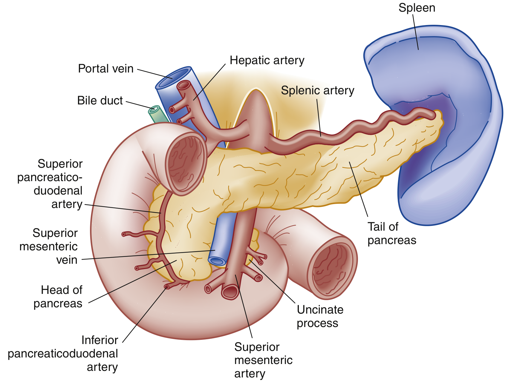
Definition
Acute pancreatitis (AP) is a sudden inflammatory condition of the pancreas involving enzymatic autodigestion and destruction of pancreatic tissue. It ranges from a mild, self-limited illness to life-threatening multiorgan failure with sepsis. It is the most common pancreatic disease worldwide and one of the top reasons for GI-related hospitalization in the US. - Rosen's Emergency Medicine, p. 1265
Diagnosis requires 2 of 3:
- Abdominal pain strongly suggestive of AP (epigastric, radiating to back)
- Serum amylase and/or lipase ≥3× upper limit of normal
- Characteristic imaging findings
- Grainger & Allison's Diagnostic Radiology, p. 677; Current Surgical Therapy 14e, p. 603
Etiology
| Category | Causes |
|---|---|
| Mechanical/Obstructive | Gallstones (40-70%), post-ERCP, tumors, pancreas divisum, ampullary stenosis |
| Toxic/Metabolic | Alcohol (25-35%), hypertriglyceridemia (TG >1000 mg/dL), hypercalcemia, uremia, scorpion venom |
| Drugs | Valproate, L-asparaginase, 6-mercaptopurine, steroids, thiazides, azathioprine |
| Infectious | Mumps, coxsackievirus, HIV, CMV, EBV, Salmonella, Mycoplasma, ascariasis |
| Trauma | Blunt abdominal trauma |
| Idiopathic | ~5-10% (many may be occult microlithiasis or genetic) |
| Genetic | SPINK1, CFTR, PRSS1 mutations (consider in recurrent/pediatric cases) |
- Rosen's Emergency Medicine, Box 77.1, p. 1266
Smoking and diabetes are independent risk factors. ALT elevation (positive predictive value 95%) is particularly specific for biliary/gallstone pancreatitis.
Pathophysiology
The central mechanism is premature intracellular activation of digestive enzymes (primarily trypsinogen → trypsin) within the pancreas itself, triggering autodigestion. This leads to:
- Local pancreatic inflammation and tissue destruction
- Release of pro-inflammatory cytokines (IL-1, IL-6, TNF-α)
- Systemic inflammatory response syndrome (SIRS)
- Increased microvascular permeability → third-spacing of fluids
- In severe cases: multiorgan dysfunction (lungs, kidneys, cardiovascular)
Enzymatic degradation of surfactant contributes to pulmonary sequelae. Coagulopathy results from cytokine-mediated activation of the coagulation cascade.
Classification (Revised 2012 Atlanta)
Morphological types:
- Interstitial edematous pancreatitis - diffuse enlargement without necrosis; most common; usually resolves within first week
- Necrotizing pancreatitis - affects ~5-10% of patients; may involve pancreatic parenchyma and peripancreatic tissue; necrosis may be sterile, liquefy, or become infected
Severity grades:
| Grade | Features | Mortality |
|---|---|---|
| Mild | No organ failure, no local/systemic complications | Very rare (<5%) |
| Moderately severe | Transient organ failure (<48h) OR local/systemic complications without persistent organ failure | ~15% of cases |
| Severe | Persistent organ failure >48h (single or multi-organ) | 36-50%; higher with infected necrosis |
- Current Surgical Therapy 14e, p. 603-604
Organ failure (Modified Marshall score - any 1 of):
- Cardiovascular: systolic BP <90 mmHg or HR >130 bpm
- Pulmonary: PaO₂ <60 mmHg
- Renal: creatinine >2.0 mg/dL
Clinical Features
Symptoms:
- Persistent epigastric or LUQ pain, typically moderate-to-severe, radiating to the back, chest, or flanks
- Pain relieved by sitting forward or leaning forward
- Nausea, vomiting, anorexia
- Note: pain intensity does NOT correlate with severity
Signs:
- Vital signs: fever, tachycardia, and sometimes hypotension in severe disease
- Jaundice suggests obstruction (gallstone or tumor)
- Epigastric tenderness ± guarding; rebound less common
- Absent bowel sounds if ileus present
- Shallow respirations (pain splinting); basilar crackles or decreased breath sounds
- Cullen sign - bluish periumbilical discoloration (hemoperitoneum) - rare, poor prognosis
- Grey Turner sign - reddish-brown flank discoloration (retroperitoneal hemorrhage) - rare, poor prognosis
- Murphy sign if gallstone etiology
Systemic complications:
-
ARDS, pleural effusions (up to 50% of patients, predominantly left-sided)
-
Cardiovascular collapse and shock (from fluid shifts)
-
Acute kidney injury (hypoperfusion + inflammatory mediators)
-
DIC/coagulopathy
-
Rosen's Emergency Medicine, p. 1265-1266
Investigations
Bloods:
- Lipase - preferred over amylase; more sensitive and specific; elevated for 1-2 weeks (amylase 3-5 days)
- Both begin rising within hours of onset; degree of elevation does NOT correlate with severity
- Amylase falsely negative in alcohol and hypertriglyceridemia-induced AP
- LFTs (ALT, AST, bilirubin) - evaluate for biliary etiology; ALT >3× ULN has PPV of 95% for gallstone pancreatitis
- FBC, BMP - assess for SIRS, organ failure
- Calcium, triglycerides - if no obvious etiology
- CRP >100 mg/L - useful marker of severity during hospitalization
- BUN - rising BUN associated with inadequate hydration and higher mortality
Imaging:
CT/MRI not routinely indicated for diagnosis. CT recommended only in:
- Diagnostic uncertainty or normal enzymes with high clinical suspicion
- Ruling out other intra-abdominal pathology
- Assessing complications when patient fails to improve after 48-72 hours
Best done 3-7 days after presentation for complication assessment (earlier CT may underestimate necrosis).
Ultrasound: first-line to assess for gallstones/biliary dilation.
Severity Scoring Systems
BISAP Score (Bedside Index of Severity in Acute Pancreatitis)
Used within first 24h. Score ≥3 = substantially increased mortality risk.
| Letter | Criteria |
|---|---|
| B | BUN >25 mg/dL |
| I | Impaired mental status (GCS <15) |
| S | SIRS (≥2 of 4 criteria present) |
| A | Age >60 years |
| P | Pleural effusion on imaging |
Ranson Criteria
At admission (non-biliary):
- Age >55 years
- WBC >16,000/mm³
- Glucose >200 mg/dL
- AST >250 IU/L
- LDH >350 IU/L
At 48 hours:
- Hematocrit drop >10%
- BUN rise >5 mg/dL
- Calcium <8 mg/dL
- PaO₂ <60 mmHg
- Base deficit >4 mEq/L
- Fluid needs >6 L
Score ≥3 = severe disease.
Other Systems
- APACHE II - can be calculated at any time; PPV 43%, NPV 89% for critically ill
- CT Severity Index (CTSI/Modified CTSI) - based on degree of necrosis, inflammation, and fluid collections on CT
- PASS (Pancreatitis Activity Scoring System) - dynamic, continuous monitoring throughout illness course
CT scoring does NOT supersede clinical scoring systems for identifying severe disease. - Grainger & Allison's, p. 685
- Rosen's Emergency Medicine, Table 77.1, p. 1267; Harrison's 22e, p. 2791; Current Surgical Therapy 14e, p. 604
Management
1. Fluid Resuscitation (cornerstone of treatment)
- Aggressive IV fluid replacement - Lactated Ringer's preferred over normal saline (lower incidence of SIRS)
- Typical rate: 250-500 mL/hr initially
- Target: urine output >0.5 mL/kg/hr, decreasing BUN and hematocrit
- A decrease in hematocrit and BUN within 12-24h confirms adequate resuscitation
- Less aggressive strategy may be sufficient for milder disease; adjust for cardiac/pulmonary/renal comorbidities
2. Pain Management
- IV opioids are safe and recommended
- Hydromorphone or morphine; meperidine no longer preferred
- NSAIDs and patient-controlled analgesia can supplement
3. Nutrition
- Mild AP: low-fat solid diet once patient can tolerate oral intake
- Severe AP: enteral nutrition preferred over TPN - initiated within 2-3 days of admission
- Enteral feeding maintains gut barrier integrity, limits bacterial translocation, lower cost, fewer complications than TPN
- Gastric feeding (nasogastric) is safe; jejunal (nasojejunal) has possible physiologic advantages but not proven superior
- Nil-by-mouth has no benefit and worsens outcomes in most cases
4. Antibiotics
- No role for prophylactic antibiotics in necrotizing pancreatitis - does not improve survival and may promote opportunistic fungal infections
- Empiric antibiotics indicated for: clinical decompensation, suspected infected necrosis, confirmed infection
- Drug-induced pancreatitis: discontinue offending agent
5. Addressing Etiology
- Gallstone pancreatitis: cholecystectomy (same admission for mild AP; after resolution for severe)
- Post-ERCP: rectal indomethacin and/or pancreatic duct stenting for prophylaxis
- Hypertriglyceridemia: insulin infusion, fibrates; adjuncts include heparin or plasmapheresis (limited evidence); outpatient lipid-lowering therapy
- Hypercalcemia: treat underlying hyperparathyroidism or malignancy
6. ICU Triage
-
Lower BISAP, normal hematocrit, low BUN → regular ward
-
Persistent SIRS at 24h or significant comorbidities → step-down unit
-
Higher BISAP, elevated hematocrit and BUN, respiratory failure, organ failure → ICU
-
Harrison's 22e, p. 2791-2792; Rosen's Emergency Medicine, p. 1268-1270
Local Complications
| Complication | Timing | Description |
|---|---|---|
| Acute Peripancreatic Fluid Collection (APFC) | <4 weeks | Homogenous, non-encapsulated, no necrosis; most resolve spontaneously |
| Pancreatic Pseudocyst | >4 weeks | Encapsulated collection with defined wall, without significant necrosis |
| Acute Necrotic Collection (ANC) | <4 weeks | Heterogeneous collection with necrotic debris |
| Walled-Off Necrosis (WON) | >4 weeks | Encapsulated necrotic collection |
| Vascular complications | Variable | Splenic/portal vein thrombosis, pseudoaneurysm |
| Bowel necrosis | Late | Rare, poor prognosis |
| Fistula | Variable | Pancreatico-pleural fistula (↑ pleural amylase) |
Suspect local complications in: prolonged/worsening symptoms, secondary rise in enzymes, fever, leukocytosis, sepsis.
Management of Necrosis
- Sterile necrosis: managed conservatively unless complications arise
- Infected necrosis (suspected): empiric broad-spectrum antibiotics (carbapenems penetrate necrosis well)
- Fine-needle aspiration (FNA) of necrosis for Gram stain/culture is no longer universally recommended - risk of contaminating sterile collections
- Drainage/debridement ("step-up approach"):
- Percutaneous/endoscopic drainage first
- Video-assisted retroperitoneal debridement (VARD) or endoscopic necrosectomy if drainage fails
- Open surgical necrosectomy (last resort - high morbidity/mortality)
- Interventions delayed until necrosis is walled off (typically >4 weeks) for better outcomes
Multidisciplinary team (gastroenterology, surgery, interventional radiology, ICU) is recommended; transfer to tertiary pancreas center if specialized services unavailable.
- Harrison's 22e, p. 2792; Current Surgical Therapy 14e, p. 604-605
Prognosis
- Overall mortality ~5% (declining over time despite rising incidence)
- Severe AP: 36-50% mortality
- Infected pancreatic necrosis: extremely high mortality
- Rising BUN during hospitalization = inadequate hydration + higher in-hospital mortality
- Persistent SIRS at 24h = predictor of organ failure and necrosis
- CRP >100 mg/L during hospitalization = marker of severity
Key Pearls
- Lipase is preferred over amylase (more sensitive, specific, and remains elevated longer)
- Degree of enzyme elevation does NOT predict severity
- CT should NOT be routine - wait 48-72h (ideally 3-7 days) to assess complications
- No role for prophylactic antibiotics in necrotizing pancreatitis
- Enteral nutrition over TPN in severe AP
- Lactated Ringer's over normal saline for fluid resuscitation
- Delay intervention on necrosis until it is walled off (>4 weeks)
- ALT >3× ULN has 95% PPV for biliary pancreatitis
Sources: Rosen's Emergency Medicine 10e | Harrison's Principles of Internal Medicine 22e (2025) | Current Surgical Therapy 14e | Sleisenger & Fordtran's GI & Liver Disease | Grainger & Allison's Diagnostic Radiology
This is a shared conversation. Sign in to Orris to start your own chat.