
Pain pathway
Reading File
The pain pathway involves nociceptors detecting stimuli, signals traveling via A-delta and C fibers to the spinal cord dorsal horn, ascending to the brain via the spinothalamic tract, and being processed in the thalamus and cortex. Modulation occurs via the descending inhibitory system (endorphins, enkephalins).
Searching sources...
Finding Sources
Finding Sources
Reading File
Reading File
Reading File
Reading File
Reading File
Loading Image
Reading File
Loading Image
Here is a thorough breakdown of the pain pathway, drawn from Harrison's Principles of Internal Medicine 22E and Neuroscience: Exploring the Brain, 5th Edition.
The Pain Pathway
Pain is both a sensation and an emotion - it combines a discriminative component (where, how intense, what quality) with an affective component (suffering, fear, urgency to act). The pathway has four functional stages: transduction → transmission → modulation → perception.
1. Peripheral Mechanisms - Transduction
Primary Afferent Nociceptors
Peripheral nerves carry three neuron types: primary sensory afferents, motor neurons, and sympathetic postganglionic neurons. The cell bodies of primary sensory afferents sit in the dorsal root ganglia (DRG).
Primary afferents are classified by diameter and myelination:
| Fiber Type | Diameter | Myelination | Conduction | Function |
|---|---|---|---|---|
| Aβ | Large | Heavily myelinated | Fast | Light touch, NOT pain normally |
| Aδ | Small | Thinly myelinated | Moderate | Sharp, well-localized ("first pain") |
| C | Smallest | Unmyelinated | Slow | Dull, burning, aching ("second pain") |
Aδ and C fibers are the primary afferent nociceptors - blocking them abolishes pain perception entirely.
Individual nociceptors are polymodal - they respond to:
- Intense heat or cold
- Intense mechanical distortion (pinch)
- Acidic pH
- Chemical irritants: ATP, serotonin, bradykinin (BK), histamine
- TRPV1 (vanilloid receptor) mediates heat and acidic stimuli and is activated by capsaicin
Sensitization
When tissues are damaged or inflamed, the threshold for activating nociceptors drops - this is sensitization, driven by inflammatory mediators (bradykinin, prostaglandins, nerve growth factor, leukotrienes):
- Peripheral sensitization: inflammatory mediators activate intracellular signaling in nociceptor terminals → increased production and insertion of ion channels → lower activation threshold
- Central sensitization: ongoing nociceptor input during inflammation raises excitability of dorsal horn neurons in the spinal cord
Neurogenic Inflammation
Activated nociceptors also act as effectors - they release substance P, CGRP, and cholecystokinin from their peripheral terminals. Substance P causes vasodilation, mast cell degranulation, leukocyte chemotaxis, and further inflammatory mediator release - amplifying the local inflammatory response.
2. Transmission - Spinal Cord
Entering the Dorsal Horn
Primary afferent nociceptors enter the spinal cord via the dorsal root and synapse in the dorsal horn of the spinal gray matter.
Key neurotransmitters released at this synapse:
- Glutamate - rapid excitation of second-order dorsal horn neurons
- Substance P and CGRP - slower, longer-lasting excitation
Referred Pain
Each spinal pain-transmission neuron receives convergent inputs from many primary afferents - both from skin and from deep structures (viscera, muscles). Because the brain normally attributes dorsal horn activity to cutaneous inputs, activation from deep structures gets mislocalized to the skin supplied by the same spinal segment. Example: diaphragmatic inflammation (C3-C4 segments) is perceived as shoulder pain because shoulder skin is innervated by the same segments.
3. Ascending Pathways
Spinothalamic Tract
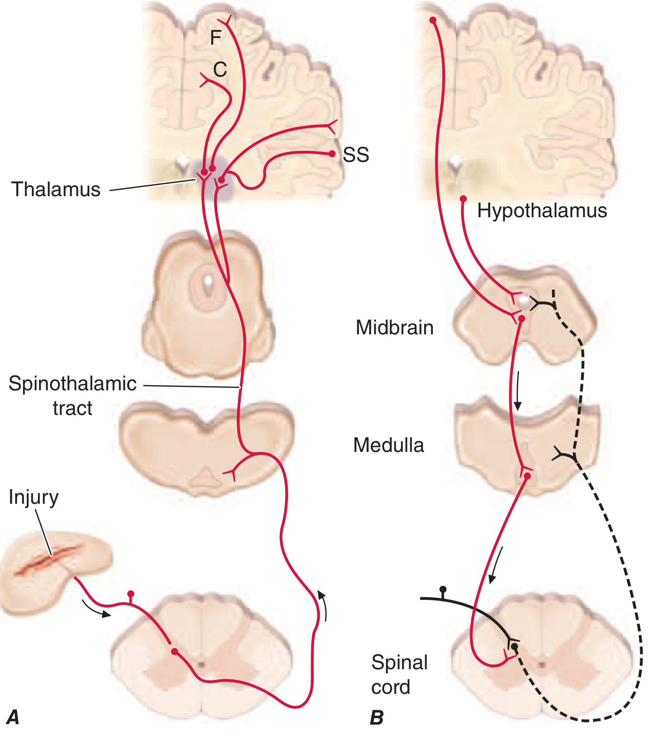
Most dorsal horn neurons activated by nociceptors send their axons contralaterally to form the spinothalamic tract (STT), which runs in the anterolateral white matter of the spinal cord through the lateral edge of the medulla and the lateral pons and midbrain. Interrupting this pathway produces permanent loss of pain and temperature discrimination.
Spinothalamic axons terminate in multiple thalamic regions:
- VP (ventral posterior) nucleus - segregated from touch fibers
- Intralaminar nuclei of the thalamus
Thalamus to Cortex
From the thalamus, pain signals diverge widely to:
- Somatosensory cortex (SS) - mediates sensory-discriminative aspects: location, intensity, quality
- Anterior cingulate cortex (C) - mediates the affective/emotional dimension (suffering)
- Frontal insular cortex (F) - also contributes to the emotional dimension
This dual cortical representation explains why frontal lobe lesions can reduce suffering from pain while the ability to localize it remains intact.
Trigeminal Pain Pathway (Face/Head)
Pain and temperature from the face travel via small-diameter fibers in the trigeminal nerve → synapse on the spinal trigeminal nucleus of the brainstem (an extension of the substantia gelatinosa) → axons cross midline and ascend as the trigeminal lemniscus → thalamus → cortex. This is the facial analogue of the spinothalamic pathway.
4. Pain Modulation
Pain perception is highly variable - the same injury can cause vastly different pain in different people or contexts (soldiers in battle, athletes, placebo effect). This is explained by descending modulatory circuits.
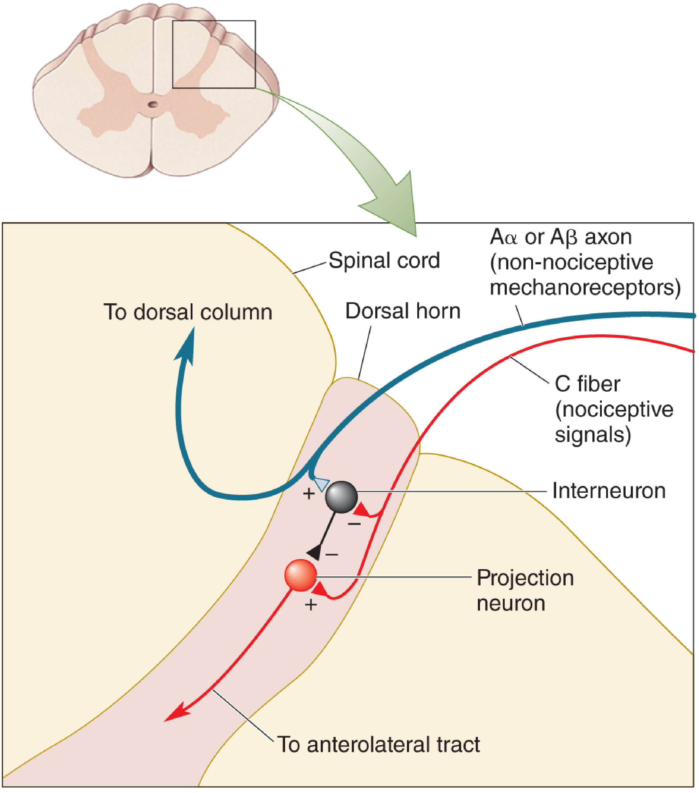
Gate Control Theory (Melzack & Wall, 1965)
The firing of dorsal horn projection neurons is determined by the balance of Aβ (large, non-nociceptive) vs. C fiber (nociceptive) input:
- High Aβ activity → excites inhibitory interneurons → "closes the gate" on C fiber signals → less pain
- High C fiber activity → inhibits interneurons → "gate stays open" → more pain
- Clinical application: TENS (transcutaneous electrical nerve stimulation) exploits this by activating large-diameter axons to reduce chronic pain
Descending Opioid Modulation
A dedicated descending inhibitory pathway runs: Frontal cortex/Hypothalamus → Periaqueductal gray (PAG, midbrain) → Rostral ventral medulla (RVM) → Dorsal horn
Key features:
- Each component contains opioid receptors and is sensitive to opioid drugs
- Contains endogenous opioid peptides: enkephalins and β-endorphin
- Activated by: attention, suggestion, intense emotion directed away from the injury, opioid analgesics
- Placebo analgesia activates this system - naloxone blocks it
- Nocebo effect (expectation of more pain) does the reverse
Summary Flow
Noxious stimulus
      ↓
Peripheral nociceptor (Aδ / C fiber) — TRANSDUCTION
      ↓
Dorsal root ganglion → Dorsal horn of spinal cord — TRANSMISSION
      ↓ (glutamate, substance P, CGRP)
Second-order neuron crosses midline → Spinothalamic tract (anterolateral)
      ↓
Thalamus (VP nucleus + intralaminar nuclei)
      ↓
Somatosensory cortex (location/quality) + Anterior cingulate/Insula (suffering)
      ↑↓
Descending modulation (PAG → RVM → dorsal horn, via enkephalins/β-endorphin)
Sources:
- Harrison's Principles of Internal Medicine 22E (2025), Chapter 14 - Pain: Pathophysiology and Management
- Neuroscience: Exploring the Brain, 5th Edition, Chapter 12 - The Somatic Sensory System
This is a shared conversation. Sign in to Orris to start your own chat.