
atrial flutter management
"atrial flutter" AND "catheter ablation"
ECG atrial flutter sawtooth pattern
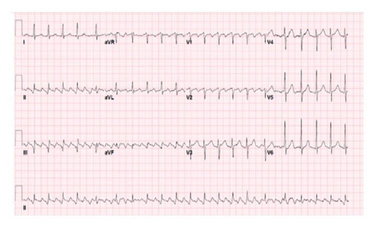
A 12-lead electrocardiogram (ECG) demonstrating typical atrial flutter with variable atrioventricular (AV) block. The tracing shows a classic 'sawtooth' baseline pattern, most prominent in the inferior leads (II, III, and aVF), representing rapid, regular macro-reentrant atrial activity (F-waves). The ventricular response (QRS complexes) is irregularly irregular because the AV node conducts atrial impulses at varying ratios (e.g., 2:1, 3:1, or 4:1 conduction). This diagnostic image is a key educational resource for cardiology, illustrating the distinction between atrial flutter and atrial fibrillation through the presence of organized, repetitive atrial waveforms. The QRS complexes appear narrow, indicating normal intraventricular conduction. This ECG provides a clinical example of supraventricular tachycardia and the importance of identifying baseline atrial morphology when assessing irregular rhythms.
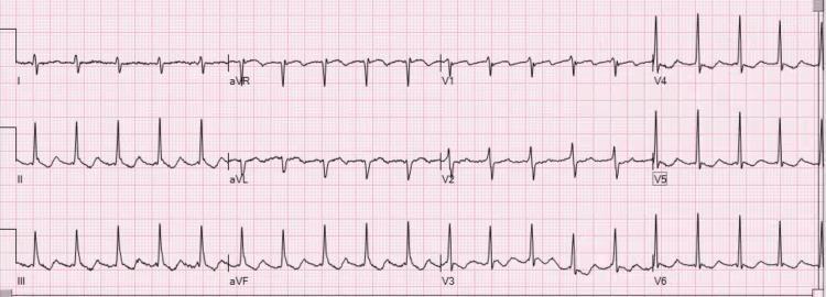
Diagnostic Image: This 12-lead electrocardiogram (ECG) demonstrates atrial flutter with a 2:1 atrioventricular (AV) conduction ratio. The tracing shows highly regular atrial activity with a characteristic sawtooth morphology. In the inferior leads (II, III, and aVF), the flutter waves are distinctly positive (upright), while in lead V1, the flutter waves exhibit a negative (inverted) polarity. This specific polarity pattern—upright flutter waves in the inferior leads and inverted in V1—is diagnostic of clockwise typical atrial flutter (atypical by counter-clockwise standards). The QRS complexes are regular and narrow, occurring at approximately half the frequency of the atrial rate (atrial rate ~300 bpm; ventricular rate ~150 bpm). In the precordial leads V4-V6, the QRS and T-wave morphologies are preserved, although discrete P waves are replaced by the undulating flutter baseline. The image illustrates a classic supraventricular tachycardia frequently encountered in cardiology and emergency medicine, serving as an educational example of macro-reentrant atrial rhythms.
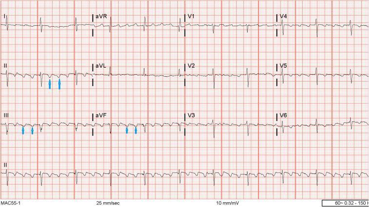
A standard 12-lead electrocardiogram (ECG) displayed on a traditional grid background (25 mm/sec, 10 mm/mV). The tracing demonstrates typical Atrial Flutter (AFL). The most prominent feature is the continuous 'sawtooth' baseline pattern, consisting of rapid, regular flutter waves (F waves). These F waves are most clearly visualized in the inferior leads (II, III, and aVF), where they exhibit a predominantly negative deflection. Blue arrows in leads II, III, and aVF highlight these recurring macro-reentrant atrial oscillations. The ventricular rhythm is regular, suggesting a fixed atrioventricular (AV) conduction ratio, likely 2:1 or 4:1 given the flutter rate. Normal QRS complexes and T waves are present across all leads but are superimposed upon the undulating flutter baseline. Precordial leads (V1-V6) show small, upright flutter waves in V1, typical for counterclockwise cavotricuspid isthmus-dependent atrial flutter. This diagnostic image serves as a classic educational example for identifying supraventricular tachyarrhythmias and understanding the morphology of counterclockwise atrial flutter.
Atrial Flutter: Management Overview

1. Acute/Hemodynamically Unstable Patients
- If flutter causes hypotension, pulmonary edema, ischemia, or severe symptoms: immediate synchronized electrical cardioversion (flutter typically responds to low energy, often 50-100 J).
2. Anticoagulation
- Use the CHA2DS2-VASc score plus bleeding risk assessment to guide anticoagulation, applying the same principles as atrial fibrillation.
- Anticoagulate before elective cardioversion (rhythm restoration) unless cardioversion is emergent for instability.
- Patients with atrial flutter carry roughly a 3% per year thromboembolic risk, so those at risk of recurrence need long-term anticoagulation similar to AF - Goldman-Cecil Medicine, p. 574; Harrison's Principles of Internal Medicine 22E.
- If CTI ablation is successful and flutter does not recur, long-term anticoagulation is generally not required.
3. Rate Control
- AV nodal-blocking agents (beta-blockers, non-dihydropyridine calcium channel blockers - diltiazem/verapamil, or digoxin in select cases) are used, but rate control in atrial flutter is often harder to achieve than in AF because the organized atrial rhythm conducts less variably through the AV node.
4. Rhythm Control
- Antiarrhythmic drugs: sotalol, dofetilide, disopyramide, ibutilide, and amiodarone can be used for chemical cardioversion or maintenance of sinus rhythm, but more than 70% of patients relapse on drug therapy alone.
- Electrical cardioversion is effective for restoring sinus rhythm acutely.
5. Catheter Ablation - First-Line Therapy
- Radiofrequency ablation of the cavotricuspid isthmus is considered first-line therapy for typical (CTI-dependent) atrial flutter because of its high success rate (>95% cure rate), low complication risk, and low recurrence - Harrison's Principles of Internal Medicine, 22E, p. 1988; Goldman-Cecil Medicine, p. 574.
- Caveat: up to half of patients who undergo successful CTI ablation later develop atrial fibrillation, so surveillance continues after the procedure.
- Atypical atrial flutter (non-CTI-dependent, often left atrial, seen after mitral valve surgery or AF ablation) is also preferentially treated with catheter ablation, but success rates are lower; antiarrhythmic drugs or intermittent cardioversion are reasonable alternatives when ablation is less feasible.
Practical Stepwise Approach
- Assess hemodynamic stability -> cardiovert immediately if unstable.
- Stratify thromboembolic risk (CHA2DS2-VASc) -> start/continue anticoagulation.
- Achieve rate control if remaining in flutter.
- Decide on rhythm control strategy: cardioversion +/- antiarrhythmics, versus proceeding directly to catheter ablation (preferred long-term strategy for typical flutter given high cure rate).
- Monitor for post-ablation atrial fibrillation; continue anticoagulation based on residual risk.
Note on evidence currency
indications of anticoagulant in a flutter
"atrial flutter" AND anticoagulation
Indications for Anticoagulation in Atrial Flutter
1. Risk-stratified long-term anticoagulation (chronic/non-cardioversion setting)
- Score 0 (men) / 1 (women, sex as the only risk factor): oral anticoagulation generally not recommended.
- Score 1 (men) / 2 (women): no anticoagulation, antiplatelet therapy, or an oral anticoagulant are all acceptable options - decision individualized.
- Score ≥2 (men) / ≥3 (women): oral anticoagulation is recommended - Tintinalli's Emergency Medicine, Table 18-13.
2. Peri-cardioversion anticoagulation (this is the indication most specific to flutter management)
- If flutter duration is unknown or ≥48 hours: anticoagulate with a direct oral anticoagulant (or heparin) for at least 3 weeks before elective cardioversion, OR obtain a transesophageal echocardiogram (TEE) to exclude atrial thrombus before proceeding sooner.
- If TEE shows thrombus, cardioversion is postponed and the patient is treated with therapeutic anticoagulation for at least 3 weeks before reattempting.
- All patients should be anticoagulated for at least 1 month after cardioversion if flutter/AF has been present ≥48 hours (or duration unknown), because atrial mechanical function ("atrial stunning") lags behind electrical recovery, keeping thromboembolic risk elevated even after sinus rhythm is restored - Goldman-Cecil Medicine; Fuster and Hurst's The Heart, 15th ed.
- Exception: if flutter onset is clearly and reliably <48 hours, cardioversion can often proceed without the 3-week pre-treatment, though many centers still anticoagulate periprocedurally, especially if any stroke risk factors are present.
- For hemodynamically unstable patients requiring emergency cardioversion, don't delay for anticoagulation - cardiovert first, then start anticoagulation as soon as possible afterward and continue for at least 4 weeks.
3. After catheter ablation
- If cavotricuspid isthmus ablation is successful and flutter does not recur, long-term anticoagulation is generally not necessary (assuming no other indication such as concomitant AF or an elevated CHA2DS2-VASc score from other risk factors).
- Anticoagulation decisions post-ablation remain somewhat debated, largely because of the substantial rate of subsequent atrial fibrillation in these patients, so ongoing risk factors still warrant reassessment - Harrison's Principles of Internal Medicine 22E, p. 1988.
4. Special note
Practical summary table
| Scenario | Anticoagulation indication |
|---|---|
| CHA2DS2-VASc ≥2 (men) / ≥3 (women) | Recommended, long-term |
| CHA2DS2-VASc 1 (men) / 2 (women) | Individualized (anticoagulant, antiplatelet, or none) |
| CHA2DS2-VASc 0 (men) / 1 (women, sex only) | Not routinely recommended |
| Flutter duration ≥48h or unknown, planned cardioversion | Anticoagulate ≥3 weeks before (or TEE to exclude thrombus) and ≥4 weeks after |
| Emergency cardioversion for instability | Anticoagulate as soon as possible after, continue ≥4 weeks |
| Successful CTI ablation, no recurrence | Long-term anticoagulation usually not needed (reassess if other risk factors) |