
Tell me everything about hematopoiesis, normal blood flow, Hermostasis and coagulation cascade both primary and secondary along with the factors and enzymes involved in great detai from basics to clinicals. Also include diagrams and illustration
Reading File
Now let me run all the library searches in parallel:
Finding Sources
Finding Sources
Finding Sources
Now let me read the most comprehensive sources in parallel:
Reading File
Reading File
Reading File
Excellent - rich content retrieved. Now let me get more detail from the coagulation cascade section, hematopoiesis growth factors, and the clinical aspects, plus fetch key diagrams:
Reading File
Reading File
Loading Image
Excellent! Now let me fetch the coagulation cascade diagrams and get more detail on anticoagulant mechanisms and platelet details:
Loading Image
Loading Image
Loading Image
Now let me read the anticoagulant mechanisms, platelet details, and the Robbins hemostasis section more fully:
Reading File
Reading File
Now let me fetch remaining key images and get the physiology blood flow section:
Loading Image
Loading Image
Finding Sources
I now have all the essential content and diagrams needed. Let me also quickly get the endothelial anticoagulant diagram and the normal blood flow physiology from Guyton:
Reading File
Reading File
Let me fetch the endothelium anticoagulant diagram and then get normal blood circulation from Guyton:
Loading Image
Finding Sources
I now have everything needed for a comprehensive response. Let me update the todos and write the final answer:Here is the complete, textbook-sourced guide from basics to clinicals.
Hematopoiesis, Normal Blood Flow, Hemostasis & Coagulation Cascade
PART 1: HEMATOPOIESIS
1.1 Definition and Sites Through Development
Hematopoiesis is the process by which all formed elements of blood - red cells, white cells, and platelets - are produced from a common precursor, the hematopoietic stem cell (HSC).
Embryonic progression of hematopoietic sites:
| Gestational Period | Primary Site |
|---|---|
| Week 3 | Yolk sac (transient - mainly embryonic red cells, plus long-lived tissue macrophages like microglia and Kupffer cells) |
| Week 5-6 | Aorta-Gonad-Mesonephros (AGM) region - definitive HSCs arise here |
| Month 3 | Liver (chief site until near birth) |
| Month 4 onward | Bone marrow (takes over progressively) |
| Birth | Marrow throughout skeleton is active; hepatic hematopoiesis dwindles |
| Post-puberty | Restricted to axial skeleton; ~50% of marrow space remains active |
The fetal placenta also harbors HSCs harvested at birth from umbilical cord blood - this is the basis for therapeutic cord blood transplantation.
- Robbins, Cotran & Kumar Pathologic Basis of Disease, p. 548
1.2 The Hematopoietic Stem Cell (HSC)
Normal adult marrow contains 50,000-200,000 HSCs, sitting at the apex of a differentiation hierarchy. HSCs have two essential properties:
- Pluripotency - a single HSC can generate ALL mature blood cell types
- Self-renewal - at least one daughter cell re-enters the stem cell pool to prevent depletion
HSCs reside in a specialized marrow niche where stromal cells and secreted factors protect them. Under stress (severe anemia, acute inflammation), HSCs mobilize into the peripheral blood - this is exploited clinically by administering G-CSF to donors to mobilize HSCs for peripheral blood collection and transplantation.
HSC surface markers (mouse model, clinically extrapolated): cKIT+, Sca-1+, LIN- (negative for lineage-specific markers)
1.3 Hematopoietic Differentiation Hierarchy
Fig. 13.1 - Differentiation of blood cells (Robbins, Cotran & Kumar):
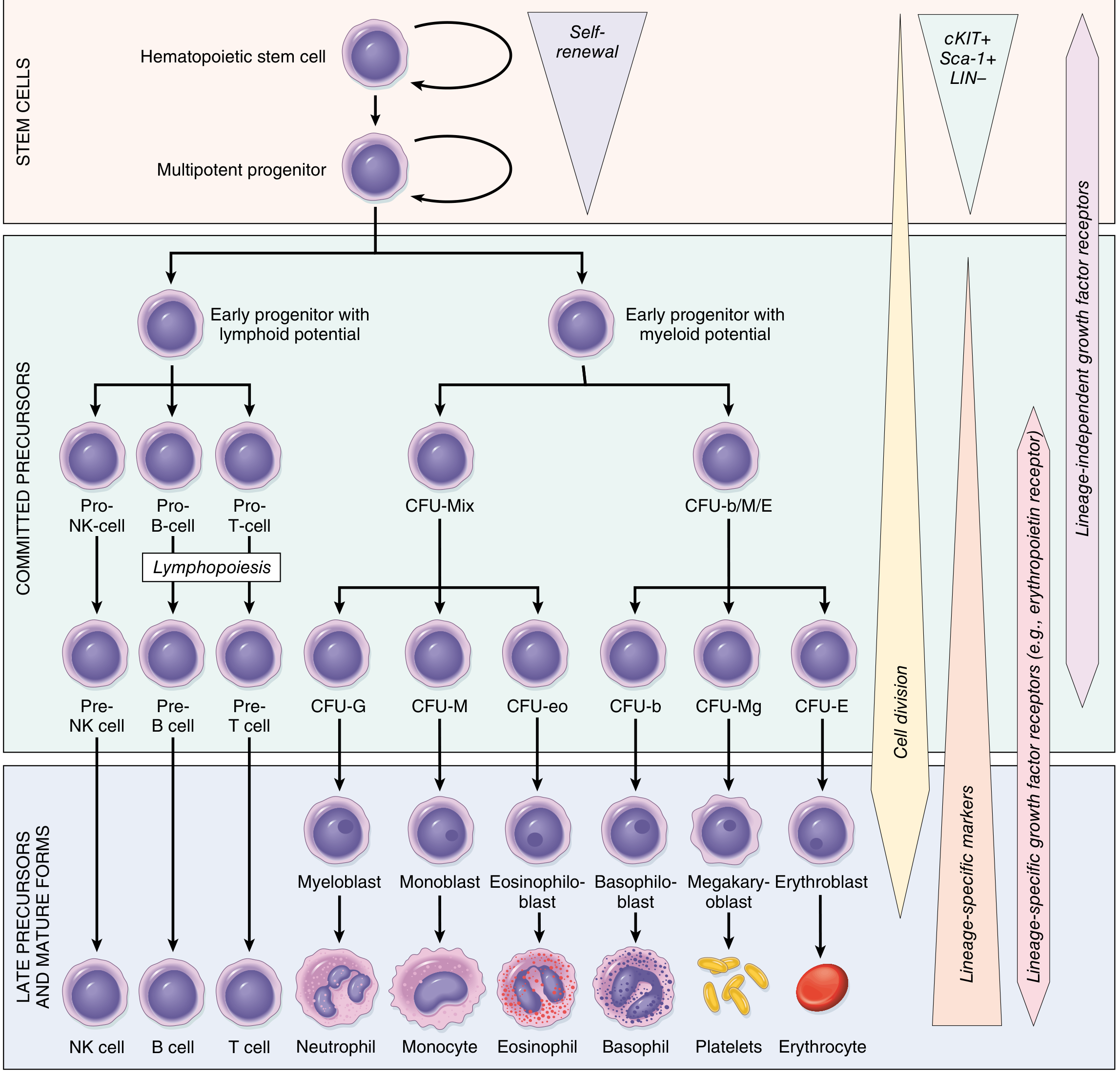
CFU = Colony Forming Unit; LIN- = lineage marker negative; NK = Natural Killer
The hierarchy in order:
HSC (self-renewing)
└── Multipotent Progenitor (high proliferative, low self-renewal)
├── Early Lymphoid Progenitor
│ ├── Pro-NK → Pre-NK → NK Cell
│ ├── Pro-B → Pre-B → B Cell
│ └── Pro-T → Pre-T → T Cell
└── Early Myeloid Progenitor
├── CFU-Mix (granulocyte-erythrocyte-monocyte-megakaryocyte)
│ ├── CFU-G → Myeloblast → Neutrophil
│ └── CFU-M → Monoblast → Monocyte
└── CFU-b/M/E
├── CFU-eo → Eosinophiloblast → Eosinophil
├── CFU-b → Basophiloblast → Basophil
├── CFU-Mg → Megakaryoblast → Megakaryocyte → Platelets
└── CFU-E → Erythroblast → Erythrocyte
1.4 Growth Factors (Colony-Stimulating Factors)
These cytokines regulate lineage-specific proliferation and differentiation:
| Growth Factor | Target Progenitor | Clinical Use |
|---|---|---|
| KIT ligand (Stem Cell Factor) | Very early progenitors (cKIT+) | HSC maintenance |
| FLT3 ligand | Very early progenitors | Mobilization, DC generation |
| GM-CSF (Granulocyte-Macrophage CSF) | Committed myeloid progenitors | Neutropenia post-chemotherapy |
| G-CSF | Committed neutrophil progenitors | Neutropenia; HSC mobilization for transplant |
| M-CSF | Monocyte progenitors | Macrophage production |
| Erythropoietin (EPO) | CFU-E (erythroid progenitors) | Anemia of CKD |
| Thrombopoietin (TPO) | Megakaryocyte progenitors | Thrombocytopenia |
| IL-3 | Broad multipotent progenitors | Experimental |
- Robbins, Cotran & Kumar Pathologic Basis of Disease, Table 13.1
1.5 Normal Adult Blood Cell Reference Ranges
| Cell Type | Normal Range |
|---|---|
| White cells | 4.8-10.8 × 10³/μL |
| Neutrophils | 1.4-6.5 × 10³/μL |
| Lymphocytes | 1.2-3.4 × 10³/μL |
| Monocytes | 0.1-0.6 × 10³/μL |
| Eosinophils | 0-0.5 × 10³/μL |
| Basophils | <0.2 × 10³/μL |
| Red cells (male) | 4.5-5.9 × 10⁶/μL |
| Red cells (female) | 4.0-5.2 × 10⁶/μL |
| Platelets | 150-400 × 10³/μL |
1.6 Clinical Relevance of Hematopoiesis
- Aplastic anemia - HSC failure; marrow becomes replaced with fat; pancytopenia
- Leukemia/Lymphoma - malignant transformation at various stages of the hierarchy (e.g., AML from myeloblast; CML from multipotent progenitor with BCR-ABL translocation)
- Myelodysplastic syndrome (MDS) - clonal disorder with ineffective hematopoiesis and dysplastic morphology
- Polycythemia vera - JAK2 V617F mutation → erythroid hyperproliferation independent of EPO
- Essential thrombocythemia - unregulated megakaryocyte proliferation
- Extramedullary hematopoiesis - when marrow is replaced (myelofibrosis, thalassemia), the spleen and liver revert to fetal hematopoietic function
- G-CSF use - mobilizes HSCs for peripheral blood stem cell collection in transplantation
PART 2: NORMAL BLOOD FLOW AND CIRCULATION
2.1 The Cardiovascular Circuit
Blood flows through two main circuits in series:
Pulmonary (right heart) circuit:
Right atrium → Right ventricle → Pulmonary artery → Pulmonary capillaries (gas exchange) → Pulmonary veins → Left atrium
Systemic (left heart) circuit:
Left atrium → Left ventricle → Aorta → Arteries → Arterioles → Capillaries (tissue exchange) → Venules → Veins → Superior/Inferior vena cava → Right atrium
2.2 Vessel Types and Their Functions
| Vessel | Wall Characteristics | Function |
|---|---|---|
| Elastic arteries (aorta, pulmonary artery) | Thick media with elastic lamellae | Pressure reservoir; Windkessel effect - store systolic energy, release in diastole |
| Muscular arteries | Prominent smooth muscle | Distribution of flow |
| Arterioles | High smooth muscle-to-lumen ratio | Resistance vessels; primary regulators of blood pressure and organ flow |
| Capillaries | Single endothelial cell layer + basement membrane | Exchange of gases, nutrients, waste |
| Venules | Thin wall | Exchange; leukocyte trafficking |
| Veins | Thin media, valves in extremities | Capacitance vessels; contain ~65% of total blood volume |
2.3 Determinants of Blood Flow
Blood flow follows principles analogous to Ohm's Law:
Q = ΔP / R
Where:
- Q = flow (mL/min)
- ΔP = pressure gradient
- R = resistance
Resistance is determined by the Hagen-Poiseuille relationship:
R = 8ηL / πr⁴
- η = viscosity
- L = vessel length
- r = vessel radius
The radius has the most powerful effect - doubling the radius decreases resistance 16-fold. This is why arteriolar smooth muscle tone is the dominant regulator of organ blood flow.
2.4 Normal Hemodynamic Values
| Parameter | Normal Value |
|---|---|
| Cardiac output | 4-8 L/min |
| Mean arterial pressure (MAP) | ~93 mmHg |
| Systemic vascular resistance | 800-1200 dynes·s/cm⁵ |
| Pulmonary artery pressure | 25/10 mmHg (mean ~15) |
| Capillary hydrostatic pressure | ~35 mmHg (arterial end), ~15 mmHg (venous end) |
| Plasma oncotic pressure | ~25 mmHg |
2.5 Regulation of Blood Flow
- Autoregulation - intrinsic ability of organs to maintain constant flow despite changes in perfusion pressure (kidney, brain, heart)
- Metabolic regulation - CO₂, H⁺, adenosine, K⁺ from active tissue cause local vasodilation
- Endothelium-derived factors - nitric oxide (NO) causes vasodilation; endothelin-1 causes vasoconstriction
- Neural control - sympathetic adrenergic tone (α₁ receptors on arteriolar smooth muscle) maintains vascular tone
- Hormonal - epinephrine, angiotensin II (vasoconstriction); ANP, bradykinin (vasodilation)
PART 3: HEMOSTASIS
3.1 Overview
Hemostasis is a precisely orchestrated process involving platelets, clotting factors, and endothelium that occurs at the site of vascular injury to form a blood clot and limit bleeding.
The four sequential steps:
- Arteriolar vasoconstriction - immediate, neurogenic reflex + local endothelin release; transient
- Primary hemostasis - platelet plug formation
- Secondary hemostasis - fibrin deposition via coagulation cascade
- Clot stabilization and limitation - factor XIII cross-links fibrin; anticoagulant mechanisms confine the clot
- Robbins & Kumar Basic Pathology, p. 84
3.2 Primary Hemostasis - The Platelet Plug
Platelet Structure
Platelets are anucleate disc-shaped fragments shed from megakaryocytes. They contain:
- α-Granules (membrane has P-selectin): fibrinogen, factor V, vWF, fibronectin, PDGF, TGF-β, platelet factor 4
- Dense (δ) Granules: ADP, ATP, polyphosphate, ionized Ca²⁺, serotonin, epinephrine
Key surface receptors:
- GpIb - receptor for vWF (absent in Bernard-Soulier syndrome)
- GpIIb-IIIa - receptor for fibrinogen/vWF; required for platelet aggregation (absent in Glanzmann thrombasthenia)
Steps of Primary Hemostasis
Step 1 - Platelet Adhesion:
Disruption of endothelium exposes subendothelial collagen. vWF (stored in Weibel-Palade bodies of endothelium and α-granules of platelets) binds to collagen and acts as a bridge to the platelet surface receptor GpIb. This tethers and arrests platelets at the injury site.
Step 2 - Platelet Activation:
Platelets undergo dramatic shape change (smooth disc → spiky "sea urchin" with pseudopods), increasing surface area. Activation triggers:
- Conformational change in GpIIb-IIIa increasing its affinity for fibrinogen
- Translocation of phosphatidylserine to outer leaflet (provides negatively charged phospholipid surface for coagulation factor assembly)
- Release of granule contents (release reaction)
Triggers of platelet activation:
- Thrombin (via PAR-1, protease-activated receptor) - most potent
- ADP from dense granules (self-amplifying loop)
- Collagen (direct contact via GPVI receptor)
- Thromboxane A₂ (TxA₂) - produced by platelet COX-1 from arachidonic acid
Step 3 - Platelet Aggregation:
Released ADP and TxA₂ recruit more platelets. Fibrinogen bridges adjacent platelet GpIIb-IIIa receptors, forming the primary hemostatic plug.
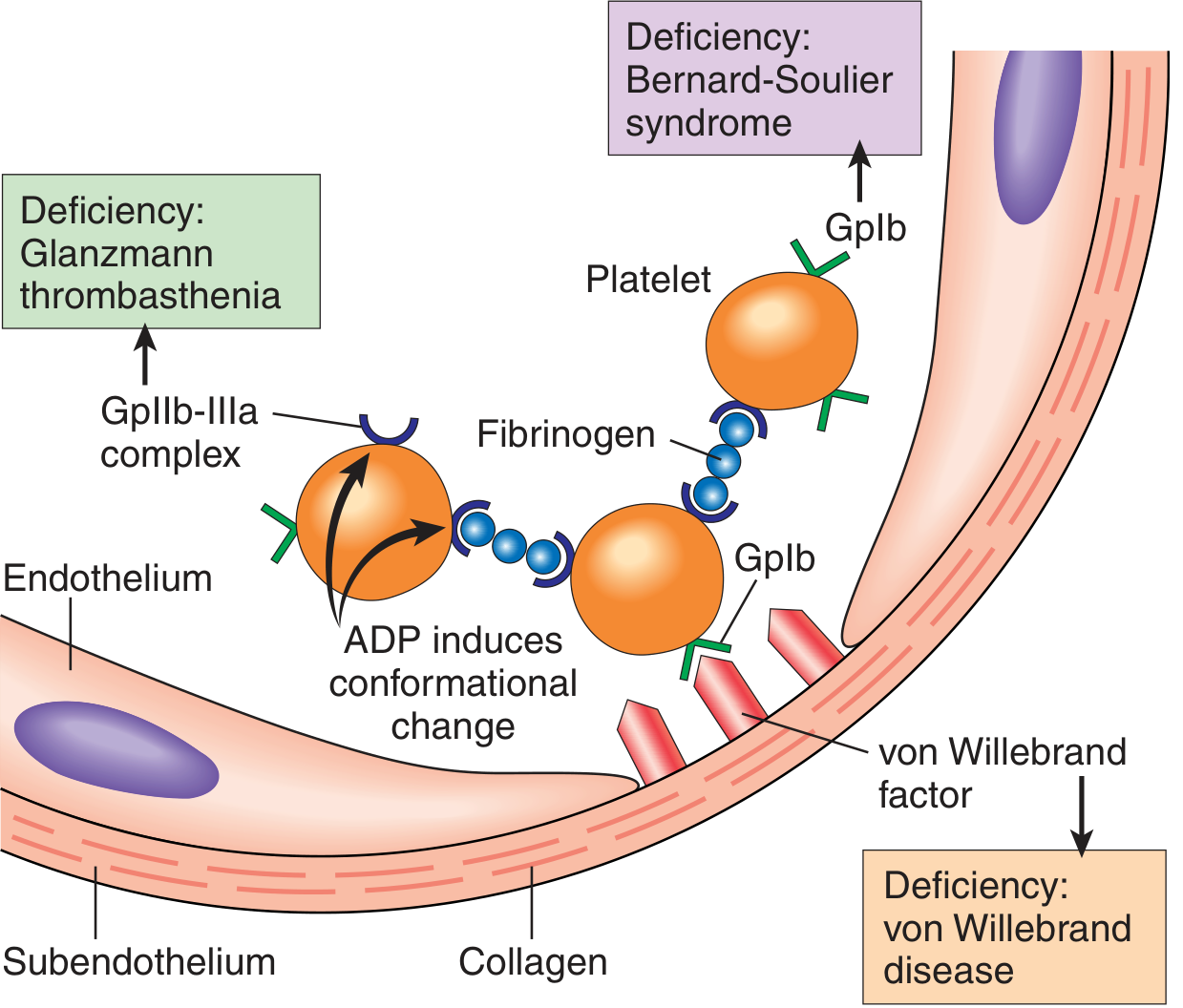
Fig. 3.6 - Platelet adhesion and aggregation. vWF bridges subendothelial collagen to platelet GpIb. Fibrinogen bridges adjacent platelets via GpIIb-IIIa. Congenital deficiencies are shown in colored boxes. (Robbins & Kumar Basic Pathology)
3.3 Secondary Hemostasis - The Coagulation Cascade
The coagulation cascade converts the loose platelet plug into a solid, covalently cross-linked fibrin clot. It is a series of amplifying enzymatic reactions - each step involves:
- An enzyme (activated coagulation factor - serine protease)
- A substrate (inactive proenzyme)
- A cofactor (reaction accelerator)
All assembled on a negatively charged phospholipid surface (provided by activated platelets) with calcium ions holding the complex together.
- Robbins, Cotran & Kumar Pathologic Basis of Disease, p. 122
3.4 Coagulation Factors - Complete List
| Factor | Name | Type | Vitamin K-dependent? |
|---|---|---|---|
| I | Fibrinogen | Substrate (converted to fibrin) | No |
| II | Prothrombin | Serine protease zymogen | Yes |
| III | Tissue Factor (Thromboplastin) | Transmembrane protein/cofactor | No |
| IV | Calcium (Ca²⁺) | Cofactor ion | No |
| V | Labile factor (proaccelerin) | Cofactor | No |
| VI | (Obsolete - Va is active form of V) | - | - |
| VII | Proconvertin | Serine protease zymogen | Yes |
| VIII | Antihemophilic factor A | Cofactor | No |
| IX | Christmas factor / Antihemophilic factor B | Serine protease zymogen | Yes |
| X | Stuart-Prower factor | Serine protease zymogen | Yes |
| XI | Plasma thromboplastin antecedent | Serine protease zymogen | No |
| XII | Hageman factor | Serine protease zymogen | No |
| XIII | Fibrin stabilizing factor | Transglutaminase | No |
| vWF | von Willebrand factor | Adhesion protein | No |
| Protein C | Natural anticoagulant | Serine protease zymogen | Yes |
| Protein S | Natural anticoagulant cofactor | Cofactor | Yes |
Vitamin K-dependent factors: II, VII, IX, X (and Proteins C and S)
- Vitamin K is required for γ-carboxylation of glutamic acid residues on these factors, which enables Ca²⁺ binding and phospholipid surface assembly
- Warfarin/coumadin blocks vitamin K recycling, inhibiting all these factors clinically
3.5 The Coagulation Cascade - Extrinsic and Intrinsic Pathways
Fig. 4.6 - The coagulation cascade in the laboratory vs. in vivo:
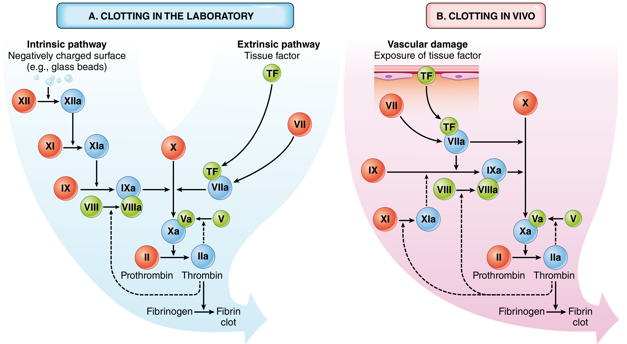
Red circles = inactive factors; Blue circles = active factors; Green circles = cofactors
3.5a The Extrinsic Pathway (Tissue Factor Pathway)
This is the primary in vivo initiator of coagulation.
- Tissue factor (TF) is a membrane-bound glycoprotein constitutively expressed on subendothelial cells (smooth muscle cells, fibroblasts). It is normally shielded from blood by the endothelium.
- Vascular injury exposes TF → Factor VII in blood binds TF → forms TF-VIIa complex
- TF-VIIa complex activates Factor X → Xa (directly)
- TF-VIIa complex also activates Factor IX → IXa (amplification arm)
Measured by: Prothrombin Time (PT)
- Tissue factor + phospholipids + Ca²⁺ added to plasma
- Tests factors VII, X, V, II, and fibrinogen (I)
- Normal PT: 11-13 seconds
- Reported as INR (International Normalized Ratio) for standardization
- Prolonged in: warfarin therapy, liver disease (decreased synthesis), vitamin K deficiency, factor VII deficiency
3.5b The Intrinsic Pathway (Contact Activation Pathway)
This pathway is critical for laboratory clotting tests but plays a lesser role in physiologic hemostasis in vivo.
- Factor XII (Hageman factor) binds to a negatively charged surface (e.g., collagen, kallikrein, glass beads in lab) → XIIa
- XIIa activates Factor XI → XIa (with high molecular weight kininogen HMWK and prekallikrein as cofactors)
- XIa activates Factor IX → IXa
- IXa + Factor VIIIa (cofactor) + phospholipid + Ca²⁺ = "Tenase complex" → activates Factor X → Xa
Measured by: Activated Partial Thromboplastin Time (aPTT)
- Negatively charged particles (kaolin/glass) + phospholipids + Ca²⁺ added to plasma
- Tests factors XII, XI, IX, VIII, X, V, II, and fibrinogen
- Normal aPTT: 25-40 seconds
- Prolonged in: heparin therapy, hemophilia A (factor VIII deficiency), hemophilia B (factor IX deficiency), factor XII deficiency, lupus anticoagulant
Important clinical note: Factor XI deficiency causes only mild bleeding (Ashkenazi Jewish population most affected), and Factor XII deficiency causes NO bleeding - demonstrating these factors are dispensable for physiologic hemostasis in vivo.
3.5c The Common Pathway
Both extrinsic and intrinsic pathways converge on Factor X activation:
- Factor Xa + Factor Va (cofactor) + phospholipid + Ca²⁺ = "Prothrombinase complex"
- Prothrombinase converts Factor II (Prothrombin) → Factor IIa (Thrombin)
- Thrombin cleaves Fibrinogen → Fibrin monomers
- Fibrin monomers spontaneously polymerize into an insoluble fibril meshwork
- Factor XIIIa (activated by thrombin) covalently cross-links fibrin → stable, permanent clot
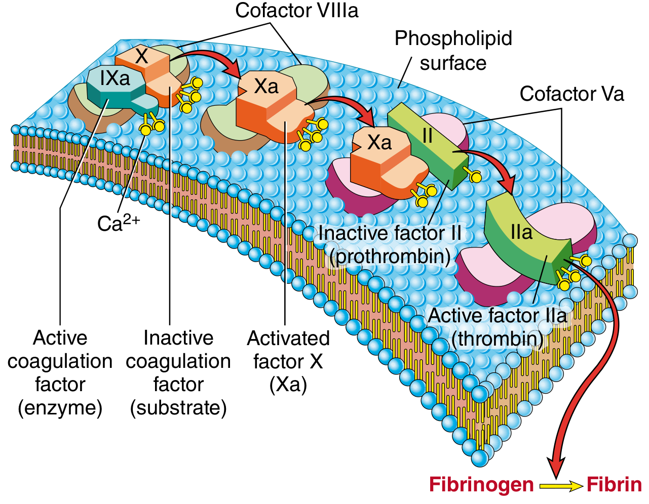
Fig. 4.7 - The prothrombinase complex. IXa (enzyme) converts substrate X to Xa with cofactor VIIIa. Then Xa (enzyme) converts substrate II (prothrombin) to IIa (thrombin) with cofactor Va. All assembled on platelet phospholipid surface with Ca²⁺.
3.6 Thrombin - The Central Enzyme
Thrombin (Factor IIa) is the most important coagulation factor, with multiple actions:
- Fibrin formation - cleaves fibrinogen into fibrin monomers
- Factor XIII activation - cross-links and stabilizes fibrin
- Amplification - activates Factors V, VIII, and XI (positive feedback loops)
- Platelet activation - activates platelets via PAR-1 (protease-activated receptor)
- Proinflammatory - activates monocytes, endothelium via PAR receptors → PDGF release, neutrophil adhesion, tissue repair, angiogenesis
- Anticoagulant switch - when thrombin reaches intact endothelium, it binds thrombomodulin and switches to anticoagulant function (activates protein C)
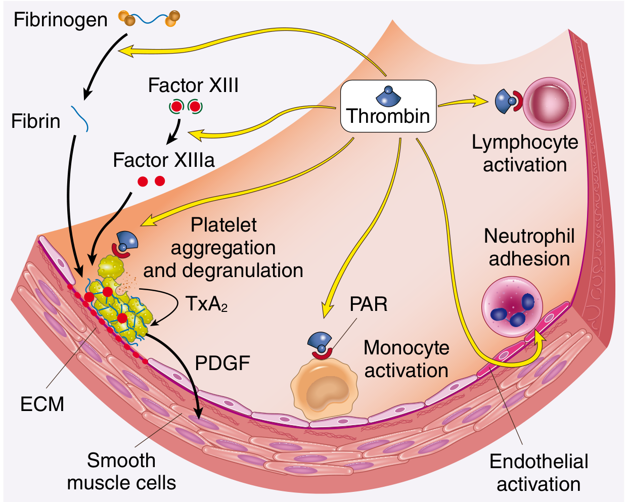
Fig. 4.8 - The multiple roles of thrombin in hemostasis and cellular activation (Robbins, Cotran & Kumar)
3.7 The In Vivo Model (Cell-Based Model of Coagulation)
The classical intrinsic/extrinsic division is a laboratory construct. In vivo, coagulation proceeds in three overlapping phases:
Phase 1 - Initiation (on TF-bearing cells):
- TF exposed by injury → TF-VIIa complex forms
- Produces small amounts of Xa, IXa, and importantly, a small amount of thrombin
- Tissue Factor Pathway Inhibitor (TFPI) quickly shuts down TF-VIIa-Xa complex
Phase 2 - Amplification (on platelet surface):
- Small amounts of thrombin activate platelets (PAR-1), Va, VIIIa, and XIa
- Factor XIa on platelet surface activates more IXa
- Platelet surface provides phospholipid scaffold
Phase 3 - Propagation (on platelet surface):
- IXa-VIIIa (tenase) complex generates large amounts of Xa
- Xa-Va (prothrombinase) generates the thrombin burst - large-scale thrombin production
- Thrombin converts fibrinogen to fibrin; activates FXIII → stable cross-linked clot
This explains why FVII, VIII, IX, X, V deficiencies cause severe bleeding (they are required for amplification/propagation) while FXI deficiency is mild and FXII deficiency causes no bleeding.
PART 4: ANTICOAGULANT MECHANISMS (Natural Inhibitors)
Once clotting is initiated, it must be confined to the injury site. Multiple countermechanisms exist:
4.1 Dilution and Clearance
Flowing blood washes activated factors away from the injury site; the liver rapidly clears them.
4.2 Requirement for Phospholipid Surfaces
Coagulation complexes only assemble on negatively charged phospholipids provided by activated platelets at the injury site - not on normal endothelium or in the circulation.
4.3 Endothelial Anticoagulant Mechanisms
Normal endothelium produces multiple anticoagulant/antithrombotic factors:
Against platelets:
- PGI₂ (prostacyclin) - raises platelet cAMP → inhibits activation (made by COX-1 in endothelium; NOT inhibited by aspirin at low doses unlike platelet COX-1)
- Nitric oxide (NO) - raises platelet cGMP → inhibits activation
- Adenosine diphosphatase (ADPase/CD39) - degrades ADP, removing a platelet activator
Against coagulation factors:
- Thrombomodulin - binds thrombin on endothelial surface; thrombin-thrombomodulin complex activates Protein C
- Endothelial Protein C Receptor (EPCR) - concentrates protein C near thrombomodulin for efficient activation
- Activated Protein C (APC) + Protein S - cleave and inactivate Factors Va and VIIIa (removes cofactors from both tenase and prothrombinase complexes)
- Heparin-like molecules (heparan sulfate) on endothelial surface - activate Antithrombin III (ATIII), which inhibits thrombin and Factors IXa, Xa, XIa, and XIIa
- Tissue Factor Pathway Inhibitor (TFPI) - synthesized by endothelium, requires protein S as cofactor; inhibits TF-VIIa-Xa complex
For fibrinolysis:
- t-PA (tissue plasminogen activator) - synthesized by endothelium; most active when bound to fibrin (hence therapeutic specificity of alteplase)
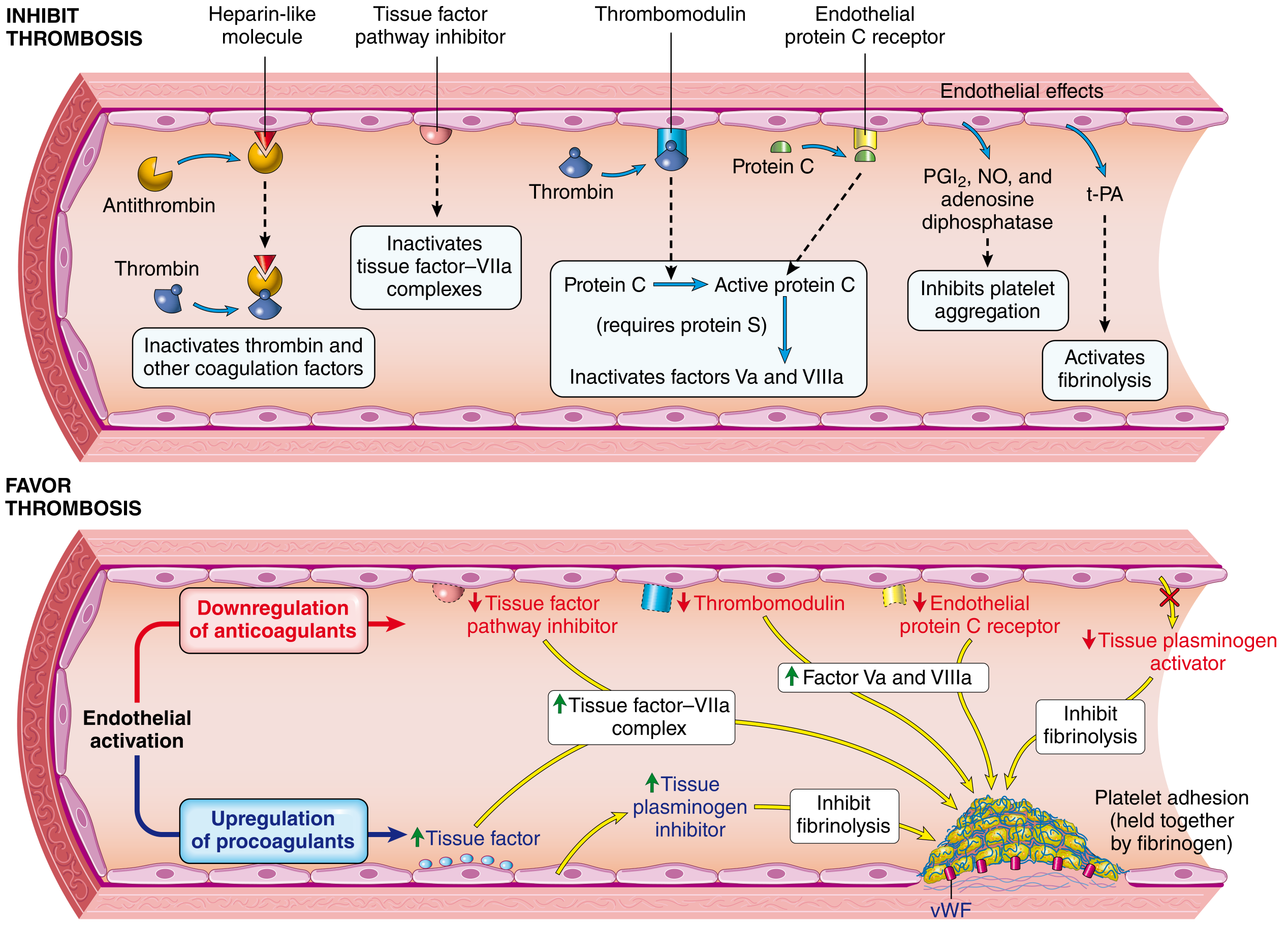
Fig. 4.10 - Anticoagulant activities of normal endothelium (top) vs. procoagulant properties of injured/activated endothelium (bottom). (Robbins, Cotran & Kumar)
PART 5: FIBRINOLYSIS
Coagulation simultaneously activates fibrinolysis to limit clot size and eventually dissolve it.
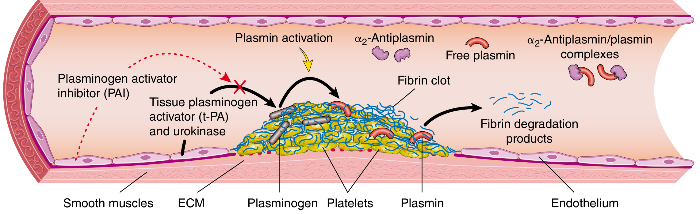
Fig. 4.9 - Fibrinolytic system (Robbins, Cotran & Kumar)
Key components:
| Component | Function |
|---|---|
| Plasminogen | Inactive precursor; circulates in plasma and associates with fibrin clots |
| t-PA (tissue plasminogen activator) | Activates plasminogen → plasmin; most active when fibrin-bound |
| Urokinase (uPA) | Alternative plasminogen activator |
| Plasmin | Active enzyme; degrades fibrin into Fibrin Degradation Products (FDPs) including D-dimers |
| α₂-Antiplasmin | Rapidly inactivates free plasmin in circulation (prevents systemic fibrinolysis) |
| PAI-1 (Plasminogen Activator Inhibitor-1) | Produced by endothelium; inhibits t-PA and uPA |
| TAFI (Thrombin Activatable Fibrinolysis Inhibitor) | Thrombin-activated; removes plasminogen-binding sites from fibrin |
Clinical relevance of D-dimers:
- D-dimers are cross-linked fibrin degradation products, specific to clot breakdown (not just fibrinogen breakdown)
- Elevated in DVT, PE, DIC, post-surgery
- Highly sensitive but not specific - used as a rule-out test (negative D-dimer essentially excludes DVT/PE)
PART 6: CLINICAL CORRELATIONS
6.1 Bleeding Disorders
| Condition | Mechanism | Lab Finding |
|---|---|---|
| Hemophilia A | Factor VIII deficiency (X-linked) | Prolonged aPTT, normal PT |
| Hemophilia B (Christmas disease) | Factor IX deficiency (X-linked) | Prolonged aPTT, normal PT |
| von Willebrand Disease | vWF deficiency/dysfunction | Prolonged bleeding time, ±prolonged aPTT (VIII may be low) |
| Bernard-Soulier Syndrome | GpIb deficiency (platelets can't bind vWF) | Thrombocytopenia, large platelets |
| Glanzmann Thrombasthenia | GpIIb-IIIa deficiency | Normal platelet count, prolonged bleeding time |
| Vitamin K Deficiency | Reduced II, VII, IX, X, Protein C & S | Prolonged PT and aPTT |
| Liver Disease | Reduced synthesis of all clotting factors except vWF | Prolonged PT and aPTT |
| DIC (Disseminated Intravascular Coagulation) | Systemic activation of coagulation → consumption of all factors and platelets | Prolonged PT, aPTT; thrombocytopenia; elevated D-dimer; low fibrinogen |
6.2 Thrombotic Disorders / Hypercoagulable States
| Condition | Mechanism |
|---|---|
| Factor V Leiden | Point mutation in Factor V (Arg506Gln) renders FVa resistant to cleavage by Protein C → most common inherited thrombophilia |
| Prothrombin G20210A | Gain-of-function mutation → increased prothrombin levels |
| Protein C Deficiency | Cannot inactivate Va and VIIIa → hypercoagulability |
| Protein S Deficiency | Protein C cannot function as cofactor |
| Antithrombin III Deficiency | Thrombin and Xa not adequately inhibited |
| Antiphospholipid Syndrome | Autoantibodies against phospholipid-binding proteins → paradoxical thrombosis despite prolonged aPTT (lupus anticoagulant) |
| Virchow's Triad | Stasis + endothelial injury + hypercoagulability → thrombosis |
6.3 Drug Targets in the Coagulation Cascade
| Drug | Target | Mechanism | Use |
|---|---|---|---|
| Heparin (unfractionated) | Antithrombin III | Potentiates ATIII → inhibits thrombin (IIa) and Xa | Acute anticoagulation |
| Low Molecular Weight Heparin (enoxaparin) | Antithrombin III | Preferentially inhibits Xa over IIa | DVT/PE prophylaxis and treatment |
| Warfarin/Coumadin | Vitamin K epoxide reductase | Inhibits recycling of vitamin K → reduced II, VII, IX, X, Protein C, S | Chronic anticoagulation (AF, mechanical valves) |
| Direct thrombin inhibitors (dabigatran, bivalirudin, argatroban) | Factor IIa (thrombin) | Direct inhibition | AF, HIT |
| Direct Xa inhibitors (rivaroxaban, apixaban, edoxaban) | Factor Xa | Direct inhibition | AF, DVT/PE |
| Aspirin | COX-1 in platelets | Irreversibly inhibits TxA₂ synthesis → reduced platelet aggregation | Antiplatelet (ACS, stroke prevention) |
| Clopidogrel/Ticagrelor | P2Y12 (ADP receptor) on platelets | Blocks ADP-mediated platelet activation | Antiplatelet (ACS, PCI) |
| Abciximab/Eptifibatide/Tirofiban | GpIIb-IIIa | Blocks fibrinogen binding → inhibits aggregation | High-risk PCI |
| Alteplase (t-PA) | Plasminogen | Converts plasminogen → plasmin → fibrinolysis | STEMI, stroke, massive PE |
| Tranexamic acid | Plasminogen (lysine binding site) | Blocks fibrinolysis | Surgical bleeding, trauma, heavy menstrual bleeding |
6.4 Laboratory Tests Summary
| Test | Pathway Tested | Normal Value | Prolonged By |
|---|---|---|---|
| PT / INR | Extrinsic + common | 11-13s / INR 0.8-1.2 | Warfarin, FVII deficiency, liver disease, vitamin K deficiency |
| aPTT | Intrinsic + common | 25-40s | Heparin, hemophilia A/B, FXII deficiency, lupus anticoagulant |
| Thrombin Time (TT) | Fibrinogen → Fibrin | 14-19s | Heparin, low/dysfunctional fibrinogen, dabigatran |
| Fibrinogen | - | 200-400 mg/dL | Low in DIC, liver disease |
| D-dimer | Fibrinolysis marker | <0.5 μg/mL | DVT, PE, DIC, post-surgery, malignancy |
| Bleeding time | Platelet function | 2-9 min | Thrombocytopenia, vWD, NSAID use, Glanzmann, Bernard-Soulier |
| Platelet count | - | 150-400 × 10³/μL | Low in ITP, HIT, DIC, aplastic anemia |
Summary Diagram: The Whole Picture
VASCULAR INJURY
│
▼
1. VASOCONSTRICTION (endothelin, neurogenic)
│
▼
2. PRIMARY HEMOSTASIS
Exposed collagen → vWF binds → GpIb (platelets adhere)
Platelet activation → shape change, granule release
ADP + TxA₂ → more platelets recruited
Fibrinogen bridges GpIIb-IIIa → PLATELET PLUG
│
▼
3. SECONDARY HEMOSTASIS (Coagulation Cascade)
EXTRINSIC: TF + VIIa → Xa (+ IXa) INTRINSIC: XII→XIIa→XI→XIa→IX→IXa
└──────────────────┬──────────────────────┘
COMMON PATHWAY
IXa + VIIIa → Xa (tenase complex)
Xa + Va → Thrombin (prothrombinase complex)
Thrombin → Fibrin (from fibrinogen)
FXIIIa → cross-linked fibrin
│
▼
STABLE FIBRIN CLOT
│
▼
4. LIMITATION & FIBRINOLYSIS
Antithrombin III (heparin cofactor) inactivates thrombin, Xa, IXa
Protein C + Protein S inactivate Va, VIIIa
TFPI inhibits TF-VIIa-Xa
t-PA activates plasminogen → plasmin → FDPs (D-dimers)
Sources:
- Robbins, Cotran & Kumar Pathologic Basis of Disease - Chapters 4 and 13 (primary source for coagulation cascade and hematopoiesis)
- Robbins & Kumar Basic Pathology - Chapter 3 (hemostasis and platelets)
- Goodman & Gilman's Pharmacological Basis of Therapeutics (hematopoiesis growth factors)
- Medical Physiology (hematopoiesis development)The full guide is complete above. Here's a quick roadmap of what was covered:
What You Just Got - A Quick Index
Part 1: Hematopoiesis
- Developmental sites (yolk sac → AGM → liver → bone marrow) with gestational timing
- HSC properties: pluripotency and self-renewal; the bone marrow niche
- Full differentiation hierarchy diagram (HSC → all 9 mature cell lineages)
- All growth factors (G-CSF, EPO, TPO, GM-CSF, KIT ligand, FLT3L) with clinical uses
- Normal adult blood cell reference ranges
- Clinical diseases: aplastic anemia, leukemia/MDS, polycythemia vera, extramedullary hematopoiesis
Part 2: Normal Blood Flow
- Pulmonary vs. systemic circuits; vessel types and their roles
- Hagen-Poiseuille law; why radius⁴ is the dominant regulator
- Normal hemodynamic values (CO, MAP, SVR, capillary pressures)
- Autoregulation, metabolic, endothelial, neural and hormonal control
Part 3-4: Hemostasis & Coagulation (Primary + Secondary)
- 4-step sequence: vasoconstriction → platelet plug → fibrin → clot limitation
- Platelet structure (α-granules vs dense granules), adhesion (vWF-GpIb), activation (ADP, TxA₂, thrombin/PAR-1), aggregation (GpIIb-IIIa-fibrinogen)
- Full coagulation factor table (all 13 factors + vWF, Protein C/S)
- Extrinsic pathway (TF-VIIa), Intrinsic pathway (XII→XI→IX), Common pathway (Xa→thrombin→fibrin)
- All 4 diagrams embedded: hematopoiesis tree, coagulation cascade lab vs. in vivo, phospholipid surface prothrombinase complex, thrombin's multiple roles, platelet adhesion/aggregation, fibrinolysis, endothelial anticoagulant vs. procoagulant states
- Cell-based in vivo model (initiation → amplification → propagation phases)
- All natural anticoagulants: ATIII, Protein C/S, TFPI, PGI₂, NO, t-PA
- Fibrinolysis: plasminogen → plasmin, D-dimers, α₂-antiplasmin, PAI-1
Part 5-6: Clinical Correlations
- Full table of bleeding disorders with lab patterns
- Thrombotic disorders: Factor V Leiden, prothrombin mutation, Protein C/S deficiency, antiphospholipid syndrome, Virchow's triad
- Drug targets table: heparin, LMWH, warfarin, DOACs (dabigatran, rivaroxaban, apixaban), aspirin, clopidogrel, GpIIb-IIIa inhibitors, alteplase, tranexamic acid
- Lab tests: PT/INR, aPTT, TT, D-dimer, bleeding time with normal ranges and clinical interpretation
This is a shared conversation. Sign in to Orris to start your own chat.