
Ottitis media externa investigations and treatment
otitis externa ear canal inflammation
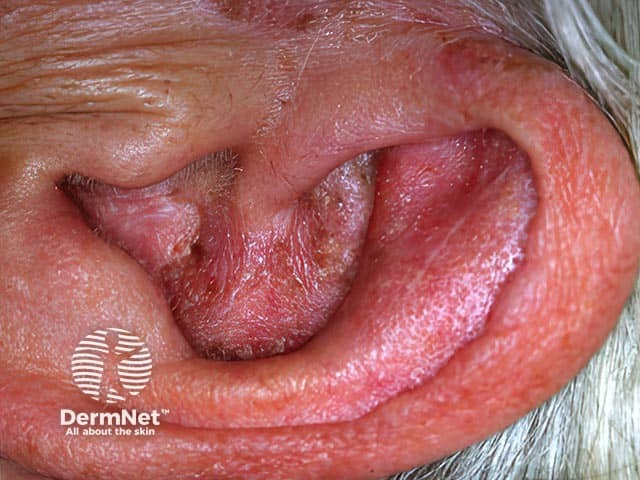
This is a high-resolution clinical photograph of the external ear (auricle/pinna) and external auditory canal region, obtained with digital photography in a frontal/anterolateral perspective. The image depicts erythema (redness) and edema (swelling) of the skin of the pinna and canal, with areas of fine to moderate desquamation and scale along the canal wall and surrounding skin. The appearance is typical of otitis externa (inflammation of the external ear canal) and may reflect concurrent superficial moisture-associated maceration. Possible etiologies include bacterial otitis externa (Pseudomonas aeruginosa, Staphylococcus species) and otomycosis (fungal infection) in humid environments; secondary bacterial infection common. The visible tissue shows no purulent discharge in this frame, though exudate can be present in other angles. The clinical significance lies in the need for targeted topical therapy (antibiotic ± steroid drops) and meticulous canal cleaning; assessment for canal edema, stenosis, or involvement of the tympanic membrane is essential; in diabetics or elderly patients malignant otitis externa should be considered if there is deep tissue involvement or systemic symptoms. Differential diagnoses include contact dermatitis, eczema, or seborrheic dermatitis of the ear. This image is relevant for education on infrared-free clinical photography of inflammatory dermatosis, diagnostic cue recognition, and treatment planning in otology and dermatology.
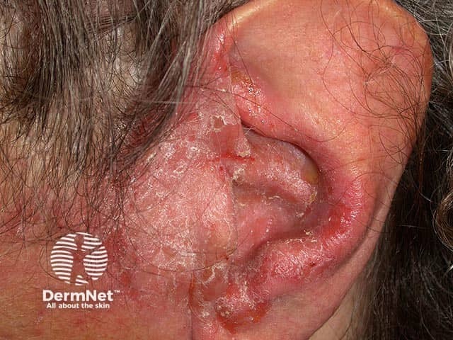
Imaging modality and orientation: this clinical photograph depicts the left auricular region, showing chronic otitis externa with extension onto the preauricular skin. Anatomical localization includes the external auditory canal entrance and surrounding preauricular soft tissue of the head and neck. Visual features include inflamed, erythematous skin with diffuse desquamation, thin adherent crusting, and fissuring around the helix and preauricular area; mild periauricular edema; no discrete mass is evident. The overall appearance is consistent with a chronic inflammatory dermatitis involving the external ear canal skin and periauricular skin. Pathophysiology entails a prolonged inflammatory process of the external ear canal skin, predisposing to fissures, excoriation, and secondary bacterial colonization; differential diagnoses include eczema/dermatitis ( seborrheic, contact, atopic ), impetigo, or cellulitis in more extensive disease. Clinical significance: the image documents disease extent, informs topical therapy choices (antibiotic plus steroid combinations), and flags potential complications such as canal stenosis or malignant otitis externa in high-risk patients. Potential clinical applications include dermatology and ENT education, tracking chronicity and response to treatment, and serving as a reference for educational case discussions and research on inflammatory skin disease of the ear. Search terms: chronic otitis externa, preauricular dermatitis, preauricular skin involvement, external auditory canal inflammation, crusting, scaling. Present.
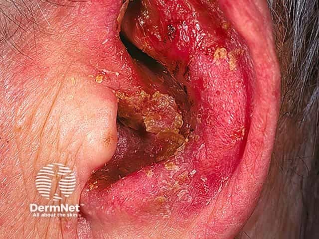
Imaging modality: Clinical photography of the external ear canal. Technique: White-light, macro close-up view capturing the external auditory canal with bright illumination to reveal mucosal color and crusting. Magnification: Macro. No contrast or staining. Acquisition parameters: Standard clinic camera settings; ambient lighting; no spectral filters. Anatomical localization: External auditory canal (EAC), part of the otic apparatus within the temporal bone; skin epithelium and mucosa lining the canal with surrounding periauricular tissues. Perspective: Close-up otologic photograph emphasizing canal lumen and canal wall margins; tympanic membrane not visualized. Clinical context: This image documents severe otitis externa with marked inflammation. Visual features include intense erythema of canal skin, edema, fissuring, and crusts or desquamation adherent to canal walls; debris may occlude the canal. Pathology involves inflammatory exudate and epithelial disruption; secondary bacterial infection is likely, and fungal infection cannot be excluded. Diagnostic significance: Confirms acute ear canal infection requiring prompt topical therapy, analgesia, and meticulous canal hygiene; urgent ENT evaluation if signs of malignant otitis externa or cranial neuropathies in high-risk patients (diabetes, immunosuppression). Differential considerations: fungal otitis externa (otomycosis), contact dermatitis, eczema, bacterial otitis externa, allergic dermatitis. Clinical correlation: Correlate with otalgia, otorrhea, hearing loss, and history of swimming or trauma; management includes culture if refractory and follow-up to ensure resolution.
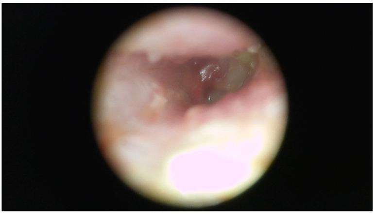
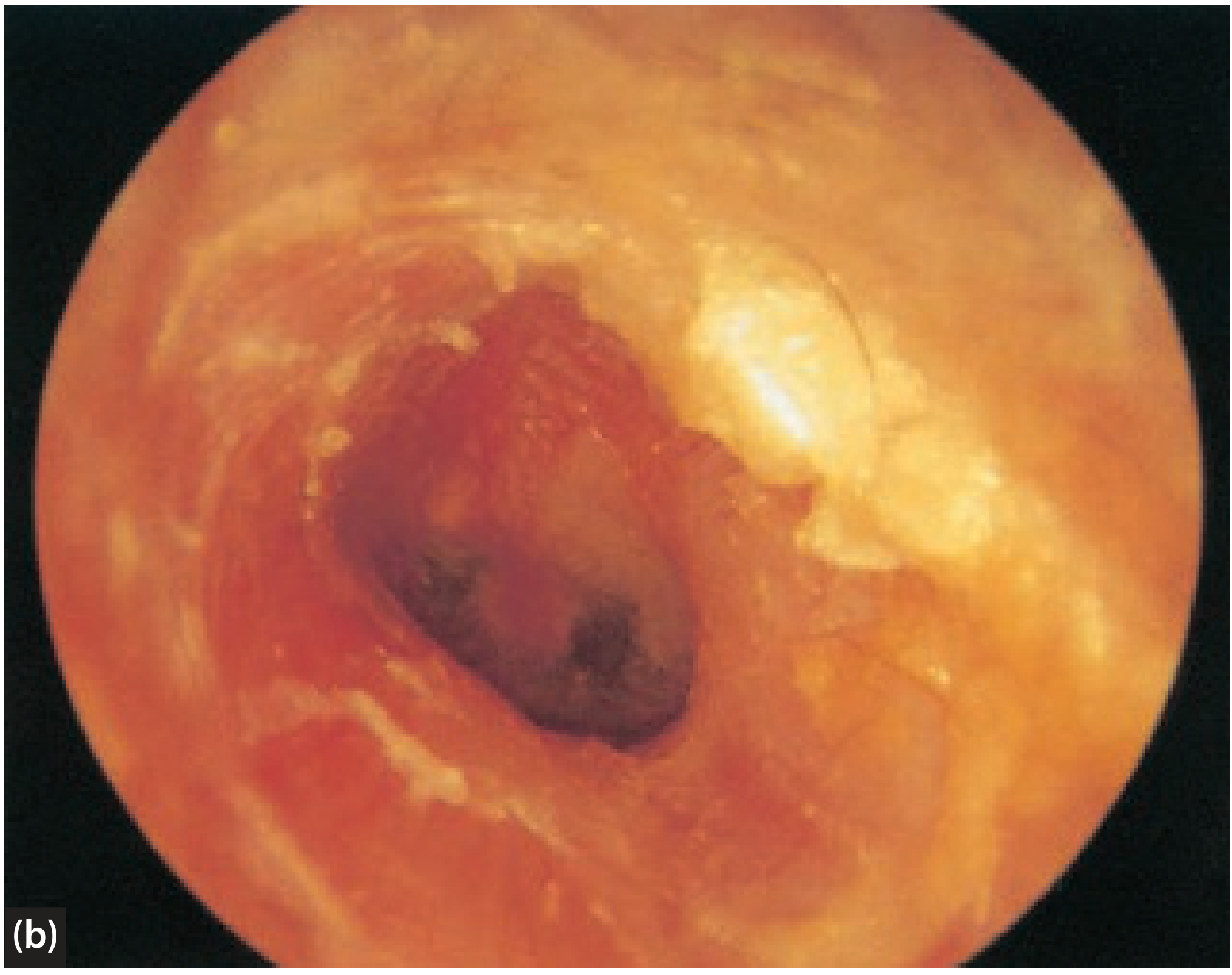
This clinical photograph is an endoscopic view (otoscopy) of the right external auditory canal (EAC). The image demonstrates significant pathological changes characterized by extensive, thick purulent discharge (pus) and whitish keratin debris that nearly fill the lumen, obstructing a clear view of the tympanic membrane. The visible portions of the external ear canal wall appear markedly erythematous and swollen, with an irregular, thickened contour indicating active inflammation and edema. Such findings are characteristic of severe otitis externa or otomastoiditis, which may be associated with chronic infections such as tuberculosis or non-tuberculous mycobacteria (NTM) in refractory cases. The clinical significance of this visual is to illustrate the physical manifestations of chronic suppurative otitis, focusing on canal stenosis due to soft tissue swelling and the accumulation of infectious exudate.
"otitis externa" AND treatment
Otitis Media Externa - Investigations & Treatment
Clinical Images
Definition & Classification
-
Stage 1 - Pre-inflammatory: Loss of lipid/acid balance (normal canal pH 4-5), blocked sebaceous/apocrine glands, pruritus
-
Stage 2 - Acute inflammatory (mild/moderate/severe): Epithelial disruption, bacterial invasion, increasing exudate and canal occlusion
-
Stage 3 - Chronic inflammatory: >6 months duration; hyperkeratosis, lichenification
-
Scott-Brown's Otorhinolaryngology Head & Neck Surgery Vol 2, p. 997
Microbiology
| Organism | Prevalence |
|---|---|
| Pseudomonas aeruginosa | 50-65% (most common) |
| Other Gram-negative organisms | 25-35% |
| Staphylococcus aureus | 15-30% |
| Streptococcus spp. | 9-15% |
| Fungi (Aspergillus, Candida) | <2% of AOE (more in chronic/post-antibiotic) |
Investigations
1. History (Key Points)
- Timing, duration, severity of symptoms
- Exposure to water (swimming, bathing) and aural toilet habits (cotton bud use)
- Periods of improvement/worsening and previous treatments
- Systemic conditions: diabetes, HIV, immunosuppression, skin disorders (eczema, psoriasis, seborrhoea)
- Topical agents used in/around the ear (hearing aids, earphones, hairsprays - allergy risk)
- Cummings Otolaryngology, p. 2650
2. Physical Examination
- Complete examination of auricle + EAC under binocular microscopy
- Check for: tragal/auricular tenderness (pathognomonic of OE), canal erythema/oedema/debris, discharge character
- Assess the tympanic membrane (integrity is critical before choosing topical drops)
- Severity grading: mild (canal erythema, no/mild oedema), moderate (sub-total canal obliteration), severe (complete obliteration)
- Check for cervical lymphadenopathy, cranial nerve palsies (VII-XII) - raises suspicion for malignant OE
- Cummings Otolaryngology, p. 2650; Scott-Brown's, p. 997
3. Microbiological Culture
- Not routinely required in uncomplicated OE
- Indicated for:
- Persistent/recurrent infection not responding to treatment
- Immunocompromised patients (diabetes, HIV)
- Suspected malignant OE
- If fungal infection is suspected
- Expert opinion reserves swabbing for resistant/high-risk cases
- Scott-Brown's, p. 998; Cummings, p. 2650
4. Imaging (for Malignant/Necrotising OE only)
- CT temporal bone - demonstrates bony erosion, soft-tissue extension, mastoid/skull-base involvement
- MRI - better soft-tissue resolution, detects intracranial extension
- Tc-99m bone scan - sensitive for osteomyelitis
- Gallium-67 scan - useful for monitoring treatment response in necrotising OE
- ROSEN's Emergency Medicine, p. 552
5. Histopathology
- Tissue biopsy if malignant OE suspected - sent to rule out neoplasm (squamous cell carcinoma can mimic OE)
- Cummings Otolaryngology, p. 2651
Treatment
Principles of Treatment (Scott-Brown's)
- Thorough and regular aural toilet
- Topical medication to the EAC (with or without a wick)
- Analgesia
- Treatment of regional/systemic complications with systemic antibiotics
- Prevention of aetiological factors (recurrence prevention)
A. Aural Toilet (Ear Cleaning)
- Most effective single treatment for OE
- Performed under microscopic guidance (suction/dry mopping) - removes debris, inflammatory exudate, and fungal elements
- Clears canal for topical drug penetration and allows visualization of the TM
- Irrigation is generally discouraged by ENT specialists (risk of complications)
- Cotton-tipped applicators should be avoided (cause local trauma, worsen inflammation)
- Scott-Brown's, p. 998; Cummings, p. 2651
B. Topical Therapy (First-Line)
Topical Antibiotic + Steroid Combinations (most common first-line)
| Preparation | Notes |
|---|---|
| Fluoroquinolone drops (ciprofloxacin, ofloxacin) | Broad spectrum, safe with TM perforation, no ototoxicity |
| Aminoglycoside drops (neomycin, gentamicin) | Effective but CONTRAINDICATED with TM perforation (ototoxic) |
| Ciprofloxacin + dexamethasone (Ciprodex) | Widely used, twice daily, excellent coverage |
| Neomycin + polymyxin B + hydrocortisone | Effective but contact hypersensitivity to neomycin is common |
Important: Fluoroquinolone drops are the preferred choice when TM integrity is uncertain - aminoglycosides must NOT be used with a perforated TM. - ROSEN's Emergency Medicine, p. 543
Acidifying Agents
- Acetic acid 2% (diluted 50% white vinegar) - effective in early/mild OE; restores normal canal pH
- Alcohol + vinegar OTC solutions - useful but no conclusive RCT evidence
- Cummings Otolaryngology, p. 2651
Wick Placement
- Used when canal is severely oedematous and drops cannot penetrate
- Options: cotton wick, Merocel sponge, Pope oto-wick, antibiotic-impregnated gauze
- Mechanically stents canal open, allows topical agents to reach proximal canal, limits oedema
- Glycerol + ichthammol (90:10) traditionally used on wick - dehydrating, anti-inflammatory, antibacterial (poor anti-pseudomonal activity)
- NSAIDs are excellent analgesics for moderate/severe cases
- Scott-Brown's, p. 998; Cummings, p. 2651
C. Treatment by Type
Acute Bacterial OE
- First-line: topical fluoroquinolone ± steroid drops × 7-10 days
- Debridement of canal (aural toilet)
- Analgesia (NSAIDs/paracetamol)
- Keep ear dry; use earplugs for swimming
Chronic OE
- Meticulous cleaning under binocular microscopy
- Topical steroid as mainstay (reduces inflammation)
- Eliminate inciting factors (stop cotton buds, hearing aid hygiene)
- Oral steroids for severe acute breakthrough episodes
- Tacrolimus topical (off-label, not widely used)
- Acidifying agents (1:1 white vinegar + rubbing alcohol)
- Contact dermatitis: identify and eliminate allergen; topical corticosteroid
- Cummings Otolaryngology, p. 2651
Fungal OE (Otomycosis)
- Debridement is the mainstay
- Clotrimazole 1% solution - broad-spectrum, available OTC
- Ketoconazole ointment, cresylate otic, or tolnaftate solution (safe with TM perforation)
- Gentian violet paint (antifungal dye)
- Acidification of canal (acetic acid)
- For Aspergillus with significant oedema or failure: oral itraconazole
- Combination cream: clotrimazole + betamethasone (Lotrisone) instilled via syringe - highly effective
- Cummings Otolaryngology, p. 2651
D. Systemic Antibiotics
- No role in uncomplicated OE limited to the EAC in immunocompetent patients
- Indicated for:
- Spreading cellulitis/perichondritis/chondritis extending beyond the EAC
- Malignant/necrotising OE
- Immunocompromised patients with progressive disease
- Coverage should include anti-pseudomonal activity (ciprofloxacin orally is first-line)
- Rosen's Emergency Medicine, p. 541; Scott-Brown's, p. 998
E. Malignant (Necrotising) OE - Special Management
- Systemic antibiotics with anti-pseudomonal activity - oral ciprofloxacin (500-750 mg BD) is first-line
- For IV: piperacillin-tazobactam, ceftazidime, or meropenem
- Duration: 6-8 weeks (guided by gallium scan normalization)
- ENT/otolaryngology input mandatory
- Surgical debridement for refractory cases
- Optimize underlying metabolic disease (glycaemic control in diabetics)
- ROSEN's Emergency Medicine, p. 554-556; Cummings Otolaryngology, p. 2652
Prevention / Recurrence Advice
- Avoid water entry into the ear canal: cotton wool + Vaseline for bathing; custom ear moulds/neoprene bandages for swimming
- Alcohol/acetic acid drops after swimming (prophylactic acidification)
- Avoid cotton buds and ear probing
- Properly fitting hearing aids; minimize use if canal is inflamed
- Scott-Brown's, p. 998
Complications
| Complication | Description |
|---|---|
| Cellulitis/perichondritis/chondritis | Extension to auricular soft tissue and cartilage; treat with oral anti-pseudomonal antibiotics |
| Malignant OE | Osteomyelitis of skull base; life-threatening; systemic IV antibiotics + ENT surgery |
| Canal stenosis/atresia | Chronic inflammation → fibrosis → medial canal fibrosis (blind-ending canal) |
| Conductive hearing loss | From canal oedema and debris |
| Meningitis, sinus thrombosis | Intracranial extension of malignant OE |
Topodiagnostic tests for facial nerve palsey
facial nerve anatomy branches course diagram
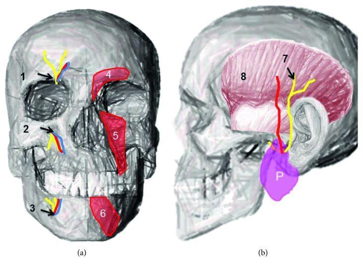
Anatomical diagrams of the human skull illustrating terminal branches of the trigeminal nerve and associated facial structures. Figure (a) is an anterior view highlighting the supraorbital notch (1), infraorbital foramen (2), and mental foramen (3), with accompanying neurovascular bundles shown in yellow (nerves) and blue/red (vessels). The diagram also depicts the overlying musculature: the corrugator supercilii (4), levator labii superioris (5), and depressor labii inferioris (6). Figure (b) provides a lateral view demonstrating the course of the auriculotemporal nerve (7) emerging from the parotid gland (P) and ascending across the temporal region. The temporalis muscle (8) is shown in pink, providing anatomical context for nerve distribution. This educational illustration demonstrates the relationship between bony landmarks, superficial facial muscles, and the sensory nerve exits essential for clinical procedures such as regional nerve blocks and the management of trigeminal neuralgias.
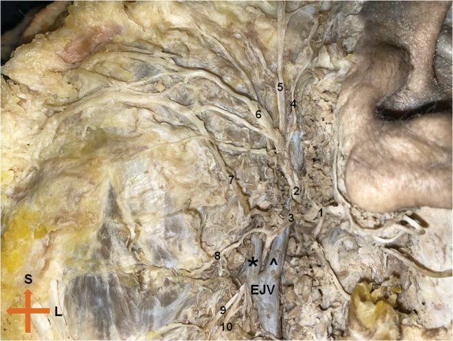
This clinical photograph displays a cadaveric dissection of the left extratemporal facial nerve (Cranial Nerve VII), highlighting its complex branching pattern and vascular relationships. The main trunk bifurcates into the temporofacial and cervicofacial divisions. The superior temporofacial division gives rise to the temporal and zygomatic branches, which course toward the upper face, and a buccal branch. The inferior cervicofacial division exhibits a rare anatomical variation where it bifurcates around the posterior facial vein; the upper branch passes superficially while the lower branch courses deep to the vein. Key anatomical landmarks include the retromandibular vein, the external jugular vein (EJV), and the masseter muscle. Labeled structures include the temporal (4), zygomatic (5), buccal (6), and marginal mandibular (8, 9) branches. This image is an essential educational resource for surgical anatomy, particularly for parotidectomy and maxillofacial procedures, demonstrating the critical proximity of neural branches to venous structures in the parotid region.
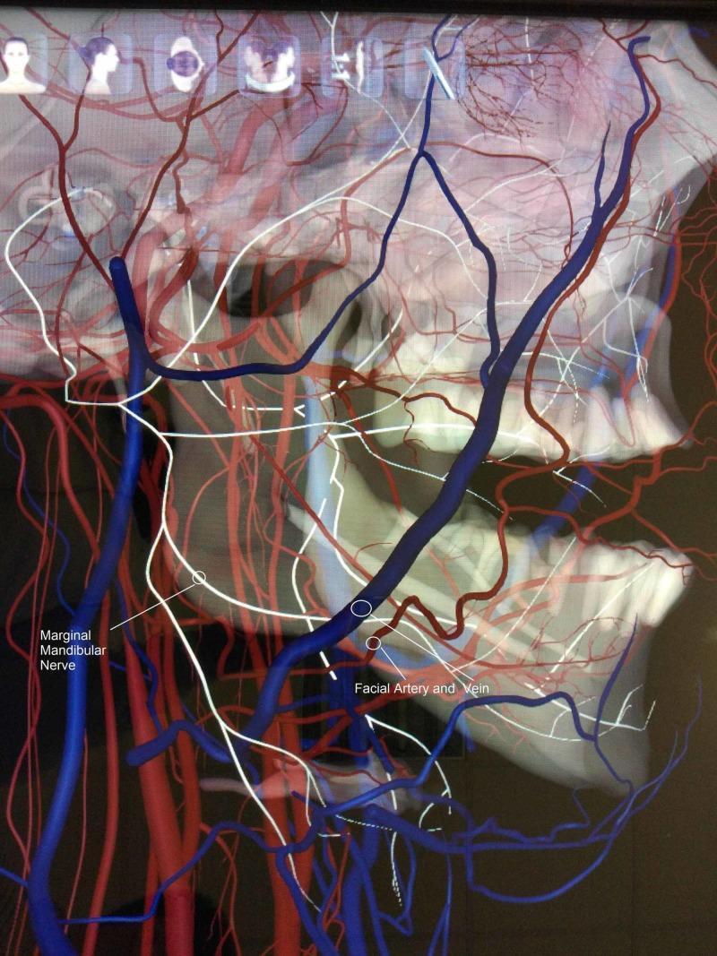
Anatomical diagram and 3D visualization illustrating a rare neurovascular anatomical variation in the maxillofacial region. This lateral view focuses on the right side of the mandible and cervical area, highlighting the spatial relationship between the marginal mandibular nerve (MMN), the facial artery, and the facial vein. The MMN is depicted as a blue tubular structure descending toward the lower border of the mandible. Crucially, the diagram demonstrates an atypical course where the MMN runs deep (medial) to the facial artery and vein (rendered in red), contrasting with the standard anatomical presentation where the nerve typically crosses superficial to these vessels. The visualization overlays these structures on a skeletal model of the skull and mandible, showing the nerve's proximity to the mandibular angle and body. This educational content is relevant for surgical anatomy, particularly in the context of neck dissections, submandibular gland excision, and rhytidectomy, where preserving the MMN is critical to prevent paralysis of the depressor muscles of the lower lip.
facial nerve intratemporal course geniculate ganglion diagram topodiagnosis
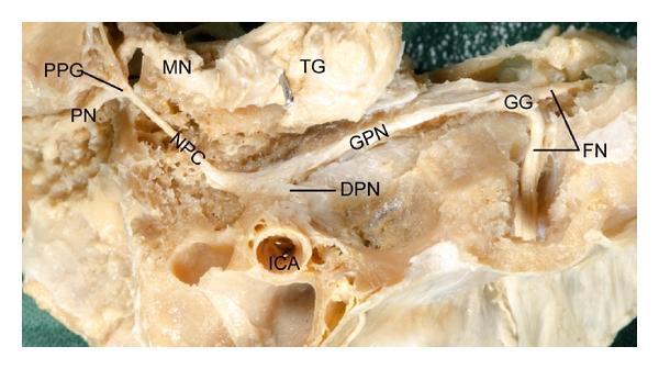
A clinical photograph of a high-resolution cadaveric dissection illustrating the complex neural connections between the geniculate ganglion (GG) and the pterygopalatine ganglion (PPG). The anatomical specimen displays the facial nerve (FN) emerging from the internal acoustic meatus and transitioning at the geniculate ganglion. From the GG, the greater petrosal nerve (GPN) is seen coursing anteriorly to join the deep petrosal nerve (DPN), which emerges near the internal carotid artery (ICA). These nerves merge to form the nerve of the pterygoid canal (NPC), also known as the Vidian nerve, which terminates in the pterygopalatine ganglion (PPG). The PPG is shown as a central hub giving rise to the palatine nerves (PN). Superior to this pathway, the trigeminal ganglion (TG) and the maxillary nerve (MN) are clearly visible, demonstrating their spatial relationship to the autonomic fibers. This educational dissection serves as a vital reference for understanding the parasympathetic and sympathetic pathways of the head, relevant for otolaryngology and neurosurgery.
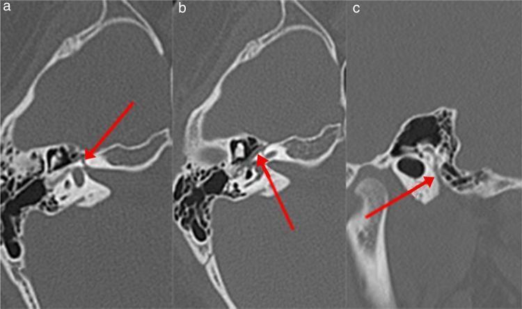
High-resolution axial computed tomography (CT) images of the left temporal bone, presented in three sections (A, B, and C) to demonstrate the course of the facial nerve (Fallopian) canal. Red arrows highlight different segments of the canal. Image A shows the labyrinthine segment, appearing as a narrow radiolucent channel within the dense petrous bone, traveling anterolaterally towards the geniculate ganglion. Image B depicts the tympanic (horizontal) segment, visualized as a thin lucency running along the medial wall of the tympanic cavity, inferior to the lateral semicircular canal. Image C illustrates the mastoid (descending) segment, where the canal assumes a vertical orientation within the mastoid portion of the temporal bone, posterior to the external auditory canal. These images are clinically relevant for evaluating anatomical variations or pathologies, such as Bell's palsy, by assessing the diameter and integrity of the bony canal across its various intratemporal segments.
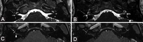
This diagnostic image set consists of four axial MRI sections (A-D) focusing on the skull base and internal auditory canals to evaluate cranial nerve VII (facial nerve). Panels A and B are axial 3D FIESTA (Fast Imaging Employing Steady-state Acquisition) images, which reveal a subtle signal hyperintensity at the tympanic segment of the right facial nerve (indicated by arrows). Panels C and D are contrast-enhanced 3D T1-weighted images demonstrating a pathological, uniformly linear enhancement involving the right geniculate ganglion (dashed arrow), canalicular segment (arrowhead), and tympanic segment (double arrow). When compared to the contralateral left side, the right facial nerve shows markedly increased contrast uptake without significant nerve thickening. These findings are characteristic of neuritis, involving multiple intratemporal segments of the facial nerve, consistent with a clinical presentation of acute peripheral facial nerve paralysis. The imaging modality provides high-resolution anatomical detail of the cisterns and nerve pathways within the petrous temporal bone.
Topodiagnostic Tests for Facial Nerve Palsy
Principle
- Greater superficial petrosal nerve (lacrimation) - leaves at the geniculate ganglion
- Nerve to stapedius - leaves in the vertical (mastoid) segment, just past the second genu
- Chorda tympani (taste, salivation) - leaves in the lower tympanic segment
- Terminal motor branches - exit at stylomastoid foramen
Anatomical Basis
Topodiagnostic Tests - Summary Table
| Test | Branch Tested | Level of Lesion if Abnormal | Normal Value |
|---|---|---|---|
| Schirmer's test | Greater superficial petrosal nerve | At or above geniculate ganglion (CPA to geniculate ganglion) | Wetted paper > 15 mm / 5 min; affected side > 50% of normal side |
| Stapedial reflex | Nerve to stapedius | At or above second genu (vertical segment) | Reflex present bilaterally |
| Taste testing / Electrogustometry | Chorda tympani | At or above chorda tympani takeoff (tympanic segment) | Symmetric taste thresholds |
| Submandibular salivary flow test | Chorda tympani (submandibular ganglion) | At or above chorda tympani takeoff | Symmetric flow rates |
| Salivary pH | Chorda tympani | At or above chorda tympani takeoff | pH 6.0-7.0; acidic with chorda tympani lesion |
1. Schirmer's Test (Lacrimation Test)
- A folded strip of sterile filter paper is placed in the conjunctival fornix at the medial canthus of each eye
- Tears wick along the paper by capillary action
- The wetted length is measured after 5 minutes
- Normally, a noxious stimulus (the paper itself) causes reflex tearing bilaterally
- Abnormal: Affected side shows less than 50% of the lacrimation on the healthy side
- Fisch's criteria: Total response (sum of both eyes) < 25 mm is abnormal
- The test is considered positive when there is asymmetry AND reduced absolute tearing
- Both sides may be reduced in Bell's palsy (subclinical contralateral involvement)
- False-negatives are common because Bell's palsy produces diffuse partial demyelination rather than a clean transection
- EGM of the soft palate may be a more sensitive indicator of GSPN involvement than Schirmer's test
- Cummings Otolaryngology Head and Neck Surgery, p. 3287
2. Stapedial (Acoustic) Reflex Test
- Standard impedance audiometry / tympanometry with reflex testing
- Ipsilateral or contralateral acoustic stimulation (threshold + 80 dB)
- Records the change in middle ear compliance
- Absent reflex or amplitude < 50% of contralateral side = abnormal
- Fisch reported the stapedius reflex is absent in 69% of Bell's palsy cases (84% when paralysis is complete)
- Hyperacusis (loudness intolerance/distortion) is the clinical correlate of stapedius dysfunction
- Patients with a positive reflex within 2 weeks of onset showed complete recovery within 12 weeks
- Recovery of the reflex within 4 weeks predicted facial recovery within 24 weeks
- Cummings Otolaryngology, p. 3288; Scott-Brown's Vol 2, p. 1425
3. Taste Testing
A. Chemical (Qualitative) Taste Testing
- Filter paper discs impregnated with aqueous solutions applied to each half of the tongue:
- NaCl - salty
- Saccharose - sweet
- Citrate / HCl - sour
- Quinine - bitter
- Patient identifies the taste on each side
- Abnormal: Loss of taste or marked asymmetry on the affected side
B. Electrogustometry (EGM) - Quantitative
- Bipolar or monopolar electrical stimulation of the tongue (current 4 µA to 4 mA)
- Elicits metallic/sour/tingling taste sensations at threshold
- Both sides of the tongue tested and compared
- Normal: Two sides differ by < 25%
- Abnormal: No response, or threshold elevation > 20 dB across sides
- Older studies showed taste abnormal in nearly all acute Bell's palsy (low discriminatory power)
- Newer evidence: of 50 patients with complete facial paralysis, 7 had no EGM response - 5 of these had incomplete long-term recovery
- Taste recovery may precede visible facial movement - normal EGM in week 2+ suggests imminent motor recovery
- EGM of the soft palate may better reflect GSPN function than Schirmer's test
- Cummings Otolaryngology, p. 3288-3289
4. Submandibular Salivary Flow Test (Magielski-Blatt Test)
- Cannulas inserted into Wharton's ducts (submandibular duct openings) bilaterally
- Saliva collected over a fixed time period (usually stimulated with citric acid/lemon)
- Volume compared between sides
- Affected side shows significantly reduced flow rate
- Used in combination with Schirmer's test and motor tests for prognostication
- May's prognostic battery: > 25% reduction in salivary flow, lacrimation, or motor response associated with 90% chance of poor recovery
- Cummings Otolaryngology, p. 3287
5. Salivary pH Testing
Lesion Localization Summary
Proximal (CPA/IAC) ←————————————————————————————→ Distal (stylomastoid foramen)

LESION SITE          TESTS ABNORMAL
─────────────────────────────────────────────────────────────
CPA / IAC            ALL tests abnormal + CN VIII involvement
                     (hearing loss, vertigo)

Geniculate ganglion  Schirmer's + stapedial reflex + taste + salivary flow
                     Herpes zoster oticus (Ramsay Hunt) classic site

Vertical segment     Stapedial reflex + taste + salivary flow
(above nerve to      Schirmer's = NORMAL
stapedius)

Tympanic segment     Taste + salivary flow
(above chorda        Schirmer's = NORMAL
tympani)             Stapedial reflex = NORMAL

Stylomastoid         Motor palsy ONLY
foramen / distal     ALL topognostic tests = NORMAL
Electrodiagnostic Tests (Motor Branch Testing)
| Test | What it measures | When useful |
|---|---|---|
| Nerve Excitability Test (NET/Hilger) | Minimum current for visible twitch; > 3.5 mA difference = degeneration | From day 3-4 onward |
| Maximal Stimulation Test (MST) | Supramaximal stimulation; compares response amplitude between sides | Similar timing to NET |
| Electroneurography (ENoG) | Peak-to-peak amplitude of evoked CMAP; compares with contralateral side | From day 4-5; most useful at 2 weeks |
| Electromyography (EMG) | Spontaneous fibrillation potentials (denervation) and voluntary motor units | Denervation potentials appear 10+ days post-onset |
| Nerve conduction velocity | 37-58 m/s normal; < 10 m/s = poor prognosis | Days to weeks post-onset |
- < 90% degeneration: excellent recovery expected
- ≥ 90% degeneration + no voluntary EMG units: surgical decompression may be considered
- < 10% of contralateral response at day 4: poor prognosis in both Bell's palsy and Ramsay Hunt syndrome
- Cummings Otolaryngology, p. 3297-3298
Limitations of Topodiagnostic Testing
- "The accuracy of localization with topodiagnostic testing has been disappointing" - Cummings
- In Bell's palsy, the lesion is diffuse demyelination throughout the nerve - tests do not localize to a single point
- Even in tumors (where a sharp lesion boundary was expected), topodiagnostic testing has been unreliable
- Tests can only identify the most proximal lesion; additional distal injuries are not detected
- Modern practice relies more on MRI with gadolinium for localization, combined with clinical examination
- Currently, these tests are used primarily for prognosis rather than strict localization
- Cummings Otolaryngology, p. 3287