
Wegeners vasculitis
Wegener's granulomatosis vasculitis histology saddle nose deformity
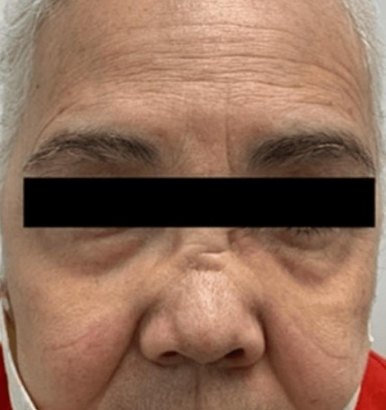
This clinical photograph shows a close-up frontal view of a patient's face, highlighting a classic saddle-nose deformity. The most prominent feature is a significant depression and collapse of the nasal bridge, creating a concave contour between the forehead and the nasal tip. The cartilaginous portion of the nose appears shortened, leading to a subtle cephalad (upturned) rotation of the nasal tip. These structural changes are characteristic manifestations of Granulomatosis with Polyangiitis (formerly Wegener's granulomatosis), where chronic inflammation and vasculitis lead to the destruction of the nasal septal cartilage. Additional visible features include moderate periorbital puffiness and age-related changes such as prominent forehead rhytids (wrinkles), nasolabial folds, and skin mottling. The image serves as a clinical example of the destructive impact of autoimmune small-vessel vasculitis on facial skeletal and cartilaginous structures.
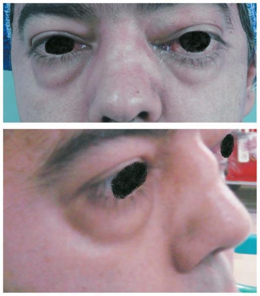
This clinical photograph consists of two panels (anterior and lateral views) showing the facial manifestations of Granulomatosis with Polyangiitis (formerly Wegener's Granulomatosis). In the upper anterior view, there is significant bilateral proptosis (exophthalmos), which is asymmetric and more pronounced in the left orbit. Accompanying this is marked bilateral periorbital and eyelid edema, along with conjunctival injection. Centrally, the nose exhibits a classic saddle nose deformity, characterized by a visible collapse and depression of the nasal bridge resulting from destruction of the underlying cartilaginous nasal septum. The lower lateral view confirms the profile of the saddle nose deformity, highlighting the depth of the bridge depression, and further illustrates the protrusion of the globe and the associated lower eyelid edema. These findings are clinically significant as classic markers of systemic granulomatous vasculitis involving the upper respiratory tract and orbits.

A clinical photograph showcasing a lateral (profile) view of a patient's face, specifically highlighting a significant nasal deformity. A red arrow points to a distinct depression and flattening of the nasal dorsum, characteristically known as a 'saddle nose' deformity. This structural collapse involves the loss of height in the cartilaginous bridge, creating a concave appearance between the nasal root and the tip, while the tip itself remains relatively defined. The image illustrates a classic physical finding associated with granulomatosis with polyangiitis (formerly Wegener granulomatosis), where chronic inflammation and destruction of the nasal septal cartilage lead to the bridge's collapse. The skin over the area remains intact, with the primary finding being the underlying skeletal/cartilaginous alteration. This visual is an essential diagnostic sign in rheumatology and otolaryngology for identifying systemic vasculitis affecting the upper respiratory tract.
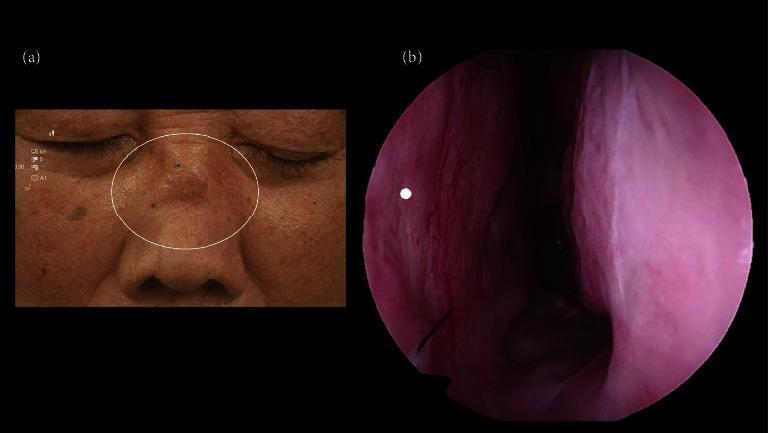
This composite image illustrates clinical and endoscopic findings associated with Granulomatosis with Polyangiitis (GPA). Figure (a) is a clinical photograph of a patient's mid-face featuring a 'saddle nose' deformity, highlighted by a white circle. This deformity is characterized by a visible depression or collapse of the nasal bridge (dorsum) resulting from cartilaginous destruction. The surrounding skin exhibits signs of chronic inflammation and texture changes. Figure (b) presents a 0-degree endoscopic view of the left nasal cavity. The nasal mucosa appears erythematous and slightly irregular. A white dot indicates the post-surgical site of a septal biopsy. Notably, the endoscopic view confirms the absence of a nasal septal perforation following the procedure. These images serve as classic educational examples of the upper respiratory manifestations of vasculitis, specifically GPA, and the importance of endoscopic monitoring to assess structural integrity and mucosal health during diagnosis and management.
"granulomatosis with polyangiitis" AND management
Granulomatosis with Polyangiitis (GPA) - Wegener's Granulomatosis
Nomenclature note: The eponym "Wegener's granulomatosis" was officially renamed Granulomatosis with Polyangiitis (GPA) in 2012 by the ACR and EULAR, after Wegener's Nazi-era activities were documented. Both names are still widely used clinically.
Definition & Pathology
- Necrotizing granulomas of the upper and lower respiratory tract
- Generalized necrotizing angiitis of small/medium vessels
- Focal necrotizing (crescentic) glomerulonephritis
- Andrews' Diseases of the Skin, p. 970
Epidemiology
| Parameter | Details |
|---|---|
| Incidence | ~3-12 per million/year |
| Peak age | 45-65 years (some sources 55-69) |
| Sex | Slight male predominance (1.3:1) |
| Race | Predilection for Caucasians |
Pathogenesis
-
c-ANCA (PR3-ANCA) is the key serological marker - present in ~90% of generalized disease
-
c-ANCA forms against proteinase-3 (PR3), which is found in neutrophil granules
-
Neutrophil activation by ANCA releases free radicals and lytic enzymes, causing endothelial damage
-
Neutrophil extracellular traps (NETs/NETosis) are increasingly recognized as key mediators
-
Staphylococcus aureus is implicated as a possible trigger - it carries a protein with a peptide sequence similar to PR3
-
Genetic risk factors: CTLA4, PTPN22, COL11A2, SERPINA1
-
Shambaugh Surgery of the Ear, p. 4801-4822
Clinical Features
Upper Respiratory Tract (90% - most common initial presentation)
- Persistent rhinorrhea, severe sinusitis, nasal mucosal ulceration
- Epistaxis, nasal septal perforation
- Failure to respond to conventional sinusitis treatment is a diagnostic clue
- Combined nasal and palatal destruction → saddle-nose deformity
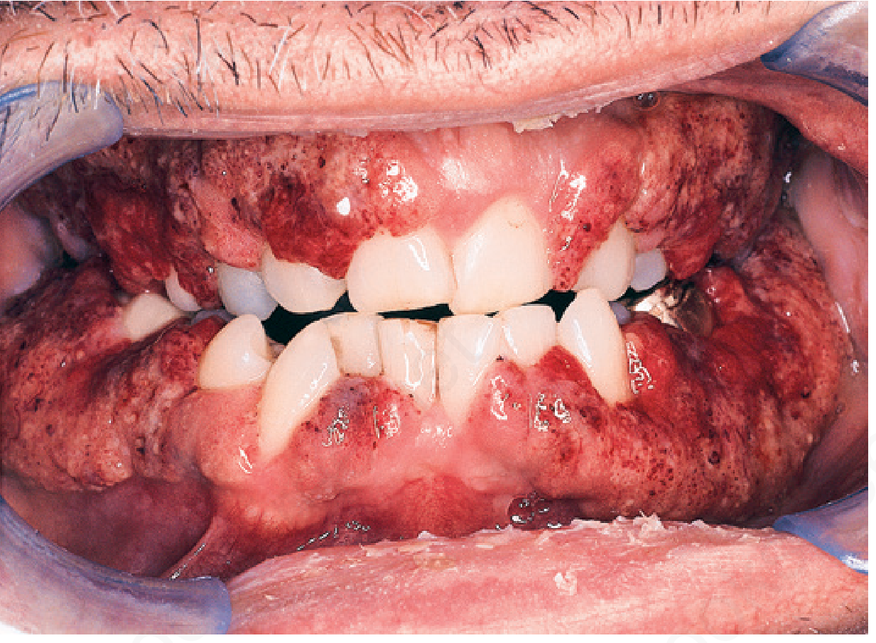
- Oral: "strawberry gums" (hypertrophic granulomatous gingivitis - pathognomonic)
- Palatal/tongue ulceration, subglottic stenosis
Pulmonary (71% have infiltrates radiographically)
- Cough, dyspnea, chest pain
- Nodules, cavities, pulmonary infiltrates, hemoptysis
Renal (85%)
- Focal crescentic necrotizing glomerulonephritis
- Hematuria, proteinuria, AKI → renal failure (historically the most common cause of death)
Ear
- Present in 19-56% of patients
- Conductive hearing loss, sensorineural hearing loss, tinnitus, vertigo, otalgia
- Draining tympanic membrane perforation; medically refractory chronic otitis media (mimics TB but with disproportionate pain)
- Facial nerve paresis
Skin (45%)
- Palpable purpura, nodules (especially extensor surfaces), ulcers
- Necrotizing angiitis → hemorrhagic pustular eruption, livedo reticularis
- Pyoderma gangrenosum-like lesions
Other Systems
- Eyes (58%): conjunctivitis, episcleritis, proptosis, orbital pseudotumor
- Joints (66%): arthralgia
- CNS (22%): cranial neuropathies (7%), mononeuritis multiplex
- Heart (12%): pericarditis, myocarditis
Clinical Images
Diagnosis
ACR/EULAR Criteria (paediatric: 3 of 6 features required)
- Renal involvement
- Upper airway involvement
- Pulmonary involvement (infiltrates, nodules, or cavities)
- Laryngo-tracheobronchial stenosis
- Granulomatous inflammation on biopsy
- ANCA positivity
Key Tests
- c-ANCA / PR3-ANCA: highly specific; positive in ~90% generalized, ~70-90% limited forms (up to 30% can be negative, especially limited disease)
- Biopsy: sinonasal biopsies have the highest specificity and sensitivity; ear biopsies often show only non-specific inflammation
- Histology: necrotizing granulomas + vasculitis; LCV pattern in skin; crescents on renal biopsy
- Urinalysis, creatinine, CXR/CT chest, ESR/CRP
Treatment
Induction of Remission
| Drug | Dose / Notes |
|---|---|
| Cyclophosphamide | 2 mg/kg/day orally OR 500-750 mg/m² IV every 2-4 weeks |
| High-dose glucocorticoids | 3 × IV methylprednisolone boluses, then prednisone 1-2 mg/kg/day |
| Rituximab (anti-CD20) | FDA approved 2011; equivalent to CYC for new cases; superior to CYC for relapsing disease; PR3-ANCA patients may respond especially well |
| Methotrexate | Alternative for mild disease (Cr <150 µmol/L, no critical organ involvement); avoid if GFR reduced |
Maintenance of Remission
- Azathioprine, methotrexate, leflunomide, MMF, or rituximab
- Slow prednisone taper; treat for at least 1 year
- Co-trimoxazole (TMP-SMX): reduces relapse by reducing nasal S. aureus carriage; consider for limited upper respiratory disease in remission
Refractory / Severe Disease
- Plasma exchange (PLEX): for diffuse pulmonary hemorrhage or RPGN near dialysis
- IVIG, anti-TNF therapy (infliximab), tacrolimus
Newer Agents
- Avacopan (C5a receptor blocker): steroid-sparing agent used alongside CYC or rituximab
Prognosis
- Untreated: mean survival 5 months, 90% mortality at 2 years
- With cyclophosphamide: complete remission in up to 93%, lasting average 4 years
- Limited form (upper respiratory tract only, no renal involvement): better prognosis
- Therapy-related adverse events (CYC toxicity, infection) now account for more deaths in year 1 than the vasculitis itself
Recent Guidelines
Summary Mnemonic: "EARs, NOSE, KIDNEY, LUNG"
- Ears - refractory otitis media, SNHL
- ANCA (c-ANCA/PR3) - key serology
- Renal - crescentic GN
- Sinusitis/saddle nose - upper airway hallmark
- Nodules/necrosis - necrotizing granulomas
- Oral - strawberry gums
- Skin - purpura, nodules
- Eyes - proptosis, episcleritis
- Kidney - crescentic GN (most dangerous)
- Induction - cyclophosphamide / rituximab + steroids
- Drug maintenance - azathioprine, MTX, MMF
- Netosis (NETs) - pathogenic mechanism
- GPA - current official name