
Recent advances in managent of fistula in ano
"fistula in ano" AND management
fistula in ano classification anatomy Parks
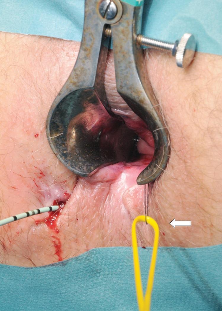
This clinical photograph captures an intraoperative view of a surgical procedure for a complex fistula-in-ano. A metallic Parks or similar anal retractor is positioned to provide exposure of the anal canal. The surgical field demonstrates the management of a fistula track using specialized instrumentation. On the left side of the image, a urological guidewire with black and white markings is seen entering the external fistula opening, which shows minor associated hemorrhage. A yellow vascular sling (indicated by a white arrow) is being threaded through the track using a 1/0 nylon suture loop. The educational focus of the image is the technique of passing a vascular sling from the internal opening to the external opening to serve as a loose seton. This procedure is critical in colorectal surgery for maintaining drainage of the fistulous track while preventing abscess formation and protecting the anal sphincter muscles. The image is intended for intermediate to advanced medical learners focusing on proctological surgical techniques.
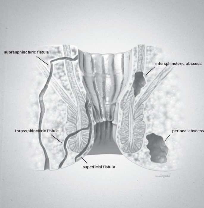
**Imaging Modality:** Medical illustration/Anatomical diagram. **Anatomical Region:** Coronal cross-section of the anorectal region, including the anal canal, rectum, internal and external anal sphincters, and surrounding perianal soft tissues. **Observed Pathology:** The illustration classifies various types of anorectal fistulas and abscesses based on their anatomical trajectory and location. **Characteristic Visual Features:** * **Fistula Tracts:** Three distinct epithelial-lined tracks are labeled. A **superficial fistula** is shown confined to the perianal skin and distal anal canal. A **transsphincteric fistula** is depicted crossing both the internal and external sphincter muscles into the ischioanal fossa. A **suprasphincteric fistula** is illustrated looping above the puborectalis muscle before descending to the skin surface. * **Abscess Formations:** Two localized collections of fluid/pus are identified. An **intersphincteric abscess** is positioned within the space between the internal and external sphincter muscles. A **perineal (perianal) abscess** is shown as a larger pocket located in the subcutaneous tissue adjacent to the anal verge. **Key Diagnostic Features:** The diagram emphasizes the relationship between inflammatory tracts and the sphincter complex, following the Parks classification system for fistula-in-ano. Suitable for clinical education on perianal sepsis and surgical planning.
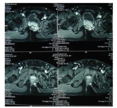
Axial pelvic Magnetic Resonance Imaging (MRI) scans demonstrate a complex suprasphincteric fistula in ano with associated abscess formation. The series of T2-weighted or contrast-enhanced images shows a high-signal-intensity fluid collection representing an abscess cavity, approximately 2 cm in diameter, located superior to the levator ani muscle. White arrows indicate the fistula tract, which presents as a distinct linear pathway of altered signal intensity extending from an internal opening in the rectum through the pelvic musculature toward the perianal region. The images highlight the relationship between the inflammatory process and key anatomical landmarks, including the rectum, levator ani, and perianal soft tissues. This diagnostic imaging is characteristic of complex perianal disease, showing both the localized suppuration (abscess) and the extensive 10 cm fistulous tract. Such findings are critical for surgical planning in cases of deep pelvic infections or complex fistula management.
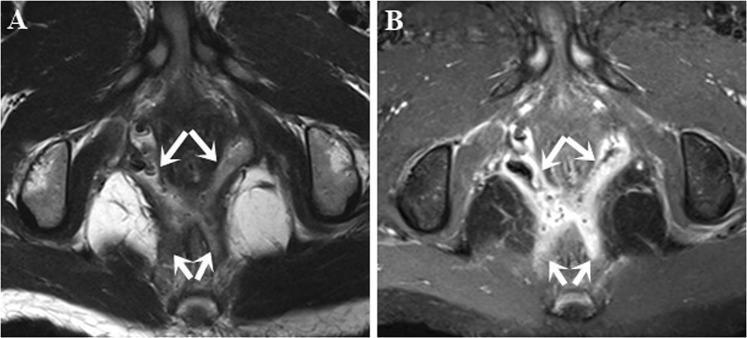
This diagnostic imaging set consists of two axial MRI slices of the pelvic region focusing on the perianal area. Image A is an axial T2-weighted turbo spin-echo (T2W TSE) sequence, and Image B is a post-contrast fat-suppressed T1-weighted turbo spin-echo (FS T1W TSE) sequence. The images demonstrate a complex high transsphincteric fistula-in-ano with associated secondary tracts. In Image A, the tracts (indicated by white arrows) appear as hyperintense (bright) linear structures against the relatively darker muscular background of the external anal sphincter and levator ani. In Image B, the secondary tracts within the ischioanal space show marked contrast enhancement, appearing significantly brighter than surrounding fat-suppressed tissues. This enhancement highlights active inflammation and the branching nature of the tracts. The visualization of these secondary extensions relative to the anal sphincter complex is critical for surgical planning and classification according to the Parks or St James's University Hospital staging systems.
PMID: 37460830
PMID: 40325545
PMID: 38091125
PMID: 40125894
"fistula in ano" AND ("LIFT" OR "laser" OR "VAAFT" OR "stem cell" OR "video-assisted")
PMID: 40392371
PMID: 42011865
Recent Advances in Management of Fistula-in-Ano
Classification (Foundation for Management)
- Type 1 - Intersphincteric (~45%): tract within the intersphincteric plane
- Type 2 - Transsphincteric (~30%): crosses both internal and external sphincters into the ischioanal fossa
- Type 3 - Suprasphincteric (~20%): loops over the puborectalis before descending
- Type 4 - Extrasphincteric (~5%): entirely outside the sphincter complex
Goals of Treatment
- Drain any active infection
- Define fistula anatomy accurately (MRI, EAUS)
- Remove or ablate the epithelialized tract
- Preserve fecal continence
- Prevent recurrence
- Sabiston Textbook of Surgery, p. 2163
Preoperative Imaging
Treatment Approaches
1. Fistulotomy (Lay-Open Technique)
- Recurrence rate: 2-8%
- Clinically significant fecal incontinence: <5% in patients with normal preoperative sphincter function
- A 2025 meta-analysis of 13 RCTs (PMID 40125894) found no significant difference between fistulotomy vs. fistulectomy in healing time, recurrence, or incontinence; fistulectomy gave lower 24h pain scores while fistulotomy had less postoperative bleeding
2. Seton Techniques
- A 2023 meta-analysis of 29 studies (PMID 38091125) found cutting seton had 6% recurrence and 16% incontinence at 6 months, with comparable recurrence to fistulotomy, advancement flap, and two-stage seton - but at the cost of greater pain and longer healing time (~14.6 weeks)
Sphincter-Preserving Techniques (Major Recent Advances)
3. LIFT Procedure (Ligation of the Intersphincteric Fistula Tract)
- Meta-analysis of 1,378 LIFT procedures (26 studies): overall success rate 76%, complication rate 14%, fecal incontinence 1.4%
- A 2023 network meta-analysis (PMID 37460830) of 52 RCTs found LIFT ranked best for minimizing bowel incontinence in both simple (99.1% of comparisons) and complex (86.2%) anal fistula
- Failure may convert the fistula to a simpler intersphincteric pattern amenable to fistulotomy; secondary healing after reintervention reaches 77-86%
- Horseshoe anatomy, Crohn's disease, and prior surgery are risk factors for failure
4. Endorectal Advancement Flap (ERAF)
- Healing rates: 66-87%
- Including internal sphincter fibers in the flap increases robustness but increases mild-moderate incontinence (up to 35%) with decreased manometric pressures
- Failure rate in the 2025 meta-analysis: 25.9%
- Continence disturbance: 7.3% (highest among sphincter-sparing techniques)
5. VAAFT (Video-Assisted Anal Fistula Treatment)
- Precisely map the tract and identify secondary branches under direct vision
- Close the internal opening with suture ligation or stapling
- Debride granulation tissue and ablate the tract with an electrode via the working channel
- Reported healing rates: 71-87% at 1-2 years
- Failure rate: 22.3% (lowest in 2025 network meta-analysis) - Sabiston, p. 2163
- Importantly, no VAAFT patients reported worsening continence in the 2025 meta-analysis
- A 2025 meta-analysis of 15 studies, 999 patients (PMID 42011865) found VAAFT associated with reduced recurrence (RR 0.94), fewer postoperative complications (RR 0.93), and improved quality of life vs. traditional techniques
6. FiLaC (Fistula Laser Closure)
- A 2025 meta-analysis reported 65% healing at median 24 months follow-up
- Failure rate: 43.9% (highest among sphincter-sparing techniques in the 2025 network meta-analysis)
- Zero continence disturbance reported in the meta-analysis
- A 2024 clinical trial (PMID 39239958) on radially emitting diode laser for transsphincteric fistulas confirmed promising results
7. Biologic Plugs and Fibrin Glue
- Bioresorbable anal fistula plugs (e.g., bioabsorbable porcine intestinal submucosa plug) are placed to obliterate the tract, providing a scaffold for native collagen deposition
- Fibrin glue injection obliterates the tract
- Neither has demonstrated reliable long-term success; these are largely reserved for Crohn's-related or highly complex fistulas where other options are unsuitable
Stem Cell Therapy (Cutting Edge)
Crohn's-Associated Fistulas
- Draining seton for initial control
- Medical optimization: anti-TNF therapy (infliximab, adalimumab), with vedolizumab and ustekinumab as alternatives
- Darvadstrocel (adipose MSC): The ADMIRE-CD trial demonstrated superiority over placebo for combined remission in perianal Crohn's fistulas
- Surgical options (LIFT, advancement flap) after medical therapy optimization
- Multiple external openings ("watering can perineum") should always raise suspicion for Crohn's disease
Emerging Technologies
| Technology | Principle | Status |
|---|---|---|
| 3D printing / modeling | Pre-op planning from MRI data | Promising, limited evidence |
| OTSC (over-the-scope clip) | Endoscopic closure of internal opening | Early studies |
| Platelet-rich plasma (PRP) | Enhances wound healing | Small studies |
| Adipose-derived MSCs | Tissue regeneration | Phase III (Crohn's approved) |
| Laser (FiLaC) | Tract ablation without sphincter division | Established, high failure rate |
| VAAFT | Endoscopic visualization + ablation | Rapidly gaining adoption |
Summary of Outcomes (2025 Network Meta-Analysis, PMID 40392371)
| Technique | Failure Rate | Continence Disturbance |
|---|---|---|
| VAAFT | 22.3% | 0% |
| LIFT | 28.6% | 1.5% |
| EAF (Endoanal Flap) | 25.9% | 7.3% |
| FiLaC | 43.9% | 0% |
- Current Surgical Therapy 14e - Fistula-in-Ano chapter
- Sabiston Textbook of Surgery - Chapter 97, p. 2163-2164
- Network meta-analysis of surgical treatments: Bhat S et al., Tech Coloproctol 2023 (PMID 37460830)
- Sphincter-sparing technique comparison: Fuschillo G et al., Tech Coloproctol 2025 (PMID 40392371)
- VAAFT meta-analysis: Wang W et al., BJS Open 2025 (PMID 42011865)
- 3D modeling review: Ruscoe Z et al., ANZ J Surg 2025 (PMID 40325545)
- Cutting seton meta-analysis: Khamar J et al., Tech Coloproctol 2023 (PMID 38091125)
- Fistulotomy vs fistulectomy: Quinn R et al., ANZ J Surg 2025 (PMID 40125894)
Advances in surgery of thyroid gland
"thyroid surgery" AND advances
thyroid surgery robotic remote access endoscopic approaches
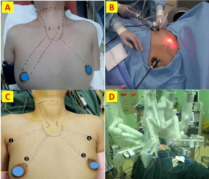
A composite of four clinical photographs demonstrating surgical approaches for remote-access thyroidectomy. Panels A and B illustrate Endoscopic Thyroidectomy via Bilateral Areola Approach (ET-BAA). Panel A shows preoperative skin markings indicating the operative tunnel from the bilateral areolar regions to the thyroid gland. Panel B displays the intraoperative setup with surgeons manually manipulating endoscopic trocars inserted via the chest wall. Panels C and D illustrate Robotic Thyroidectomy via Bilateral Axillo-Breast Approach (RT-BABA). Panel C depicts the specific four-port marking scheme: two axillary incisions (labeled 1 and 4) and two areolar incisions (labeled 2 and 3), with dashed lines outlining the dissection planes towards the neck. Panel D shows the intraoperative environment featuring a da Vinci surgical system, with multiple robotic arms draped and docked into the patient's chest and axillary ports. The images contrast the manual endoscopic technique requiring areolar access with the more complex robotic setup that utilizes four ports (axillo-breast) to facilitate a broader range of motion and 3D visualization.
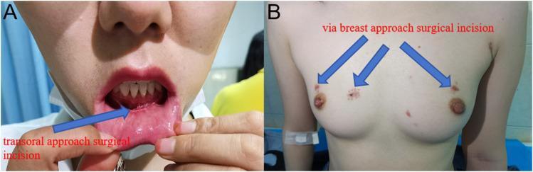
This clinical photograph set displays postoperative outcomes one month after endoscopic thyroid surgery using two distinct minimally invasive techniques. Panel A illustrates the transoral approach, showing a horizontal linear scar on the internal mucosal surface of the lower lip, positioned below the teeth and above the labial frenulum. Panel B depicts the breast approach, showing three small, symmetrical surgical incisions on the chest wall. Two incisions are located superior-laterally to each nipple, with a third central incision positioned between the breasts in the parasternal region. The images demonstrate the cosmetic advantages of extracervical approaches in thyroidectomy, specifically the Transoral Endoscopic Thyroidectomy Vestibular Approach (TOETVA) and the Breast Approach, which avoid visible scarring on the neck. These visuals serve as educational material for surgical oncology and endocrine surgery regarding patient aesthetic outcomes and port placement in remote-access thyroid surgery.
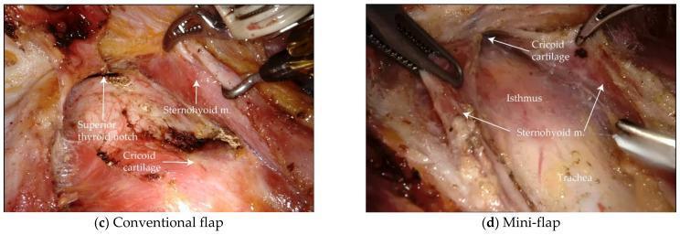
Two side-by-side intraoperative endoscopic photographs comparing conventional flap (left) and mini-flap (right) approaches during Bilateral Axillo-Breast Approach (BABA) thyroidectomy. The conventional flap image demonstrates a wider surgical exposure, with clear labeling of the superior thyroid notch, cricoid cartilage, and sternohyoid muscle. In contrast, the mini-flap image shows a more restricted surgical field focused on the central neck, highlighting the cricoid cartilage, thyroid isthmus, sternohyoid muscle, and the underlying trachea. Both images show robotic surgical instruments (forceps and retractors) positioned within the surgical plane for tissue manipulation. These images illustrate the differences in anatomical visualization and flap boundaries between the two techniques, emphasizing the reduced dissection area of the mini-flap approach while maintaining access to critical structures. The labels and visible landmarks serve to orient the viewer to the laryngeal and tracheal anatomy during the initial phase of endocrine surgery.
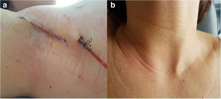
Two-part clinical photograph demonstrating surgical results of a robotic-assisted transaxillary thyroidectomy. Image 'a' provides a close-up of the surgical access point in the axilla at the end of the procedure. It shows a linear, approximately 5 cm incision closed with intermittent sutures. A semi-transparent, reddish surgical drain is visible exiting through a separate stab incision, secured with a purse-string or anchoring suture. Minimal localized erythema is present at the incision margins. Image 'b' shows the anterior neck of the same patient on the first postoperative day. The skin surface is intact with no visible cervical incisions, scars, or significant ecchymosis, highlighting the cosmetic benefit of the remote-access transaxillary approach. Visible landmarks include the suprasternal notch and clavicles. The pair illustrates the concealment of surgical scarring in natural body folds to achieve superior aesthetic outcomes in thyroid surgery.
"thyroidectomy" AND ("robotic" OR "endoscopic" OR "transoral" OR "nerve monitoring" OR "parathyroid fluorescence")
PMID: 38944497
PMID: 41182840
PMID: 38603661
PMID: 39973303
"thyroidectomy" AND ("near-infrared fluorescence" OR "autofluorescence" OR "parathyroid" OR "IONM" OR "continuous nerve monitoring")
PMID: 37536854
PMID: 37684153
PMID: 39588333
Advances in Surgery of the Thyroid Gland
1. Evolution of the Conventional Open Approach
- Smaller incisions: The "mini-incision" technique (3 cm) with minimal flap creation and video assistance has reduced wound morbidity
- Ligament of Berry dissection: Precise identification of the RLN as it passes through the ligament of Berry remains the most important step - bleeding here must be controlled with gentle pressure, never electrocautery
- Energy-based vessel sealing: Devices such as the Harmonic scalpel (ultrasonic), LigaSure, and bipolar vessel sealers have replaced silk ligatures, reducing operative time, improving haemostasis, and decreasing the risk of thermal spread to the RLN compared to monopolar cautery
- Schwartz's Principles of Surgery, p. 1040
2. Minimally Invasive Video-Assisted Thyroidectomy (MIVAT)
- Suitable for thyroid nodules <35 mm and total thyroid volume <25 mL
- Similar complication rates to conventional surgery in experienced hands
- Still results in a (small) cervical scar
- Cummings Otolaryngology, p. 2348
3. Remote-Access Thyroidectomy (No Cervical Scar)
3a. Endoscopic Remote-Access Approaches
| Approach | Access Point | Notes |
|---|---|---|
| Transaxillary | Single axillary incision | Most widely used remote approach; gasless or CO₂ insufflation |
| Anterior chest/breast | 3 trocar sites on chest wall | Bilateral areola approach (BABA) |
| Transoral vestibular (TOETVA) | 3 ports through oral vestibule | No external scar anywhere; gaining rapid adoption |
| Retroauricular (facelift) | Postauricular incision | Scar concealed in hair and ear fold |
| Bilateral axillo-breast (BABA) | 2 axillary + 2 areola | Gives bilateral neck access |
3b. Robot-Assisted Remote-Access Thyroidectomy (RATS)
- The gasless transaxillary robotic approach (Kang et al., 2009) uses a rigid retractor to maintain the operative pocket without CO₂ insufflation. Large Korean series reported low complication rates, but attempts at replication in the West revealed higher rates of brachial plexopathies, tracheo-oesophageal injuries, and RLN injury - dampening enthusiasm outside Asia
- The robotic facelift thyroidectomy (Terris et al., 2011) uses a postauricular incision along the SCM plane. A multiinstitutional study in 90 patients showed 3.9% temporary RLN weakness, 2.9% haematoma, and no permanent complications - most managed as outpatients without drains
- The transoral robotic thyroidectomy has also been reported with excellent cosmetic results
- The American Thyroid Association recommends RATS be limited to high-volume thyroid centres with appropriate patient selection
4. Intraoperative Nerve Monitoring (IONM)
Types of IONM
| Type | Method | Advantage |
|---|---|---|
| Intermittent IONM | Probe stimulation + EMG response | Standard; identifies nerve location |
| Continuous vagal IONM (vagal-CIONM) | Continuous biphasic stimulation of vagus | Real-time monitoring; alerts before injury occurs |
| Laryngeal adductor reflex CIONM | Detects motor response | Emerging, no tube required |
- ATA 2015 guidelines recommend visual identification of the RLN in all cases; IONM may be used to facilitate identification and confirm function, especially before proceeding with contralateral thyroidectomy
- A 2009 RCT (Barczynski) of 2,000 nerves at risk showed IONM reduced transient RLN injury rates vs visual identification alone - especially in high-risk cases. No significant reduction in permanent injury rates was demonstrated
- Continuous vagal IONM represents the most significant advance: if signal loss is detected, the surgeon can pause, allowing the nerve to recover before proceeding - preventing conversion of a transient to a permanent injury
- A 2024 review (PMID 37684153) thoroughly reviews CIONM subtypes and standardization of outcomes reporting
5. Parathyroid Identification and Preservation
Near-Infrared Autofluorescence (NIRAF)
- Allows distinction between parathyroid and surrounding fat/lymph nodes
- Enables the surgeon to assess whether a gland has been inadvertently removed (by examining the specimen)
- A 2024 symposium (PMID 38966217) highlighted emerging evidence for NIRAF reducing hypoparathyroidism rates
Indocyanine Green (ICG) Fluorescence Angiography
Intraoperative PTH (ioPTH) Assay
- Intraoperative Adjuncts in Thyroid Surgery, Surg Clin North Am 2024 (PMID 38944497)
- New Techniques for Intraoperative Parathyroid Localization, Adv Surg 2023 (PMID 37536854)
6. Non-Surgical Ablative Therapies for Benign Nodules
Ultrasound-Guided Thermal Ablation
| Technique | Mechanism | Notes |
|---|---|---|
| Radiofrequency ablation (RFA) | Heat from electrical current | Approved and widely used; >50% volume reduction |
| Microwave ablation (MWA) | Electromagnetic energy | Faster than RFA; similar results |
| High-intensity focused ultrasound (HIFU) | Focused ultrasound waves | No needle required; early adoption phase |
| Laser ablation (LA) | Near-infrared laser energy | Well-established for solid nodules |
- Overall complication rate: 23% (mostly minor: 21%)
- Major complications: 2% (rare)
- Dysphonia (voice change): 2%, almost always transient
- LA is a safe, effective therapy for symptomatic benign solid thyroid nodules
Ethanol Ablation
7. Extent of Surgery - Shifting Indications
Thyroid Lobectomy vs Total Thyroidectomy
- Lobectomy is now acceptable (and may be sufficient) for unifocal papillary thyroid cancer <4 cm, no extrathyroid extension, no node metastases
- Total thyroidectomy is reserved for: bilateral disease, tumors >4 cm, extrathyroid extension, aggressive histology, or need for radioiodine ablation
- This shift reduces the risk of permanent hypoparathyroidism and bilateral RLN injury in patients with low-risk disease
Active Surveillance for Low-Risk PTC
8. Molecular Diagnostics Refining Surgical Decisions
| Test | Principle | Sensitivity/Specificity |
|---|---|---|
| Afirma GSC (gene sequencing classifier) | mRNA expression profiling | >91% sensitivity; NPV 96% |
| ThyroSeq v3 | Next-generation sequencing (DNA + RNA) | Sensitivity 94%, specificity 82% |
| RosettaGX Reveal | microRNA classifier | Used in cytologically indeterminate nodules |
9. 3D Printing and Surgical Planning
- Pre-operative visualization of substernal goitres
- Training and simulation
- Complex reoperative cases with distorted anatomy
10. Outpatient (Ambulatory) Thyroidectomy
Summary of Key Advances
| Domain | Advance | Clinical Impact |
|---|---|---|
| Access | TOETVA, robotic transaxillary, facelift | No neck scar; cosmesis |
| Nerve protection | Continuous IONM (vagal-CIONM) | Reduces transient RLN injury |
| Parathyroid | NIRAF autofluorescence, ICG angiography | Reduces hypoparathyroidism |
| Haemostasis | Harmonic scalpel, LigaSure | Faster, safer dissection |
| Non-surgical | RFA, MWA, LA, HIFU for benign nodules | Avoids surgery entirely |
| Molecular Dx | Afirma GSC, ThyroSeq v3 | Avoids unnecessary surgery |
| Extent | Lobectomy for low-risk PTC; active surveillance | Reduces operative morbidity |
| Planning | 3D reconstruction/printing | Better pre-op planning |
| Recovery | ERAS + outpatient thyroidectomy | Same-day discharge |
- Cummings Otolaryngology Head and Neck Surgery - Thyroid Surgery, Chapter 124
- Schwartz's Principles of Surgery 11e - Chapter 38
- Intraoperative adjuncts review: Huang BL, Surg Clin North Am 2024 (PMID 38944497)
- Robotic axillary vs open meta-analysis: Zhang X, PLoS One 2024 (PMID 38603661)
- RLN monitoring review: Kirke DN, Otolaryngol Clin North Am 2024 (PMID 37684153)
- Parathyroid localization techniques: Kiernan CM, Adv Surg 2023 (PMID 37536854)
- Laser ablation meta-analysis: Scappaticcio L, J Clin Endocrinol Metab 2025 (PMID 39973303)
- Gasless endoscopic thyroidectomy: Cheng X, Front Endocrinol 2024 (PMID 39588333)
- Endoscopic/robotic advances: Zhou Q, J Craniofac Surg 2026 (PMID 41182840)
Recent advances in management of colorectal cancer
"colorectal cancer" AND ("advances" OR "immunotherapy" OR "targeted therapy" OR "total neoadjuvant")
colorectal cancer staging treatment algorithm molecular markers
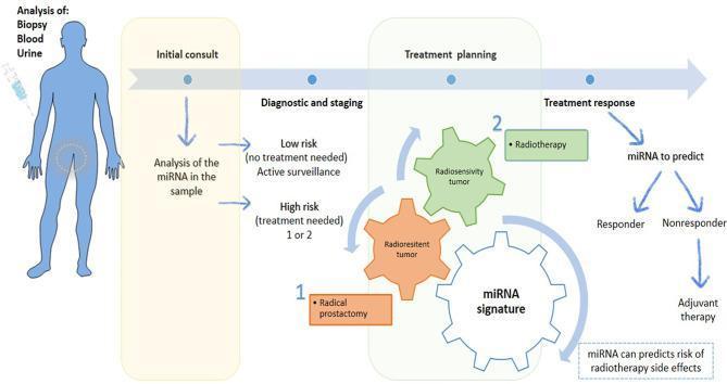
This medical flowchart illustrates the clinical pathway for personalized prostate cancer management utilizing microRNA (miRNA) analysis. The diagnostic sequence begins with an 'Initial Consult' where biological samples (biopsy, blood, or urine) are collected for miRNA profiling. These molecular markers guide 'Diagnostic and Staging,' stratifying patients into 'Low Risk' for active surveillance or 'High Risk' requiring intervention. During 'Treatment Planning,' the miRNA signature helps differentiate between 'Radiosensitive' and 'Radioresistant' tumor phenotypes. This distinction dictates the therapeutic approach: radical prostatectomy (Option 1) for radioresistant tumors or radiotherapy (Option 2) for radiosensitive cases. Post-treatment, miRNAs serve as biomarkers for 'Treatment Response,' predicting if a patient is a 'Responder' or 'Nonresponder.' In the latter case, adjuvant therapy is indicated. Additionally, the algorithm highlights the utility of miRNA signatures in predicting the risk of radiotherapy-related side effects, facilitating precision oncology by optimizing therapeutic efficacy while minimizing toxicity.
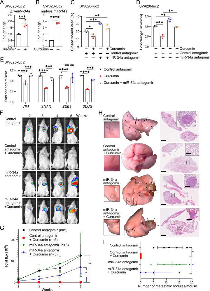
This composite educational graphic illustrates the molecular and physiological effects of curcumin on colorectal cancer (CRC) metastasis using SW620-Luc2 cell models. Panels A-E present quantitative data including qPCR results showing upregulation of pri-miR-34a and mature miR-34a by curcumin, and bar graphs demonstrating inhibition of cell migration (wound healing), invasion (Boyden chamber), and Epithelial-Mesenchymal Transition (EMT) markers like Vimentin, SNAIL, and ZEB1. Panels F-I provide clinical-level evidence through animal xenografting. Panel F shows longitudinal bioluminescence imaging (BLI) of NOD/SCID mice over five weeks, where luciferase signals indicate the progression of lung metastases. The signal is nearly abolished in the curcumin-treated group but partially restored when miR-34a is inhibited with antagomirs. Panel H contains gross anatomical photographs of resected murine lungs alongside corresponding H&E-stained histopathology sections. Black arrows and magnified inserts highlight metastatic tumor nodules. Panel I provides statistical quantification of these nodules, establishing a clear link between curcumin treatment, miR-34a induction, and the suppression of metastatic colorectal cancer progression.
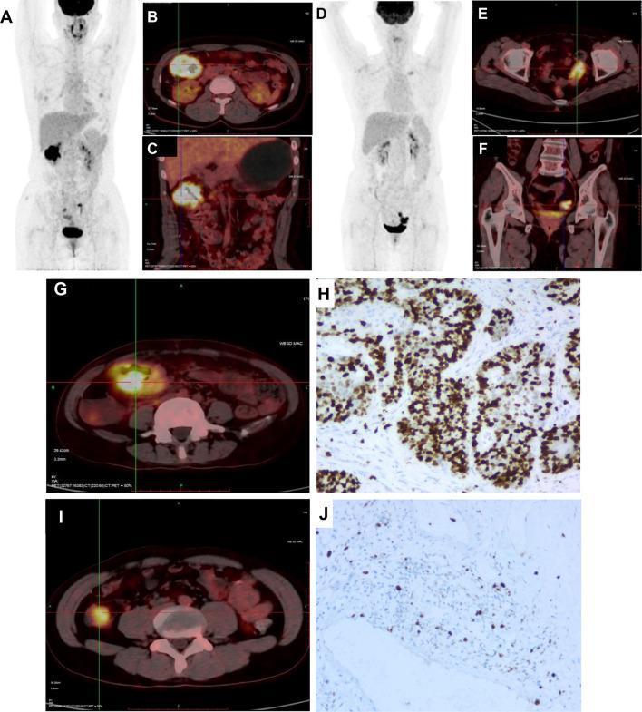
This composite image illustrates diagnostic findings in colorectal cancer (CRC) patients, focusing on 18F-FDG PET/CT imaging and immunohistochemistry (IHC). Panels A, B, and C display a patient with mutated-type KRAS, featuring a Maximum Intensity Projection (MIP) whole-body scan (A) and cross-sectional PET/CT fusions (B, axial; C, coronal) showing intense radiotracer accumulation in the hepatic flexure (SUV 20.0). Panels D, E, F, G, and I represent CRC patients with wild-type KRAS or general sigmoid involvement, showing MIP views and axial/coronal slices with 18F-FDG uptake in the sigmoid colon (SUVs ranging from 12.4 to 21.3). The radiologic images highlight the localization of primary lesions using functional metabolic activity. Panels H and J provide corresponding histological evidence via Ki-67 IHC staining. Panel H demonstrates a high proliferation index (77.8%) with dense, dark brown nuclear staining in tumor cells, while Panel J shows a lower proliferation index (22.7%) with sparse staining. This educational visual correlates metabolic imaging intensity (SUVmax) with molecular markers (KRAS status) and cellular proliferation (Ki-67) in the context of oncological staging and pathology.
PMID: 38945765
PMID: 41355781
PMID: 38309719
"colorectal cancer" AND ("robotic" OR "transanal TME" OR "TaTME" OR "organ preservation" OR "watch and wait" OR "ctDNA")
PMID: 37499673
PMID: 39284954
"rectal cancer" AND ("organ preservation" OR "watch and wait" OR "complete clinical response" OR "TaTME" OR "transanal")
"colorectal cancer" AND ("HER2" OR "NTRK" OR "KRAS G12C" OR "RAS" OR "targeted therapy")
PMID: 38194231
PMID: 39848335
PMID: 37742544
"robotic" AND "colorectal surgery" AND outcomes
"transanal total mesorectal excision" OR "TaTME" AND outcomes
PMID: 38590019
PMID: 38898341
Recent Advances in Management of Colorectal Cancer
Overview
1. Surgical Advances
1a. Total Mesorectal Excision (TME) - The Gold Standard for Rectal Cancer
| Technique | Key Advantage | Status |
|---|---|---|
| Open TME | Gold standard benchmark | Comparator for all studies |
| Laparoscopic TME (L-TME) | Less blood loss, faster recovery | Well-established |
| Robotic TME (R-TME) | Superior dexterity in narrow pelvis; better 3D vision | Rapidly growing adoption |
| Transanal TME (TaTME) | Bottom-up approach; ideal for narrow/obese pelvis, low tumors | Established in specialist centres |
1b. Transanal TME (TaTME)
- Superior visualization in a narrow male pelvis or in obesity
- Better ability to achieve distal resection margins for very low rectal tumors
- Potentially improved circumferential resection margin (CRM) positivity rates
1c. Robotic Colorectal Surgery
- Wristed instrumentation with 7 degrees of freedom (superior to straight laparoscopic instruments)
- 10x magnified 3D vision
- Tremor filtration
- Ergonomic advantage for surgeon
1d. Self-Expanding Metallic Stent (SEMS) for Obstruction
1e. CRS + HIPEC for Peritoneal Metastases
- The Dutch RCT (Verwaal) demonstrated near doubling of survival with CRS + HIPEC over systemic therapy
- The PRODIGE 7 French RCT showed median OS of 41 months (vs historical ~12 months with systemic therapy alone) with CRS alone - with HIPEC adding no additional benefit when oxaliplatin was used for only 30 minutes
- A Peritoneal Carcinomatosis Index (PCI) score >20 is generally a contraindication for CRS
- Mitomycin C HIPEC (60-90 min) is preferred over the short oxaliplatin used in PRODIGE 7
- Sabiston Textbook of Surgery, p. 1247
2. Total Neoadjuvant Therapy (TNT) for Rectal Cancer
TNT Sequences
- Induction TNT (iTNT): Chemotherapy → Chemoradiation → Surgery
- Consolidation TNT (cTNT): Chemoradiation → Chemotherapy → Surgery
- Both cTNT and iTNT significantly improved complete response (CR) rates vs standard CRT alone
- cTNT OR 2.36 (best for CR, SUCRA 0.90)
- iTNT OR 1.99 (best for 3-year DFS and OS, SUCRA 0.87)
- No significant difference in DFS or OS among the three strategies
- cTNT ranked best for organ preservation (higher CR rates); iTNT ranked best for survival
3. Watch-and-Wait (Organ Preservation Strategy)
- 77% 3-year organ preservation rate in CCR patients
- 40% 3-year organ preservation rate in near-complete response patients
- Clinical tumor response grade predicted disease-free, local recurrence-free, and overall survival
- A 3-tier grading schema (CCR / NCR / ICR) can guide decision-making
4. Targeted Systemic Therapy
4a. Anti-EGFR Therapy (Cetuximab, Panitumumab)
- Effective only in RAS/KRAS wild-type tumors (~55% of mCRC)
- KRAS G12C mutations (~3-4% of CRC) are now specifically targetable with sotorasib (AMG 510) and adagrasib (MRTX849)
- Left-sided primary tumors predict better response to anti-EGFR therapy
4b. Anti-VEGF Therapy (Bevacizumab, Ramucirumab, Aflibercept)
- Used across RAS mutation status
- Bevacizumab remains part of standard first-line FOLFOX or FOLFIRI-based regimens
- Ramucirumab and aflibercept are used in second-line disease
4c. BRAF V600E Mutations (~8-12% of mCRC)
- DCT-anti-EGFR/BRAF (doublet chemo + anti-EGFR + BRAF inhibitor) associated with best first-line OS (HR 0.49 vs DCT-anti-VEGF)
- In second/later line: anti-EGFR/BRAF ± MEK inhibitor ranked highest
- BRAF inhibition + anti-EGFR is now the backbone of BRAF-mutated CRC treatment
4d. HER2 Amplification (~2-5% of mCRC)
4e. NTRK Fusions (rare, ~0.2%)
5. Immunotherapy (Immune Checkpoint Inhibitors)
5a. MSI-H / dMMR Tumors (~5% of mCRC, ~15% of stage II-III colon)
- Pembrolizumab and nivolumab ± ipilimumab are now approved for MSI-H/dMMR mCRC
- Neoadjuvant pembrolizumab for dMMR locally advanced rectal cancer (NICHE-2 trial): 67% pathological complete response rate - this is transformative for a subset of patients
5b. MSS (Microsatellite Stable) Tumors - The Challenge
- ICI monotherapy in MSS CRC: ORR <10% - essentially ineffective
- ICI + TKI combinations: ORR 10-40%, particularly in patients without liver metastases
- ICI + anti-VEGF ± chemotherapy: modest ORRs, especially earlier lines
- Active research into overcoming MSS resistance via:
- MEK inhibitors to "inflame" the tumor
- Anti-VEGF to modulate immunosuppressive microenvironment
- Bispecific antibodies
- CAR-T cell therapy (investigational)
6. Liquid Biopsy and ctDNA Monitoring
- Molecular residual disease (MRD) detection post-resection: ctDNA positivity post-surgery identifies patients at high recurrence risk before imaging detects it
- Adjuvant chemotherapy guidance: ctDNA-positive Stage II patients benefit from adjuvant chemotherapy; ctDNA-negative patients may safely avoid it (DYNAMIC trial concept)
- Early recurrence detection: ctDNA rises months before radiological recurrence
- Treatment response monitoring: ctDNA clearance during therapy indicates response
- Resistance mutation tracking: serial ctDNA can identify emerging resistance mutations to targeted therapy
- ctDNA positivity post-resection: HR 11.99 for inferior DFS, HR 9.68 for inferior OS
- ctDNA clearance in response to adjuvant chemotherapy predicted superior outcomes (24-month DFS: 89% vs 3.3% for transient vs sustained clearance)
- These findings validate ctDNA as a prognostic biomarker and potential therapeutic guide
7. Metastatic Disease: Liver-First and Staged Approaches
- Staged approach: resect primary first, then liver after chemotherapy
- Simultaneous resection: primary + liver in one operation for selected patients with limited disease
- Liver-first approach: treat liver metastases first (chemo ± surgery), then address primary - especially valuable when primary is asymptomatic and liver disease dominates
- Conversion chemotherapy: FOLFOXIRI + bevacizumab can convert ~30-40% of initially unresectable liver metastases to resectable
-
Radiofrequency ablation (RFA)
-
Microwave ablation (MWA)
-
Stereotactic body radiotherapy (SBRT) for oligometastases
-
Irreversible electroporation (IRE) for lesions near major vessels
-
Mulholland and Greenfield's Surgery, p. 4086
8. Screening and Early Detection Advances
| Test | Key Feature | Latest Development |
|---|---|---|
| FIT (Fecal Immunochemical Test) | High-sensitivity fecal occult blood | Recommended primary screening tool |
| Colonoscopy | Gold standard; allows polypectomy | AI-assisted polyp detection improving ADR |
| Stool DNA (Cologuard) | FIT + methylated DNA markers | FDA approved; sensitivity 92% for CRC |
| Blood-based ctDNA screening | SHIELD test | FDA approved 2024 (Guardant Health) - first approved blood-based CRC screening |
| AI-assisted colonoscopy | CADe systems | Reduces adenoma miss rate by 30-50% in RCTs |
9. Molecular Subtyping (Consensus Molecular Subtypes - CMS)
| CMS | Molecular Features | Prognosis | Clinical Implication |
|---|---|---|---|
| CMS1 (MSI-immune) | MSI-H, hypermutated, inflamed | Good but poor post-relapse | ICI-sensitive |
| CMS2 (Canonical) | WNT/MYC activated | Good | Anti-EGFR benefit |
| CMS3 (Metabolic) | KRAS mutant, mixed MSI | Intermediate | Investigational |
| CMS4 (Mesenchymal) | TGF-β activated, stromal | Worst prognosis | Bevacizumab benefit |
10. Artificial Intelligence in Colorectal Cancer
- AI-assisted colonoscopy (CADe systems): computer-aided detection of polyps reduces adenoma miss rates significantly
- AI in pathology: automated grading of tumor budding, CRM assessment, MMR status prediction from H&E slides
- Radiomics: MRI-based texture analysis predicts treatment response, nodal status, and local recurrence risk
- A 2023 systematic review (PMID 36805890) reviewed AI applications across colorectal surgery
Summary of Key Advances
| Domain | Advance | Impact |
|---|---|---|
| Surgery | TaTME, Robotic TME | Better visualization in challenging pelvis |
| Rectal cancer | Total Neoadjuvant Therapy (TNT) | Higher CR rates; enables organ preservation |
| Organ preservation | Watch and Wait (OPRA, CAO-ARO) | ~77% organ preservation in CCR patients |
| Peritoneal disease | CRS + HIPEC | Median OS 41 months in selected patients |
| BRAF mCRC | Encorafenib + cetuximab ± binimetinib | Doubled OS vs chemotherapy |
| MSI-H | Pembrolizumab 1st line | Superior PFS/OS over FOLFOX |
| MSS | ICI + TKI combos | ORR 10-40%, especially without liver mets |
| Liquid biopsy | ctDNA (GALAXY study) | MRD detection, adjuvant therapy guidance |
| Blood screening | SHIELD test | First FDA-approved blood-based CRC screen |
| Oligometastases | SBRT, MWA | Ablation of unresectable metastases |
- Total neoadjuvant therapy network meta-analysis: Bedrikovetski S, Clin Colorectal Cancer 2024 (PMID 38945765)
- OPRA trial organ preservation: Thompson HM, JAMA Netw Open 2024 (PMID 38194231)
- WW vs mandatory TME pooled analysis: Williams H, Ann Oncol 2025 (PMID 39848335)
- BRAF-targeted therapy network meta-analysis: Qin BD, BMJ 2025 (PMID 41355781)
- ICI in MSS CRC systematic review: Guven DC, Oncologist 2024 (PMID 38309719)
- ctDNA and MRD - CIRCULATE-Japan: Nakamura Y, Nat Med 2024 (PMID 39284954)
- ctDNA clinical applications review: Loft M, Lancet Gastroenterol Hepatol 2023 (PMID 37499673)
- TaTME oncological outcomes: Neary E, Colorectal Dis 2024 (PMID 38590019)
- TME technique functional outcomes: Geitenbeek RTJ, Surg Endosc 2024 (PMID 38898341)
- Targeted therapy and ICI systematic review: Ruff SM, Surg Oncol 2023 (PMID 37742544)
- CRS + HIPEC: Sabiston Textbook of Surgery, Chapter 70
- Treatment selection in rectal cancer: Mulholland and Greenfield's Surgery 7e, Chapter 68
Near horizons of MIS