
Steps of management of tia and stroke
"transient ischemic attack" AND "stroke" AND management
ischemic stroke management algorithm thrombolysis tPA
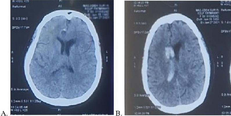
This diagnostic image consists of two axial non-contrast brain CT scans (labeled A and B) demonstrating the progression of an acute cerebrovascular event. Scan A (pre-thrombolysis) shows a subtle area of hypodensity in the left cerebral hemisphere, specifically involving the left external capsule and the anterior horn of the lateral ventricle, consistent with early ischemic infarction. Scan B (post-thrombolysis) reveals a significant change in pathology: high-attenuation (hyperdense) areas are now visible within the ventricular system, particularly in the right lateral and third ventricles. This hyperdensity indicates acute intraventricular hemorrhage (IVH), representing a hemorrhagic transformation following the administration of intravenous tissue plasminogen activator (tPA). The comparison illustrates the potential complication of intracranial bleeding during reperfusion therapy for ischemic stroke. This material is suitable for neurology and radiology education regarding stroke management and associated risks.
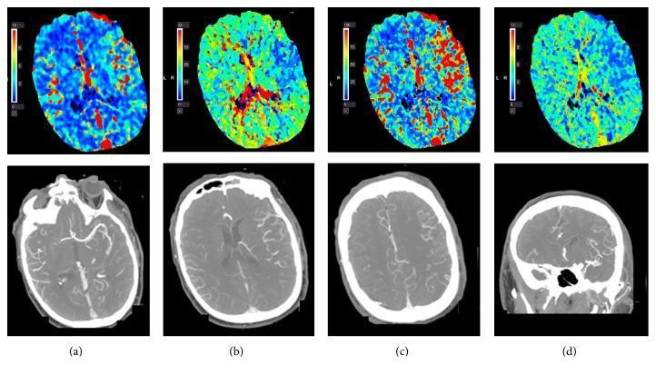
This composite diagnostic image illustrates cerebral hyperperfusion in a 61-year-old patient following intravenous thrombolysis (tPA). The top row consists of four axial Computed Tomography Perfusion (CTP) maps. Subfigure (a) Cerebral Blood Volume (CBV) and (c) Cerebral Blood Flow (CBF) demonstrate increased values in the left middle cerebral artery (MCA) territory, visualized as prominent warm red/orange areas compared to the contralateral side. Subfigure (b) Time to Peak (TTP) and (d) Mean Transit Time (MTT) maps show corresponding areas of shortened transit times, indicated by cooler blue/green hues. The bottom row presents CT Angiogram (CTA) images in axial (a-c) and coronal (d) planes. These images show a distinct dilation of the left MCA and its cortical branches compared to the right side, with marked contrast enhancement. This combination of findings—increased CBF/CBV and shortened transit times alongside arterial dilation—is indicative of post-ischemic hyperperfusion syndrome or reactive hyperemia in the context of acute stroke management. The images are highly relevant for neuroradiology education focusing on vascular imaging and perfusion parameters after reperfusion therapy.
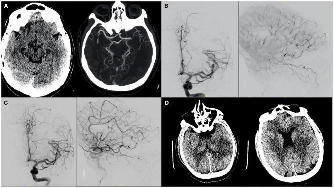
This composite figure displays diagnostic and interventional neuroimaging of a left middle cerebral artery (MCA) ischemic stroke. Panel A consists of a non-contrast axial head CT and CT angiography (CTA) Maximum Intensity Projection (MIP) showing an acute occlusion in the left M1 segment. Panels B and C are Digital Subtraction Angiography (DSA) images in AP and lateral projections. Panel B illustrates the cerebral vasculature after intravenous thrombolysis (IV tPA), showing migration of the thrombus into distal MCA branches with persistent distal filling defects. Panel C shows improved vessel patency and distal perfusion in the left MCA territory following targeted intra-arterial tPA administration. Panel D presents follow-up axial CT scans demonstrating the evolution of the infarct, visualized as regions of decreased parenchymal density (hypodensity) in the left putamen, temporal, and parietal lobes, consistent with established infarction in a large vessel occlusion territory. The series illustrates the clinical pathway from diagnostic imaging to mechanical/pharmacological intervention and final radiologic outcome.
Management of TIA and Stroke
PART 1: TRANSIENT ISCHEMIC ATTACK (TIA)
Definition
Step 1 - Risk Stratification: ABCD² Score
| Risk Factor | Points |
|---|---|
| Age > 60 years | 1 |
| BP > 140/90 mmHg | 1 |
| Clinical: Unilateral weakness | 2 |
| Clinical: Speech disturbance without weakness | 1 |
| Duration 10-59 min | 1 |
| Duration ≥ 60 min | 2 |
| Diabetes | 1 |
- 0-3 = Low risk (1% stroke risk in 48 h)
- 4-5 = Moderate risk (4.1% at 48 h)
- ≥6 = High risk (8% at 48 h; 17-22% at 3 months)
Step 2 - Immediate Evaluation
- Non-contrast CT head - to rule out hemorrhage
- MRI brain (DWI preferred) - detects infarction in TIA mimics
- CT/MR angiography - evaluate carotid and intracranial vessels
- ECG - detect atrial fibrillation
- Echocardiogram - evaluate for cardioembolic source
- Carotid Doppler/imaging
- Labs: CBC, coagulation screen, glucose, lipids, HbA1c - Adams and Victor's Principles of Neurology
Step 3 - Disposition (Admit vs. Outpatient)
- Admit to hospital if: ABCD² ≥ 4, DWI-positive lesion, suspected cardioembolism, high-grade carotid stenosis, or first TIA within 24-48 hours
- Rapid outpatient TIA clinic within 24 hours may be used for low-risk patients - ROSEN's Emergency Medicine
Step 4 - Antithrombotic Therapy
- Dual antiplatelet therapy (DAPT): Aspirin + Clopidogrel started within 24 hours, continued for 21 days, followed by single antiplatelet long-term
- Based on pooled analysis of POINT and CHANCE trials - DAPT reduced major ischemic events vs. aspirin alone
- Major hemorrhage risk increases slightly: 0.2% (aspirin alone) vs. 0.9% (DAPT)
- Alternative: Aspirin + Ticagrelor (180 mg load, then 90 mg BID) showed similar benefit with less genetic heterogeneity in platelet inhibition - Harrison's 22E; ROSEN's Emergency Medicine
Note on CYP2C19: ~30% of people (especially Asians) carry a CYP2C19 loss-of-function variant causing poor clopidogrel metabolism. Ticagrelor avoids this issue. - Harrison's 22E
- Anticoagulation (NOAC preferred; warfarin if mechanical valve)
- Immediate anticoagulation considered except when bleeding risk exceeds benefit or CHA₂DS₂-VASc = 0 (men) / 1 (women) with self-terminating AF - ROSEN's Emergency Medicine
Step 5 - Revascularization
- Carotid endarterectomy (CEA) or carotid artery stenting (CAS) if ipsilateral high-grade carotid stenosis (>70%) is the likely cause
- CEA is most beneficial when performed within 2 weeks of the TIA - Bradley & Daroff's Neurology; Adams & Victor's
Step 6 - Secondary Prevention (Long-term)
- Antiplatelet therapy (single agent after 21 days of DAPT)
- Statin therapy (target LDL < 70 mg/dL)
- Antihypertensive therapy
- Diabetes management (tight glycemic control)
- Smoking cessation, weight reduction, exercise
- Anticoagulation if AF is identified
PART 2: ACUTE ISCHEMIC STROKE
Step 1 - Prehospital Rapid Response
- ABC stabilization; O₂ only if SpO₂ < 95% (avoid routine supplementation in normoxic patients)
- IV access, cardiac monitor
- Avoid dextrose-containing fluids in normoglycemic patients (hyperglycemia worsens ischemia)
- Check blood glucose (hypoglycemia mimics stroke)
- Rapid transport; notify receiving ED early ("stroke alert")
- Target door-to-imaging ≤ 20 minutes - ROSEN's Emergency Medicine
Step 2 - Emergency Department Time Targets (NINDS Guidelines)
| Management Component | Target |
|---|---|
| Door to doctor | ≤ 10 min |
| Door to CT scan | ≤ 25 min |
| Door to CT results | ≤ 45 min |
| Door to drug (tPA) | ≤ 60 min |
| Door to monitored bed | ≤ 3 h |
Step 3 - Immediate Investigations
- Non-contrast CT head (mandatory - rules out hemorrhage before thrombolysis)
- MRI with DWI (if available, more sensitive)
- CT Angiography - identify large vessel occlusion (LVO) for thrombectomy eligibility
- CT Perfusion - assess penumbra (salvageable tissue)
- Labs: CBC, PT/INR, aPTT, glucose, BMP, cardiac enzymes, type & screen
- ECG
- Chest X-ray
Step 4 - Blood Pressure Management
- BP must be ≤ 185/110 mmHg before tPA administration
- Agents to lower BP if needed:
- Labetalol 10-20 mg IV over 1-2 min (may repeat once)
- Nicardipine infusion 5 mg/h (titrate up to 15 mg/h)
- Clevidipine 1-2 mg/h IV
- Monitor BP every 15 min during infusion, then every 30 min for 6 h, then hourly for 16 h
- Target BP < 180/105 mmHg
- Systolic 180-230 or diastolic 105-120: Labetalol 10 mg IV q10-20 min (max 300 mg) or infusion
- Systolic > 230: Labetalol or Nicardipine; consider nitroprusside if uncontrolled
- Permissive hypertension - do not aggressively lower BP unless > 220/120 mmHg (to maintain penumbral perfusion) - ROSEN's Emergency Medicine
Step 5 - Blood Glucose Control
- Treat hyperglycemia to target 140-180 mg/dL
- Avoid hypoglycemia (closely monitor)
- Persistent in-hospital hyperglycemia in the first 24 hours is associated with worse outcomes - ROSEN's Emergency Medicine
Step 6 - IV Thrombolysis (IV Alteplase / tPA)
- Standard: within 3 hours of symptom onset (or last known well)
- Extended: 3-4.5 hours in selected patients
- Hemorrhage on CT
- Symptom onset > 4.5 hours (or unknown onset)
- BP > 185/110 after treatment attempts
- Platelet count < 100,000
- INR > 1.7 or anticoagulant use
- History of prior ICH, intracranial neoplasm
- Recent major surgery (< 14 days) or GI bleed (< 21 days)
- Recent stroke or head trauma (< 3 months)
Tenecteplase (0.25 mg/kg IV, max 25 mg, single bolus) is an emerging alternative - some guidelines now consider it non-inferior to alteplase and easier to administer.
Step 7 - Mechanical Thrombectomy (Endovascular Treatment)
- ICA, M1, M2, basilar artery occlusions
- NIHSS ≥ 6 (functionally significant deficit)
- Pre-stroke mRS 0-1 (good baseline function)
- 0-6 hours: Standard window (based on DAWN, DEFUSE-3 trials)
- 6-24 hours: Extended window in selected patients with favourable imaging (perfusion mismatch)

Step 8 - Antiplatelet Therapy (Non-cardioembolic stroke)
- Start Aspirin 325 mg within 24-48 hours of stroke onset (after ruling out hemorrhage and 24 h after tPA)
- For minor ischemic stroke (NIHSS ≤ 3) not receiving tPA: DAPT (aspirin + clopidogrel) for 21 days, then aspirin alone for long-term
Step 9 - Anticoagulation (Cardioembolic stroke / AF)
- Atrial fibrillation: NOAC preferred (dabigatran, rivaroxaban, apixaban)
- Timing of anticoagulation after stroke is individualized based on stroke size and hemorrhagic risk
- For AF with minor stroke: may start anticoagulation within days; larger strokes: delay 2-4 weeks
Step 10 - Admission and Monitoring
- All significant strokes: admit to stroke unit (evidence shows reduced mortality and dependency)
- ICU monitoring for ≥ 24 h if:
- Large hemispheric stroke (risk of herniation/malignant edema)
- Posterior fossa stroke
- Post-tPA treatment
- Continuous cardiac monitoring for at least 24-48 hours to detect AF
Step 11 - Management of Complications
| Complication | Management |
|---|---|
| Malignant cerebral edema | Osmotherapy (mannitol, hypertonic saline); decompressive hemicraniectomy within 48 h for large MCA territory infarct in patients < 60 yrs |
| Seizures | AEDs only if clinical seizures occur (prophylaxis not recommended) |
| Dysphagia | Screen before oral intake; NG tube if needed |
| DVT/PE prevention | Pneumatic compression stockings; early mobilization; LMWH when safe |
| Fever | Treat aggressively (paracetamol) - fever worsens outcome |
| Aspiration pneumonia | Early mobilization, head-of-bed 30°, swallow screen |
Step 12 - Hemorrhagic Stroke (ICH) Management
- No tPA (contraindicated)
- Strict BP control: Target SBP < 140 mmHg (INTERACT2 trial)
- Reverse any coagulopathy: FFP/PCC for warfarin, idarucizumab for dabigatran, andexanet alfa for factor Xa inhibitors
- Neurosurgical consultation:
- Cerebellar hemorrhage > 3 cm: surgical evacuation (life-saving)
- Hydrocephalus: EVD (external ventricular drain)
- Lobar/basal ganglia ICH: surgical benefit is limited (STICH trials)
- ICP monitoring for large bleeds
- Avoid anticoagulants; restart antiplatelet/anticoagulant therapy only after ≥ 4 weeks based on risk-benefit
Step 13 - Rehabilitation and Secondary Prevention
- Start early mobilization (within 24-48 h if hemodynamically stable)
- Multidisciplinary team: physiotherapy, OT, speech therapy
- Address all vascular risk factors:
- Statins (high-intensity): atorvastatin 40-80 mg daily
- Antihypertensives: ACE inhibitor + thiazide diuretic combination preferred
- Antiplatelet or anticoagulant maintenance
- Diabetes control, smoking cessation, alcohol reduction, diet, exercise
- Evaluate for depression (post-stroke depression is common)
Summary: Key Time Targets
| Intervention | Target Time |
|---|---|
| CT scan | ≤ 25 min from door |
| IV tPA (ischemic stroke) | ≤ 60 min from door |
| Mechanical thrombectomy | ≤ 90 min from door (groin puncture) |
| Antiplatelet therapy (TIA/minor stroke) | Within 24 h |
| Carotid endarterectomy (TIA) | Within 2 weeks of TIA |