
Rash on inner thigh near groin region for a female that comes in hot weather and goes away by applying candid cream. But this time around the same rash is not going away and the skin has peeled a bit from that area which makes it really difficult for the person to walk
tinea cruris groin fungal rash inner thigh
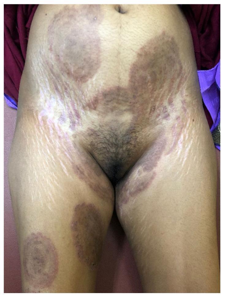
This clinical photograph shows the lower trunk, groin, and upper thighs of a patient exhibiting extensive dermatophytosis and secondary skin changes. Several large, erythematous, and hyperpigmented plaques are visible across the abdomen and inner thighs, characteristic of tinea corporis and tinea cruris. Notably, on the right thigh, a plaque displays a 'tinea pseudoimbricata' pattern, characterized by multiple concentric annular rings. Other areas show 'tinea recidivans,' where active inflammatory margins appear within previously healed sites. Superimposed on these fungal lesions are prominent cutaneous signs of corticosteroid misuse, specifically striking striae albae (white linear stretch marks) and patchy hypopigmentation, particularly dense along the lateral abdomen and inguinal folds. These atrophic changes indicate chronic application of high-potency topical fixed-dose combinations (FDCs). The image demonstrates the complex presentation of steroid-modified tinea (tinea incognita) caused by Trichophyton indotineae, highlighting the visual intersection of recalcitrant fungal infection and iatrogenic skin damage.
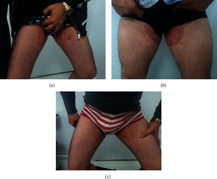
This set of clinical photographs illustrates the progression and treatment of resistant tinea cruris (groin dermatophytosis). Panels (a) and (b) represent the baseline clinical state in two different patients, showing large, well-demarcated, erythematous, and brownish plaques localized to the bilateral groin and upper inner thighs. The lesions exhibit central clearing in some areas but are characterized by active, scaly, and crusted borders, indicative of a persistent fungal infection. Panel (c) provides a comparison chart showing the clinical resolution of the same patients following systemic antifungal therapy with voriconazole. In this follow-up image, the previously visible inflammatory plaques have significantly faded, leaving behind clear skin with a more uniform texture and color, and an absence of active scaling or crusting. This series demonstrates the clinical manifestation of Trichophyton-induced dermatophytosis that was initially resistant to standard terbinafine and fluconazole treatments, and highlights the visual evidence of therapeutic success after second-line antifungal intervention.
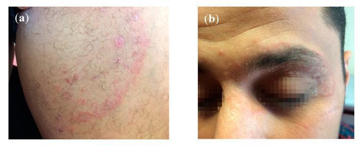
This composite of clinical photographs illustrates a case of multi-focal dermatophytosis caused by T. mentagrophytes Type VIII in a 28-year-old male. Panel (a) displays tinea cruris, characterized by a large, annular (ring-shaped) erythematous lesion on the groin or thigh. The lesion features a prominent, raised, red, and scaly advancing border with evidence of central clearing and smaller adjacent satellite papules. Panel (b) depicts tinea faciei with periocular involvement. An erythematous, scaly, ring-like lesion is visible affecting the left upper and lower eyelids, as well as the supraorbital (eyebrow) region. The plaques demonstrate classic fungal morphology with peripheral inflammation and a dry, flaky texture. These images illustrate the clinical presentation of chronic, recalcitrant dermatophytosis, which may be resistant to standard terbinafine treatment. The case highlights the importance of recognizing fungal skin infections in different anatomical regions (cruris vs. faciei) within the same patient.
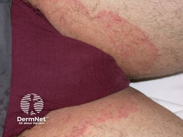
Clinical imaging modality: Digital clinical photography of the groin region showing a dermatophyte infection consistent with tinea cruris. An inguinal plaque is illustrated, presenting as an annular, erythematous lesion bordered by a scaling, active margin with a central area of clearing. The lesion localizes to the upper medial thigh and inguinal fold, with mild adjacent maceration from intertriginous skin. Color and texture emphasize pink to red erythema and peripheral scale; occasional vesiculation may be observed at the advancing border in inflamed areas. The image depicts a unilateral involvement, though tinea cruris can be bilateral in many patients. Surrounding skin shows mild pruritus and scratching. Clinically, this pattern aligns with dermatophyte infection of the groin; differential diagnoses include candidal intertrigo, erythrasma, inverse psoriasis, and contact dermatitis. Diagnostic significance lies in recognizing a common, transmissible fungal dermatosis that typically responds to topical antifungals or oral therapy if extensive. Correlative microbiology (KOH preparation, fungal culture) strengthens confirmation. This photograph is valuable for clinical education on groin anatomy, lesion morphology, and treatment planning, illustrating the classic annular configuration with an active advancing border and central clearing, consistent with tinea cruris and dermatophytosis in intertriginous skin. Educationally, this image supports case-based learning and dermatitis differential.
tinea cruris treatment resistant terbinafine
Trichophyton indotineae India resistant tinea cruris clotrimazole treatment 2024 2025
Tinea Cruris with Treatment Failure - What's Happening and What to Do
What This Most Likely Is
- Appears in hot/humid weather (heat + sweat = ideal fungal environment)
- Inner thigh near the groin
- Responds to antifungal cream (Candid = clotrimazole)
- Recurring and now not responding - this is a significant warning sign
Why It's Not Going Away This Time
- Clotrimazole and other azoles (like in Candid cream)
- Terbinafine (the standard oral antifungal)
- Sometimes even itraconazole
- Andrews' Diseases of the Skin, p. 15 (block4)
- Dermatology 2-Volume Set 5e, p. 1606
Differential Diagnoses to Keep in Mind
| Condition | Distinguishing Feature |
|---|---|
| Erythrasma | Copper-brown color, coral-red fluorescence under Wood's lamp |
| Candidal intertrigo | Moist, satellite pustules, involves skin folds deeply, may involve labia |
| Inverse psoriasis | Well-defined, non-scaly plaques, may have psoriasis elsewhere |
| Seborrheic dermatitis | Also involves chest/axillae |
| Contact dermatitis | History of new soap/detergent/pad use |
What Should Be Done Now
Immediate Steps:
- Stop Candid cream alone - it is likely insufficient at this point
- Keep the area dry - loose cotton underwear, dry thoroughly after bathing, avoid synthetic fabrics
- Do NOT use any cream that contains a steroid (check for betamethasone, clobetasol, dexamethasone in the ingredient list - avoid Quadriderm, Panderm, Lobate-GM, etc.)
Medical Workup the Dermatologist Will Do:
- KOH (potassium hydroxide) scraping - confirms fungal infection under microscope
- Fungal culture - identifies the species and tests drug sensitivity (especially important for T. indotineae)
- Wood's lamp exam - rules out erythrasma
Treatment the Doctor May Prescribe:
- Switch from clotrimazole to luliconazole 1% cream or sertaconazole - newer azoles with better activity
- Or terbinafine cream (allylamine class - fungicidal, not just fungistatic, faster acting)
- Itraconazole 100-200 mg/day for 2-4 weeks (preferred if T. indotineae suspected)
- Terbinafine 250 mg/day for 2-4 weeks (but may fail if T. indotineae)
- Voriconazole has been used in severe resistant cases
- Goodman & Gilman's Pharmacological Basis of Therapeutics, p. 1506
- Textbook of Family Medicine 9e, p. (block11)
Preventive Measures (After Cure)
- Wash and dry the groin area thoroughly after sweating or bathing
- Wear breathable, loose cotton underwear; change daily
- Dust with antifungal powder (like Abzorb or Candid Dusting Powder) in summer months
- Treat any concurrent athlete's foot (tinea pedis) - it's a common source of re-infection spread through clothing
- Do not share towels, clothing, or bedding
- Weight loss if overweight (skin folds trap moisture)