
Classification of pancreatic tumors radiology easily
Reading File
Now let me search the medical library and web simultaneously for pancreatic tumor classification from a radiology perspective.
Finding Sources
Finding Sources
Searching the Web
pancreatic tumors classification radiology CT MRI features easy summary 2024
Finding Sources
Reading Web Page
https://radiopaedia.org/articles/pancreatic-neoplasms?lang=u…
Finding Sources
Finding Sources
Reading File
Finding Sources
Reading File
Reading File
Reading File
Reading File
I now have rich material from the textbooks. Let me also fetch a couple of the key images and get information on solid tumors (PDAC, PNETs) from another section.
Reading File
Loading Image
Loading Image
I now have comprehensive material. Let me compile the full answer with all the key radiology-oriented classification details.
Classification of Pancreatic Tumors - Radiology Made Easy
The big-picture split is solid vs. cystic, then you refine by vascular behavior on CT (hypo- vs. hypervascular) and cyst morphology (micro- vs. macrocystic, communicating vs. non-communicating duct). Here is the full framework.
Step 1 - The Master Split
PANCREATIC TUMORS
├── SOLID
│   ├── Hypovascular (most common)
│   │   ├── Pancreatic Ductal Adenocarcinoma (PDAC) ← 85% of all
│   │   └── Acinar Cell Carcinoma (ACC)
│   └── Hypervascular
│       └── Pancreatic Neuroendocrine Tumor (pNET)
│           (also: metastases from RCC, HCC, melanoma)
└── CYSTIC
    ├── Benign (almost always)
    │   └── Serous Cystadenoma (SCA)
    ├── Premalignant / Mucinous
    │   ├── Mucinous Cystic Neoplasm (MCN)
    │   └── IPMN (main-duct / branch-duct / mixed)
    └── Mixed Solid-Cystic
        └── Solid Pseudopapillary Neoplasm (SPN)
SOLID TUMORS
1. Pancreatic Ductal Adenocarcinoma (PDAC) - 85%
| Feature | Finding |
|---|---|
| CT appearance | Hypodense/hypoattenuating mass - arterial AND portal venous phase (due to desmoplastic stroma = hypovascular) |
| Margins | Ill-defined, infiltrative |
| Location | 70% pancreatic head |
| Secondary signs | Pancreatic duct dilation ("double duct sign" = dilated CBD + PD), upstream pancreatic atrophy, vascular encasement (SMA, celiac, portal/SMV) |
| Modality of choice | Multiphasic CT (pancreatic protocol); MRI/MRCP for small tumors not visible on CT |
| Key sign | Double duct sign = dilated common bile duct + dilated pancreatic duct; strongly suggests head PDAC |
"Typical imaging features of pancreatic cancer on dynamic CT include a hypoattenuating mass compared with the adjacent pancreatic parenchyma in the late arterial and portal venous phases due to decreased vascularity within the tumoral tissue and desmoplastic stroma." - Current Surgical Therapy 14e
Resectability on CT (Varadhachary/Katz System):
- Resectable: no vascular contact, no distant mets
- Borderline resectable: <180° contact with SMA/CA, short segment SMV/PV involvement
- Unresectable: >180° SMA/celiac encasement, solid aortic contact, distant metastases
2. Pancreatic Neuroendocrine Tumors (pNETs)
| Feature | Finding |
|---|---|
| CT appearance | Hypervascular - bright arterial enhancement (opposite of PDAC) |
| Location | Any part; functional tumors tend to be small |
| Functional vs. non-functional | Functioning = insulinoma, gastrinoma, VIPoma, glucagonoma; Non-functioning = usually large at diagnosis |
| Insulinoma | Smallest, most common functional pNET; usually benign; intense arterial blush |
| Gastrinoma | May be in "gastrinoma triangle"; associated with MEN-1 |
| WHO 2017 Grade | G1 (Ki-67 <3%), G2 (3-20%), G3 (>20%), NEC (poorly diff.) |
CYSTIC TUMORS
Quick Memory Table
| Feature | SCA | MCN | IPMN | SPN |
|---|---|---|---|---|
| Who gets it | Older F (postmenopausal) | Perimenopausal F | Older M or F | Young F (<40) |
| Location | Anywhere | Body/tail | Head (main-duct); Anywhere (branch) | Body/tail |
| CT/MRI appearance | Microcystic honeycomb; central scar/calcification | Macro-cystic, unilocular/multilocular; NO duct communication | Duct dilation (main-duct) or "grape-like" cluster (branch) | Mixed solid + cystic; hemorrhagic |
| Malignant potential | None (nearly) | Yes (5-15%) | Yes (main > branch) | Low (15%) |
| Key differentiator | Honeycomb + low CEA | Ovarian stroma; elevated CEA; does NOT communicate with PD | Communicates with PD (MRCP key!) | Mixed solid/cystic in young woman |
| Management | Observe if <4 cm, asymptomatic | All resect (lifetime malignancy risk) | Main-duct: resect; Branch-duct: Fukuoka criteria | Resect (curative) |
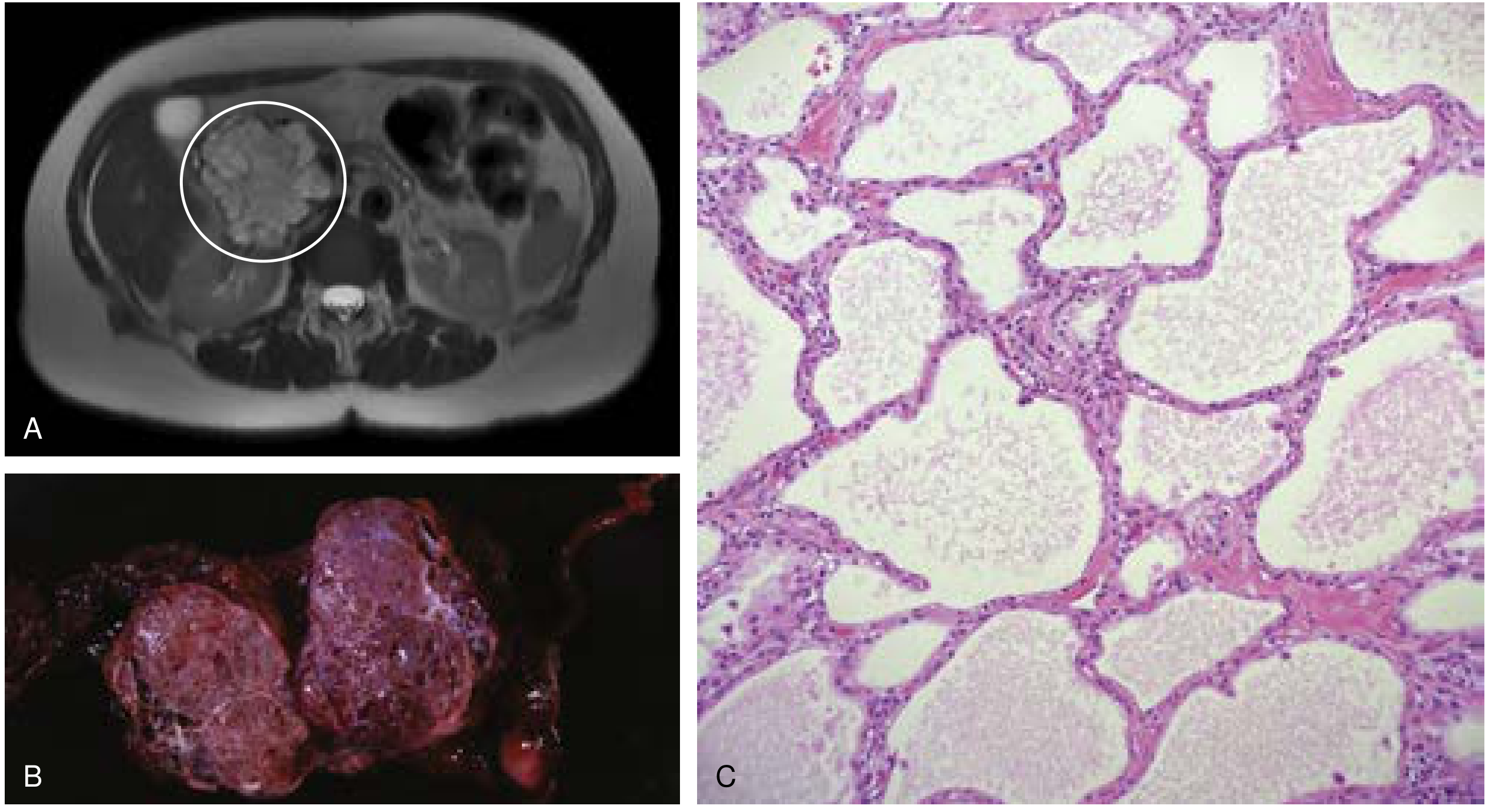
3. Serous Cystadenoma (SCA) - Benign
- Accounts for 33% of pancreatic cysts; almost universally benign
- CT/MRI: classic "honeycomb" or microcystic pattern (many tiny cysts <2 cm each) with a central stellate scar and sunburst calcification
- Fluid: very low CEA (<5 ng/mL) and low amylase - this is diagnostic
- Associated with Von Hippel-Lindau syndrome (lifetime risk ~40%)
4. Mucinous Cystic Neoplasm (MCN) - Premalignant
- 25% of pancreatic cysts; almost exclusively perimenopausal females
- Located in body/tail
- CT/MRI: well-encapsulated, unilocular or multilocular cyst; does NOT communicate with pancreatic duct (important!)
- Elevated CEA in cyst fluid; elevated CEA + low amylase = MCN
- Malignancy risk: 5-15%; red flags = mural nodule, solid component, calcifications, wall thickening
- Path hallmark: ovarian-type stroma underneath the epithelium (required for diagnosis)
5. IPMN (Intraductal Papillary Mucinous Neoplasm)
Three subtypes by location:
| Type | Imaging | Malignancy Risk | Action |
|---|---|---|---|
| Main-duct IPMN | Diffuse or segmental PD dilation (>5 mm) | High (60-70%) | Resect |
| Branch-duct IPMN | "Cluster of grapes" cysts; communicates with PD on MRCP | Lower (~25%) | Fukuoka guidelines (observe vs. resect) |
| Mixed | Both features | High | Resect |
- Key test: MRCP - shows communication between cyst and pancreatic duct (pathognomonic for IPMN vs. MCN)
- Worrisome features (Fukuoka): mural nodule, thickened/enhancing walls, PD 5-9 mm, abrupt PD caliber change
- High-risk stigmata: obstructive jaundice, enhancing mural nodule >5 mm, PD ≥10 mm - resect
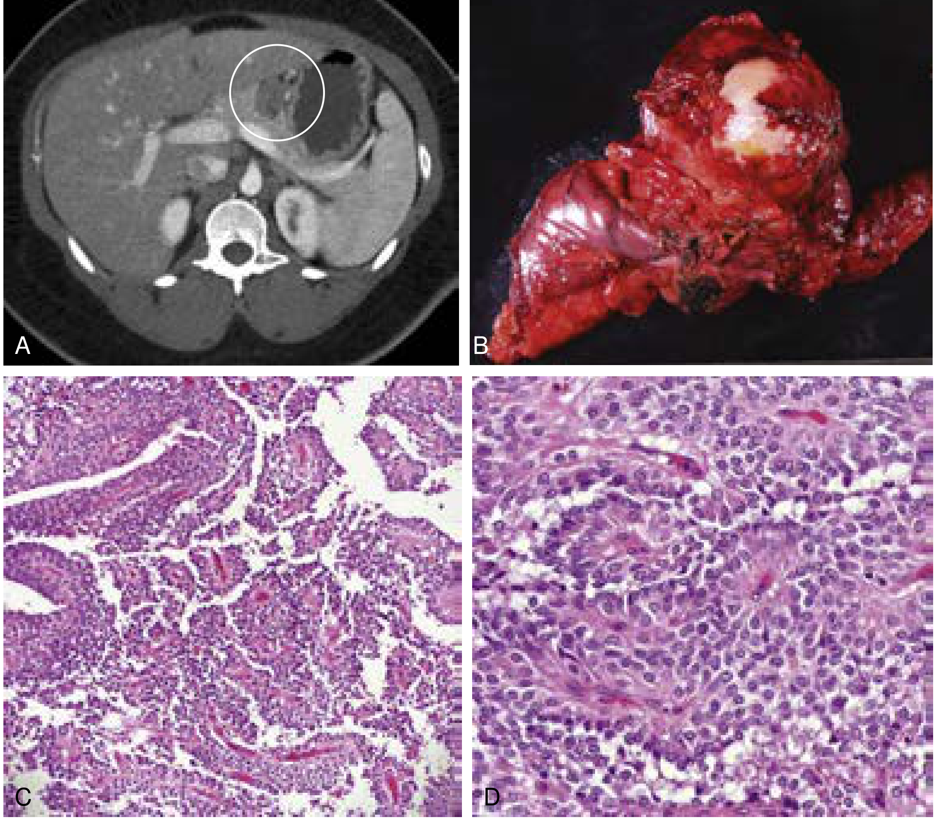
6. Solid Pseudopapillary Neoplasm (SPN) - Low Malignant Potential
- Young females (~20s-30s); rare (5% of resected cysts; 1% of all pancreatic tumors)
- CT: mixed solid + cystic mass; hypoenhancing; often hemorrhagic with fluid-fluid levels; well-circumscribed
- MRI: heterogeneous T1/T2 signal due to hemorrhage; fibrous capsule
- Pathognomonic: "floating debris" from internal hemorrhage and necrosis
- WNT/beta-catenin pathway mutations (90%)
- Management: resection is usually curative; ~4.4% recurrence rate
Rare Solid Tumors (Know for Completeness)
| Tumor | Key Radiology Clue |
|---|---|
| Acinar Cell Carcinoma (ACC) | Large exophytic solid mass; 1-2% of pancreatic tumors; males; can be cystic variant |
| Pancreatoblastoma (PBA) | Children; solid mass with calcifications; pancreatic head; elevated AFP; associated with Beckwith-Wiedemann |
| PEComa | Hypoenhancing mass, well-circumscribed; HMB-45 positive |
| Lymphoma | Bulky, homogeneous, hypovascular mass; no duct obstruction despite large size ("silent" obstruction) |
| Metastases | Most common from RCC (hypervascular), lung, melanoma; may be multiple |
The "Radiology First Look" Decision Tree
Is it CYSTIC or SOLID?
│
├── SOLID
│   └── Is it HYPERVASCULAR on arterial phase?
│       ├── YES → pNET (also consider RCC met)
│       └── NO (hypodense) → PDAC (most likely)
│
└── CYSTIC
    ├── Microcystic/honeycomb → SCA (benign, observe)
    ├── Macrocystic, body/tail, no duct comm., F only → MCN (resect all)
    ├── Communicates with pancreatic duct (MRCP) → IPMN
    │       └── Main duct dilated? → High risk (resect)
    │       └── Branch-duct? → Fukuoka criteria
    └── Mixed solid/cystic, young woman → SPN (resect)
Key Imaging Protocols
- Pancreatic protocol CT: Non-contrast + arterial phase (35-40 s) + portal venous phase (70 s) + sometimes delayed. Arterial phase is key for hypervascular tumors (pNET) and for vascular involvement assessment
- MRI/MRCP: Best for cystic lesions, duct communication, and small tumors missed on CT
- EUS + FNA: For cyst fluid analysis (CEA, amylase, cytology) when CT/MRI are equivocal
Sources: Current Surgical Therapy 14e; Sleisenger and Fordtran's Gastrointestinal and Liver Disease; Maingot's Abdominal Operations; Harrison's Principles of Internal Medicine 22E (2025)
This is a shared conversation. Sign in to Orris to start your own chat.