

26169.jpg
Explain all these thoroughly for first year mbbs
Reading File
Now let me search the medical library for all 5 topics in parallel.
Finding Sources
Finding Sources
Finding Sources
Finding Sources
Finding Sources
Finding Sources
Now let me read the relevant sections in parallel:
Reading File
Reading File
Reading File
Reading File
Reading File
Reading File
Reading File
Reading File
Reading File
Now let me fetch the key diagrams and get the properties of synapse content from Guyton:
Loading Image
Loading Image
Loading Image
Now let me get the synaptic properties content and the nerve regeneration image:
Reading File
Loading Image
I now have excellent content from the textbooks. Let me compile the comprehensive answer for the 1st year MBBS student.
SGD Topics - 1st Year MBBS Physiology
Sahiwal Medical College | Tutor: Dr. Aroona
1. Properties of Synapse
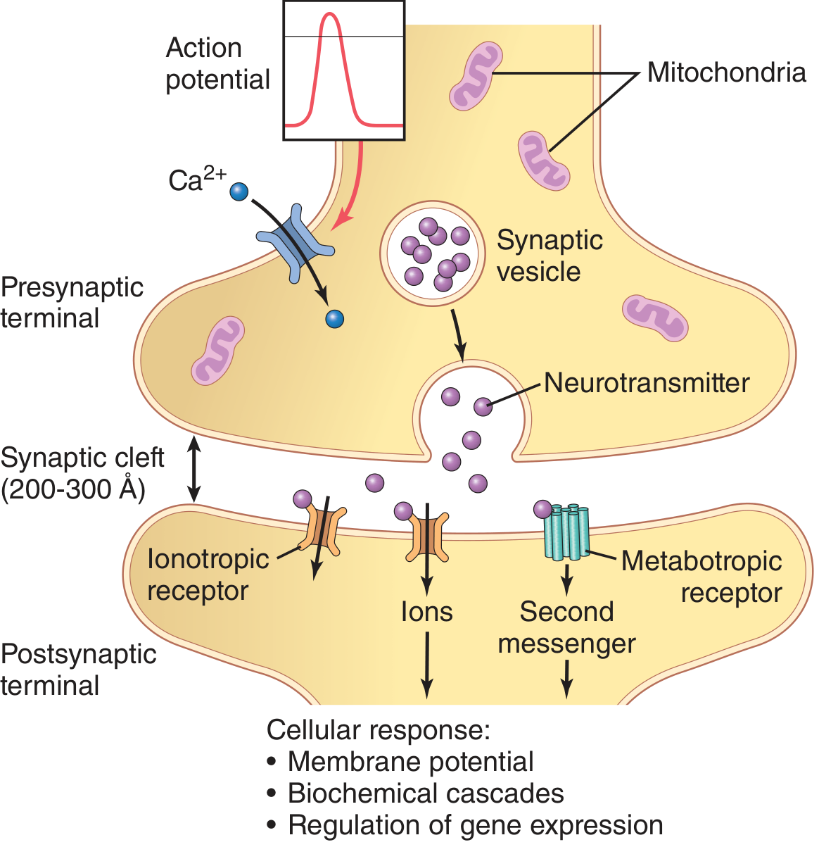
A synapse is the specialized junction between two neurons (or between a neuron and an effector) where information is transmitted. It has two sides: the presynaptic terminal (sending side) and the postsynaptic membrane (receiving side), separated by a synaptic cleft of 200-300 Angstroms (Å).
Types of Synapses
| Feature | Chemical Synapse | Electrical Synapse |
|---|---|---|
| Transmission medium | Neurotransmitter | Gap junctions (ion current) |
| Direction | One-way only | Bidirectional |
| Synaptic cleft | 200-300 Å | Very narrow / absent |
| Speed | Slight delay (1ms) | Instantaneous |
| Main role | Most CNS synapses | Synchronizing neuronal groups |
Key Properties of Chemical Synapses
1. One-way (Unidirectional) Transmission
Chemical synapses only transmit from the presynaptic to the postsynaptic neuron. This is because only the presynaptic terminal contains synaptic vesicles and only the postsynaptic membrane has receptors. This directionality allows precise signal routing.
2. Synaptic Delay
There is a delay of about 0.5 ms at each synapse. This time accounts for: Ca²⁺ entry into the terminal → vesicle docking → exocytosis → neurotransmitter diffusion → receptor binding. Pathways with many synapses have longer total delays.
3. Summation (Spatial and Temporal)
A single presynaptic terminal discharge does NOT produce enough EPSP to fire the postsynaptic neuron. About 40-80 terminals must fire simultaneously. There are two types:
- Spatial summation: Multiple presynaptic terminals fire at the same time
- Temporal summation: A single terminal fires in rapid succession, building up EPSP
4. Excitatory Postsynaptic Potential (EPSP)
When an excitatory transmitter (e.g., glutamate) binds, Na⁺ rushes in, raising the membrane potential from -65 mV toward 0 mV (to about -45 mV). If EPSP reaches the threshold at the axon hillock (initial segment), an action potential fires. The axon hillock has 7x more voltage-gated Na⁺ channels than the soma, making it the site of AP generation.
5. Inhibitory Postsynaptic Potential (IPSP)
Inhibitory transmitters (e.g., GABA, glycine) open K⁺ or Cl⁻ channels, making the membrane MORE negative (e.g., -65 → -70 mV) - called hyperpolarization. This makes it harder to reach firing threshold.
6. Fatigue
With repeated high-frequency stimulation, the synapse fatigues - synaptic vesicles deplete, the postsynaptic response diminishes. This is a protective mechanism preventing excessive nervous system stimulation.
7. Effect of Calcium
Ca²⁺ entry through voltage-gated calcium channels in the presynaptic terminal is absolutely necessary for vesicle exocytosis. Without Ca²⁺, no neurotransmitter is released. The SNARE protein complex (synaptobrevin, syntaxin, SNAP-25) mediates vesicle docking and fusion.
8. Neurotransmitters
- Small-molecule, fast-acting: Acetylcholine, glutamate, GABA, glycine, dopamine, norepinephrine, serotonin
- Neuropeptides, slow-acting: Substance P, enkephalins, endorphins (cause longer-lasting effects, act on second messenger systems)
9. Post-tetanic Potentiation
After a burst (tetanus) of presynaptic stimulation, subsequent stimuli produce a larger than normal postsynaptic response for a period - due to residual Ca²⁺ in the presynaptic terminal.
10. Sensitization and Habituation
Synaptic responses can increase (sensitization) or decrease (habituation) with repeated stimulation - this forms the cellular basis of learning and memory.
2. Axonal Transport
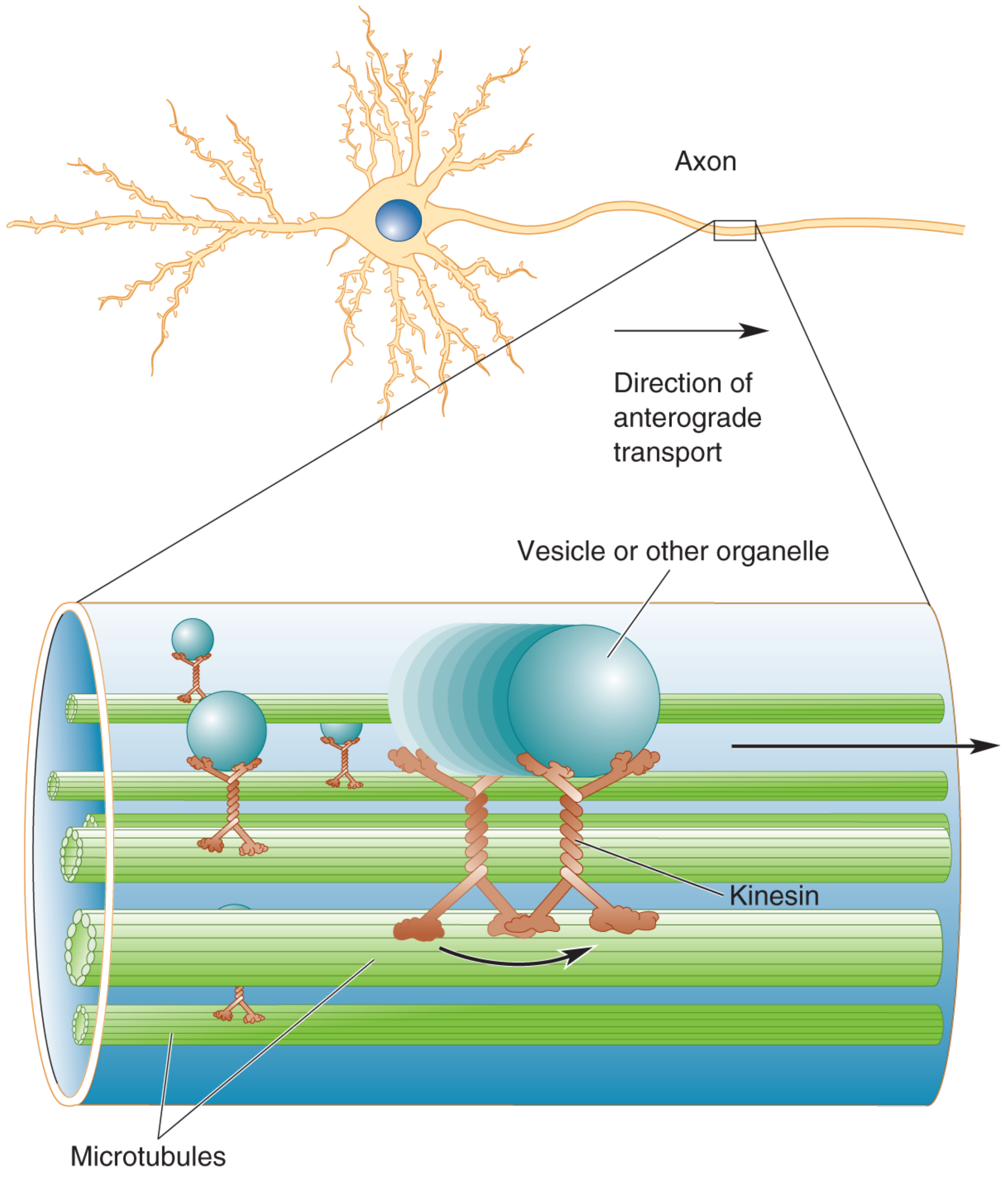
The cell body (soma) is the metabolic engine of the neuron. All proteins are synthesized there (since axons lack ribosomes). Yet the axon terminal can be 1 meter or more away - so a transport system is needed to ship proteins down (and return signals up).
Types of Axonal Transport
| Type | Direction | Speed | Motor Protein | What is Carried |
|---|---|---|---|---|
| Fast anterograde | Soma → Terminal | ~400 mm/day | Kinesin | Synaptic vesicles, membrane organelles, enzymes |
| Slow anterograde | Soma → Terminal | 0.5-10 mm/day | Kinesin | Cytoskeletal proteins (actin, tubulin), metabolic enzymes |
| Retrograde | Terminal → Soma | ~200 mm/day | Dynein | Used vesicles, NGF, viral particles, toxins |
Mechanism
- The axon contains microtubules running along its length like railway tracks
- Kinesin is the motor protein for anterograde transport - it "walks" toward the (+) end of microtubules (toward the terminal), using ATP
- Dynein is the motor protein for retrograde transport - it "walks" toward the (-) end (toward the soma), also using ATP
- Material is packaged in membrane-bound vesicles
Clinical Importance
- Herpes zoster and polio virus travel by retrograde transport to reach the cell body
- Tetanus toxin travels retrogradely to the spinal cord
- Nerve Growth Factor (NGF) travels retrogradely to signal the soma about conditions at the terminal
- Interrupting axonal transport (as in axonal injury) leads to Wallerian degeneration (see below)
- This is why the cell body maintains the functional integrity of the entire axon
Source: Ganong's Review of Medical Physiology, 26th Ed.; Neuroscience: Exploring the Brain, 5th Ed.
3. Nerve Injury
Peripheral nerve injuries are classified by the Seddon Classification (clinical) and the Sunderland Classification (anatomical):
Seddon Classification
| Grade | Name | What is Damaged | Recovery |
|---|---|---|---|
| 1st | Neurapraxia | Myelin only (conduction block) | Full, within weeks |
| 2nd | Axonotmesis | Axon cut, endoneurium intact | Good, Wallerian degeneration occurs |
| 3rd | Neurotmesis | Entire nerve (including connective tissue) severed | Poor without surgery |
Sunderland Classification
| Grade | Injury | Recovery |
|---|---|---|
| I | Focal demyelination (=neurapraxia) | Complete |
| II | Axon + myelin disrupted, endoneurium intact (=axonotmesis) | Good |
| III | Axon, myelin, endoneurium disrupted; perineurium intact | Variable |
| IV | Only epineurium intact | Poor |
| V | Complete severance (=neurotmesis) | None without surgery |
Segmental Demyelination (Grade I / Neurapraxia)
- Only a focal segment of the myelin sheath is damaged
- The axon itself is intact
- Results in slowing of conduction velocity or conduction block across that segment
- Recovery occurs by remyelination within weeks to months
- New myelin is thinner with more internodes
4. Wallerian Degeneration
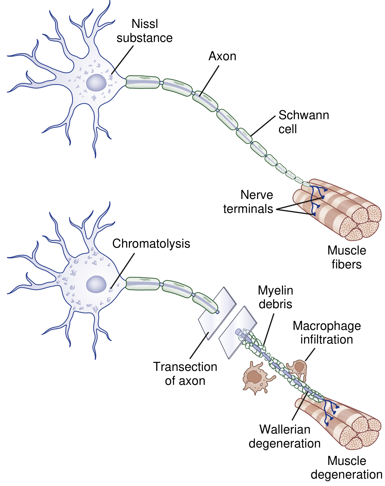
Named after Augustus Waller (1850), who observed that cutting a nerve causes degeneration of the segment distal to the cut.
What Triggers It?
Disruption of normal anterograde axonal transport from the soma to the distal axon. Without the soma's supply, the distal axon degenerates.
Changes Distal to the Injury (Days 1-7)
- Day 1-2: Axon becomes irregular and granular; rapid influx of Ca²⁺ and Na⁺ through the disrupted membrane activates calcium-dependent proteases
- Day 3: Schwann cells retract from nodes of Ranvier; activated Schwann cells and macrophages begin digesting the myelin - forming myelin ovoids
- Day 7: The entire distal axon degenerates; macrophages (recruited by cytokines) phagocytose myelin debris - this entire process takes approximately 1 week
- After cleanup: Schwann cells proliferate and form Bands of Büngner - longitudinal tubes that serve as guides for regenerating axons
Changes Proximal to the Injury
- A limited degree of axon breakdown extends proximally up to the first node of Ranvier
- The cell body undergoes CHROMATOLYSIS:
- Nissl substance (rough endoplasmic reticulum) breaks up and disperses
- Cell nucleus shifts to an eccentric (peripheral) position
- Cell body swells
- This represents a metabolic shift from axon maintenance to protein synthesis - preparing for regeneration
- In very proximal injuries (e.g., root avulsion), the cell body itself may undergo apoptosis
Changes at the Target Organ
- If the nerve does not regenerate in time, the denervated muscle undergoes atrophy and collagen deposition in endomysium/perimysium
- Target organs must remain viable for successful reinnervation
5. Nerve Regeneration
How Regeneration Begins
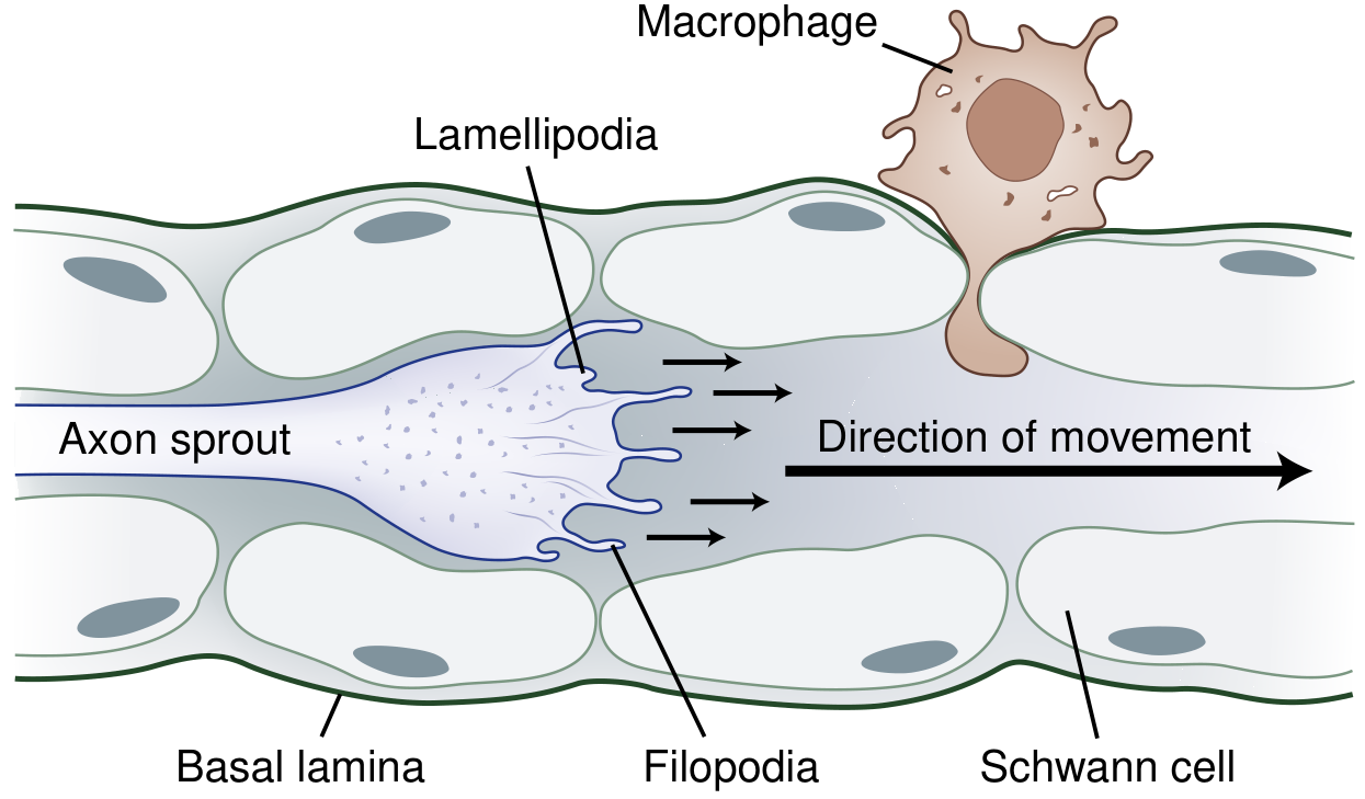
After Wallerian degeneration clears the distal segment, the proximal stump sprouts new axon buds. This process depends on the type of injury:
1. Remyelination (Grade I / Focal Demyelination Only)
- Schwann cells divide and remyelinate the damaged segment
- Recovery of function within weeks to months
- New myelin is thinner with more internodes per original internode
2. Collateral Sprouting (Partial Nerve Injury)
- When some (not all) axons to a muscle are damaged
- Intact motor axons produce sprouts from:
- Nodes of Ranvier → nodal sprouts
- Nerve terminals → terminal sprouts (as early as 4 days after injury)
- These sprouts adopt denervated muscle fibers, increasing motor unit size
- Clinical recovery takes 3-6 months
3. Proximal-to-Distal Regeneration (Severe/Complete Injury, Grades II-V)
The step-by-step sequence:
- Axon sprouting from the proximal stump begins (stimulated by chromatolysis)
- A growth cone forms at the tip of each sprout - it has:
- Filopodia (finger-like projections that sense the environment)
- Lamellipodia (sheet-like projections)
- The growth cone is guided through Bands of Büngner (the tunnels left by Schwann cells in the distal stump) toward the target
- Neurotropism: Guidance molecules (semaphorins, ephrins, netrins, slits) attract or repel the growth cone
- Neurotrophic factors that promote regeneration: NGF, BDNF, NT-3, NT-4, GDNF, CNTF, FGF, IGF
- Growth cone secretes plasminogen activators to dissolve cellular debris blocking the path
Rate of Regeneration
- Proximal lesions: 2-3 mm/day
- Distal lesions: ~1 mm/day
- Average: ~1 inch (~25 mm) per month (classic teaching point)
Factors Affecting Regeneration
| Factor | Effect |
|---|---|
| Intact endoneurial tube | Improves success significantly |
| Disrupted endoneurial tube | Axons may stray, forming neuroma (source of neuropathic pain) |
| Delay >4 months to cross gap | Distal tube shrinks to <3 μm, impedes recovery |
| Age | Younger patients regenerate better |
| Proximal vs distal injury | Proximal injuries have poorer outcome (longer time to target) |
| Denervated muscle >2 years | Irreversible fibrosis, no reinnervation possible |
Clinical Note on Neuroma
When regenerating axons stray into surrounding connective tissue (due to disrupted endoneurium), they form a disorganized tangle called a neuroma. Sodium channels accumulate at this site, generating ectopic firing and neuropathic pain.
Summary Table for Quick Revision
| Topic | Key Numbers / Points to Remember |
|---|---|
| Synaptic cleft width | 200-300 Å |
| Presynaptic terminals on motor neuron | 10,000-200,000 |
| Terminals needed for EPSP→AP | 40-80 |
| Synaptic delay | ~0.5 ms |
| Fast anterograde transport | ~400 mm/day; motor: kinesin |
| Slow anterograde transport | 0.5-10 mm/day |
| Retrograde transport | ~200 mm/day; motor: dynein |
| Wallerian degeneration completes | ~1 week distal to injury |
| Chromatolysis | Cell body response to axon injury |
| Collateral sprouting onset | 4 days after injury |
| Nerve regeneration rate | 1-2 mm/day (~1 inch/month) |
| Max time for reinnervation | ~2 years |
Sources: Guyton & Hall Textbook of Medical Physiology; Ganong's Review of Medical Physiology, 26th Ed.; Neuroscience: Exploring the Brain, 5th Ed.; Bradley & Daroff's Neurology in Clinical Practice
This is a shared conversation. Sign in to Orris to start your own chat.