
A-a gradient
Reading File
Quick answer: The A-a (alveolar-arterial) gradient is the difference between the partial pressure of oxygen in the alveoli (PAO2) and in the arterial blood (PaO2). It reflects how efficiently the lungs transfer oxygen from alveolar gas to pulmonary capillary blood. A normal gradient is roughly 5-15 mmHg in a young healthy adult (increases with age). An elevated gradient points to a V/Q mismatch, diffusion impairment, or intrapulmonary shunt as the cause of hypoxemia - rather than hypoventilation alone.
Searching sources...
Finding Sources
Finding Sources
Reading File
Reading File
Reading File
Reading File
Reading File
Loading Image
Loading Image
Reading File
Loading Image
Reading File
Reading File
I now have comprehensive information from multiple authoritative sources. Let me put together a thorough answer.
Alveolar-Arterial (A-a) Gradient
Definition
The A-a gradient is the difference between the partial pressure of oxygen in the alveoli (PAO2) and the partial pressure of oxygen in arterial blood (PaO2):
A-a gradient = PAO2 - PaO2
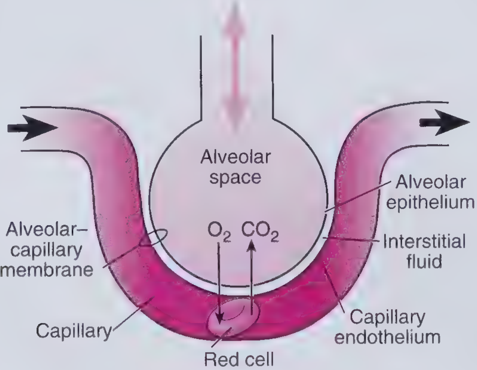
It quantifies how efficiently gas transfer occurs across the alveolar-capillary membrane. In a perfect lung, alveolar and arterial PO2 would be equal and the gradient would be zero. In reality, even healthy lungs have a small gradient due to physiological V/Q mismatch and a tiny anatomical shunt through the bronchial and thebesian veins.
How to Calculate It
Step 1 - Calculate PAO2 using the Alveolar Gas Equation
Full form:
PAO2 = (FiO2 × [PB - PH2O]) - (PaCO2 / R)
Where:
- FiO2 = fraction of inspired oxygen (0.21 on room air)
- PB = barometric pressure (760 mmHg at sea level)
- PH2O = water vapor pressure at body temperature (47 mmHg)
- PaCO2 = arterial CO2 (assumed equal to alveolar CO2)
- R = respiratory exchange ratio (typically 0.8)
Simplified at sea level on room air:
PAO2 = 150 - (1.25 × PaCO2)
Step 2 - Obtain PaO2 from the arterial blood gas
Step 3 - Subtract
A-a gradient = PAO2 - PaO2
Example: PaCO2 = 40 mmHg → PAO2 = 150 - 50 = 100 mmHg. If PaO2 = 95 mmHg, the A-a gradient = 5 mmHg (normal).
- Comprehensive Clinical Nephrology, 7th ed.
- Medical Physiology (Boron & Boulpaep)
Normal Values
The normal A-a gradient increases with age and with increasing FiO2.
Upper limits of normal at room air by age (from Frameworks for Internal Medicine):
| Age (years) | A-a gradient (mmHg) |
|---|---|
| 20 | 17 |
| 30 | 21 |
| 40 | 24 |
| 50 | 27 |
| 60 | 31 |
| 70 | 34 |
| 80 | 38 |
A commonly used formula for the age-adjusted upper limit:
Normal A-a gradient ≈ (Age + 10) / 4
This gives a range of roughly 5-25 mmHg across adult life. The gradient also rises by 5-7 mmHg for every 10% increase in FiO2.
Why the A-a Gradient Matters: Diagnosing the Cause of Hypoxemia
The A-a gradient is the single most useful step in narrowing down the mechanism of hypoxemia.
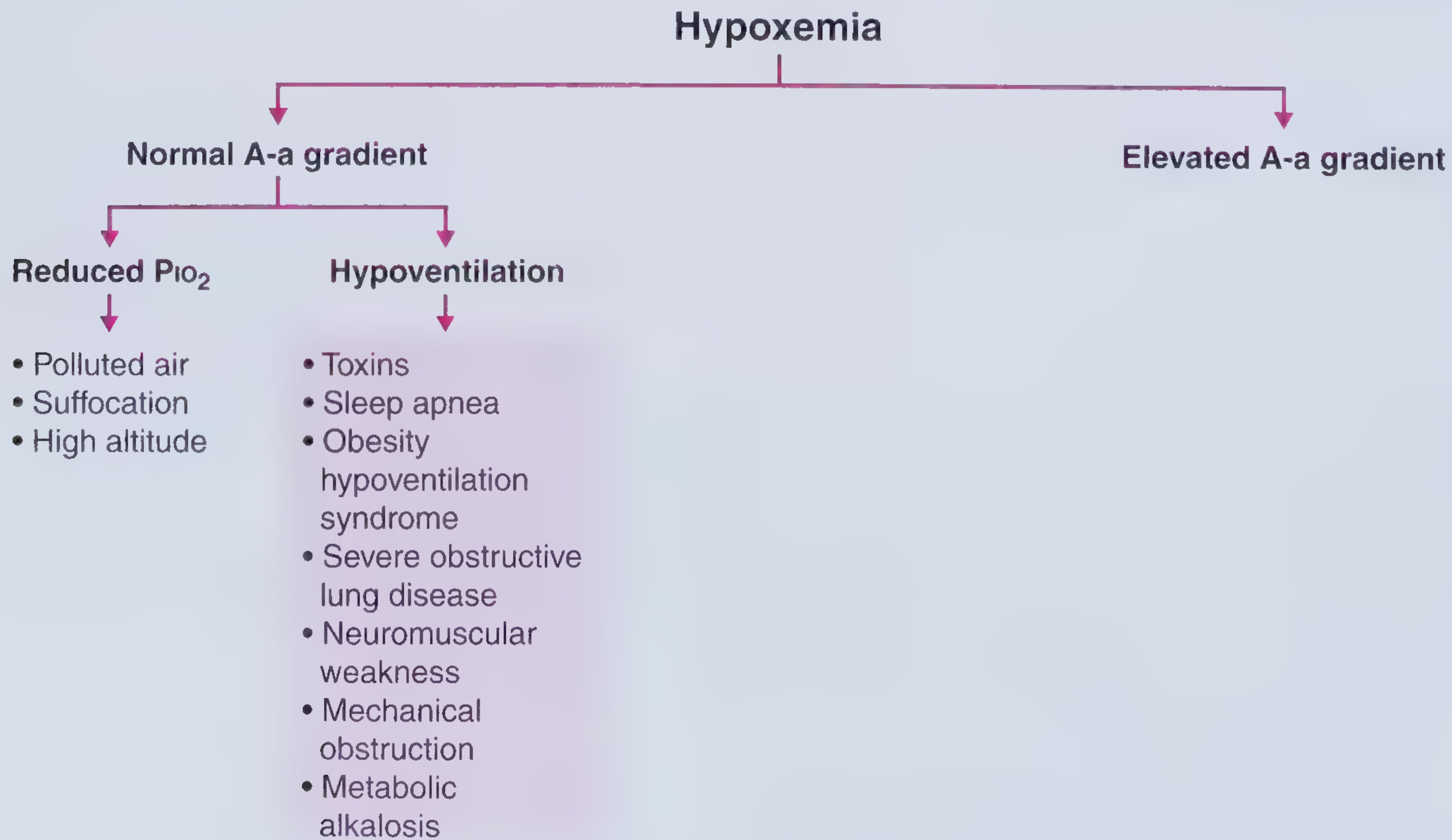
Normal A-a Gradient with Hypoxemia
The problem is "upstream" of the lungs - the lungs are transferring oxygen normally, but there is simply less oxygen to work with.
1. Reduced PiO2 (inspired oxygen)
- High altitude (lower barometric pressure, PiO2 falls)
- Enclosed/poorly ventilated spaces, suffocation
- Low FiO2 environments
2. Hypoventilation (elevated PaCO2 displaces alveolar O2)
- Narcotic/benzodiazepine toxicity
- Obstructive sleep apnea
- Obesity hypoventilation syndrome (Pickwickian syndrome)
- Neuromuscular weakness (e.g., Guillain-Barré syndrome)
- Kyphoscoliosis
- Metabolic alkalosis (compensatory)
- Severe obstructive lung disease
Key point: in pure hypoventilation, the A-a gradient is normal because the lungs are working fine - there is just less alveolar O2 to exchange. Supplemental O2 corrects this effectively.
Elevated A-a Gradient with Hypoxemia
The problem is in the lungs themselves - impaired transfer of O2 across the alveolar-capillary interface.
1. V/Q Mismatch
- Regions with low V/Q (perfusion > ventilation): COPD, asthma, pneumonia, pulmonary edema
- Regions with high V/Q (ventilation > perfusion / dead space): pulmonary embolism, pulmonary hypertension
- The most common cause of an elevated A-a gradient overall
- Responds to supplemental O2
2. Right-to-Left Shunt (true shunt)
- Intrapulmonary: consolidated/atelectatic lung, ARDS, large AVM
- Intracardiac: ASD/VSD with right-to-left flow, patent foramen ovale (especially with elevated right-sided pressures from PE)
- Blood completely bypasses ventilated alveoli
- Hallmark: does NOT correct with 100% O2 (PaO2 fails to rise above ~500 mmHg on FiO2 1.0)
3. Diffusion Impairment
- Interstitial lung disease, emphysema, pulmonary fibrosis, pulmonary hypertension
- Thickened alveolar-capillary membrane impairs O2 transfer
- Usually responds partially to supplemental O2
4. Low Mixed Venous O2 Tension
- Low cardiac output, severe anemia, high O2 consumption (fever, sepsis)
- Worsens the A-a gradient by delivering more desaturated blood to the lung
Distinguishing Shunt from Other Causes: The 100% O2 Test
In V/Q mismatch without true shunt, breathing 100% O2 raises PaO2 substantially (because even poorly-ventilated alveoli eventually deliver O2 to the blood). In true shunt, blood never contacts alveolar gas, so PaO2 remains low regardless of FiO2.
- Medical Physiology (Boron & Boulpaep); Morgan & Mikhail's Clinical Anesthesiology, 7e
A practical tool for managing shunt is the iso-shunt diagram (Benatar et al., 1973), which maps expected PaO2 against FiO2 at different shunt fractions - useful for titrating O2 in ventilated patients. It is most reliable for FiO2 between 35-100%.
Key Clinical Worked Examples
| PaO2 | PaCO2 | PAO2 | A-a Gradient | Interpretation |
|---|---|---|---|---|
| 70 | 40 | 100 | 30 (elevated) | V/Q mismatch or shunt |
| 55 | 60 | 75 | 20 (mildly elevated) | Hypoventilation + some V/Q |
| 50 | 80 | 50 | 0-5 (normal) | Pure hypoventilation |
Summary
| Feature | Normal A-a | Elevated A-a |
|---|---|---|
| Mechanism | Low PiO2 or hypoventilation | V/Q mismatch, shunt, diffusion defect |
| Lungs transferring O2? | Yes (normal) | No (impaired) |
| Responds to O2? | Yes, rapidly | Partially (V/Q) or poorly (shunt) |
| PaCO2 | Often elevated (hypoventilation) | Normal or low (hyperventilation) |
The A-a gradient is a fast, bedside-available tool derived entirely from the ABG. It directs the clinician toward the correct mechanism of hypoxemia before any imaging or advanced testing is needed.
Sources: Medical Physiology (Boron & Boulpaep); Comprehensive Clinical Nephrology, 7e; Frameworks for Internal Medicine; Morgan & Mikhail's Clinical Anesthesiology, 7e
This is a shared conversation. Sign in to Orris to start your own chat.