
17 year old female with fever of unknown since 1 month with CBC, esr, crp,lft,rft,ure,ra factor, procalcitonin,ferritin,LDH,serum protein electrophoresis,ana profile all are negative what is the next step for diagnosis
"fever of unknown origin" AND adolescent AND diagnosis
FUO in a 17-Year-Old Female: Next Diagnostic Steps
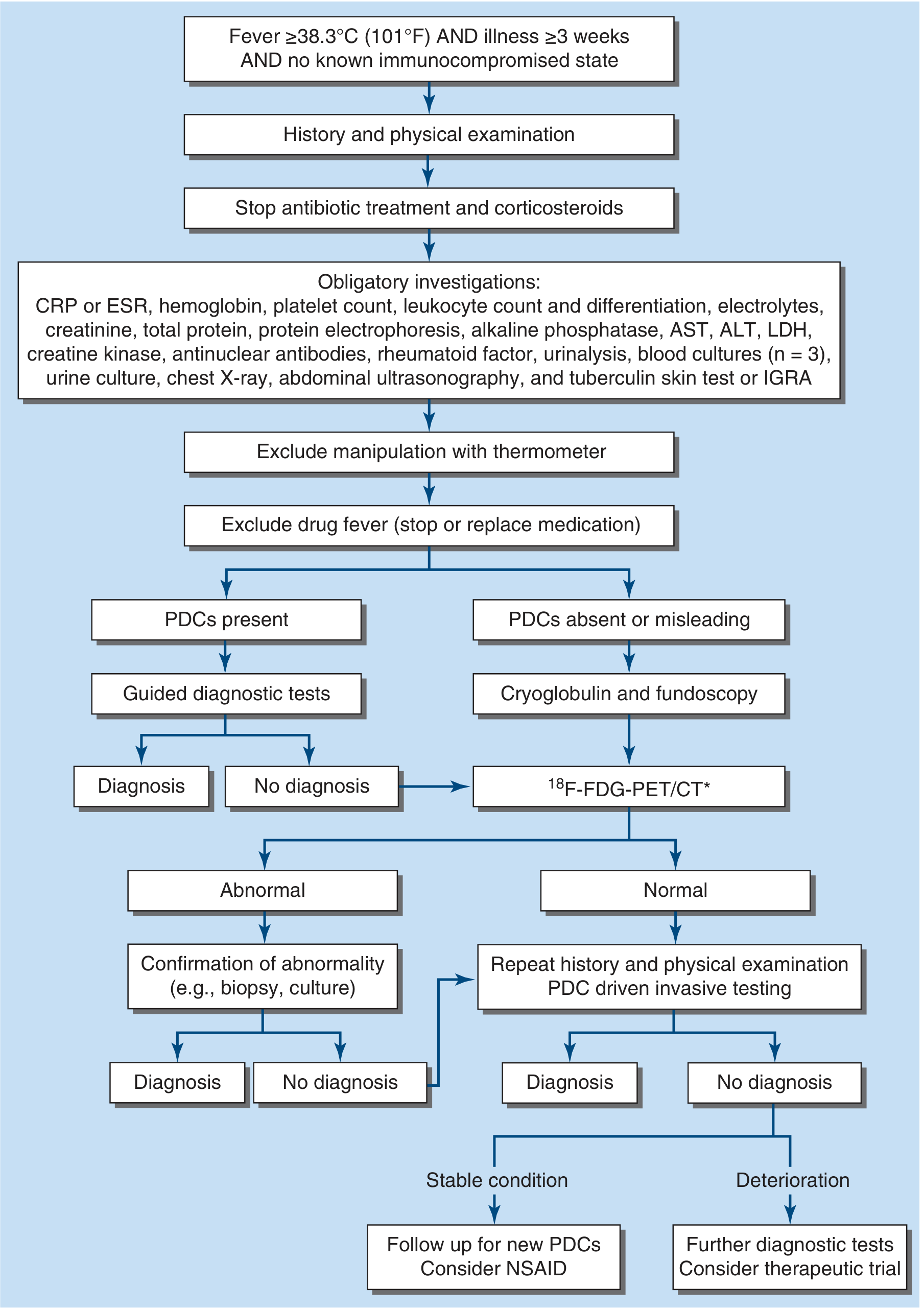
Step 1: Identify Potentially Diagnostic Clues (PDCs) - MOST CRITICAL STEP
- Fever pattern - quotidian (double daily spike = AOSD), Pel-Ebstein (Hodgkin), remittent, continuous
- Travel history - endemic areas for TB, typhoid, malaria, brucellosis, visceral leishmaniasis (kala-azar)
- Animal contact - cats (bartonellosis/cat scratch disease), farm animals (Q fever/brucellosis)
- Salmon-colored evanescent rash with fever spikes - strongly suggests Adult-onset Still's Disease (AOSD) even in a 17-year-old
- Lymphadenopathy - any tender or non-tender nodes (lymphoma, EBV, cat scratch, TB)
- Arthralgia/arthritis - AOSD, JIA systemic type, reactive arthritis
- Medications - drug fever (sulfonamides, phenytoin, allopurinol) can occur even with long-term use
- Family history - periodic fever syndromes (FMF especially if Mediterranean descent, TRAPS, HIDS)
- Oral ulcers, genital ulcers - Behcet's disease
- Weight loss, night sweats - lymphoma, TB
- Sexual history, IV drug use - endocarditis, HIV
Step 2: Investigations NOT Yet Done (Complete the Obligatory Panel)
| Test | Rationale |
|---|---|
| Blood cultures x3 (aerobic + anaerobic, ideally off antibiotics) | Endocarditis, occult bacteremia, brucellosis (prolonged incubation) |
| Tuberculin skin test (TST) / IGRA (QuantiFERON-TB Gold) | Extrapulmonary TB is a common FUO cause in South Asia; IGRA is more specific |
| Chest X-ray | Miliary TB, hilar lymphadenopathy (lymphoma, sarcoid), cardiomegaly (endocarditis) |
| Abdominal ultrasound | Occult abscess, hepatosplenomegaly, lymph nodes, biliary disease |
| Urine culture + urinalysis | Occult UTI/pyelonephritis |
| Urine examination | Pyuria, hematuria |
| Creatine kinase | Inflammatory myopathy |
| Alkaline phosphatase | Hepatic TB, granulomatous disease |
| HIV serology (4th gen Ag/Ab combo) | Acute HIV can cause prolonged FUO |
| EBV / CMV serology (IgM + IgG) | Viral causes of FUO - often missed |
| Monospot / EBV VCA IgM | EBV is a leading viral FUO cause in teenagers |
| Widal/blood culture for Salmonella Typhi | In South Asian settings |
| Malaria smear / RDT | If any travel history or endemic region |
| Cryoglobulins + fundoscopy | Recommended when PDCs are absent (per Harrison's algorithm) |
Step 3: Imaging
- Detects occult abscesses (liver, splenic, subphrenic, pelvic, perinephric)
- Detects retroperitoneal/mesenteric lymphadenopathy (lymphoma, TB)
- Detects occult malignancy
Step 4: ¹⁸F-FDG-PET/CT - Key Next-Level Investigation
- Establishes a diagnosis in >50% of unresolved FUO cases (Rheumatology textbook, Elsevier 2022)
- Detects occult lymphoma, vasculitis, granulomatous disease (sarcoid, TB), osteomyelitis, occult abscess - all in one scan
- Replaces Gallium-67 scintigraphy (higher diagnostic yield)
- Do not give glucocorticoids before the scan - they suppress FDG uptake and falsely normalize it
- If PET/CT is unavailable: whole-body MRI (comparable yield in recent studies) or Gallium-67 scintigraphy (yield ~21-54%)
Step 5: Age/Gender-Specific Diagnoses to Prioritize in This Patient
High priority given this demographics:
- Adult-onset Still's Disease (AOSD) / Systemic JIA - quotidian fever with salmon rash, arthralgia; note that ferritin may be strikingly elevated in active disease but can be normal in early stages; requires dedicated IL-18 and glycosylated ferritin measurement (glycosylated ferritin <20% is highly suggestive of AOSD)
- Lymphoma (especially Hodgkin's) - young females, B symptoms (fever + night sweats + weight loss); Pel-Ebstein fever pattern; requires CT chest/abdomen/pelvis + PET scan; excisional lymph node biopsy if lymphadenopathy present
- Tuberculosis (extrapulmonary) - miliary TB, hepatic TB, TB lymphadenitis; IGRA + CT + bone marrow biopsy
- Visceral Leishmaniasis (Kala-azar) - if from endemic region; splenomegaly, pancytopenia; rK39 antigen, splenic/bone marrow aspirate
- EBV/CMV mononucleosis - common in teens; atypical lymphocytes, hepatosplenomegaly; serology + viral PCR
- Enteric fever (Typhoid/Paratyphoid) - negative Widal is not sufficient; blood culture is gold standard
- Systemic Lupus Erythematosus - ANA may be negative early; repeat anti-dsDNA, anti-Sm, complement (C3, C4); note ANA can be negative in up to 5% of SLE
- Periodic fever syndromes (FMF, TRAPS, HIDS) - especially if fever is episodic and patient has Mediterranean/Middle Eastern ancestry; genetic testing (MEFV gene)
- Infective endocarditis - culture-negative endocarditis (HACEK, Bartonella, Coxiella, Brucella); serial blood cultures + echocardiogram
- Drug fever - rule out systematically
Step 6: If All Above Are Negative - PDC-Driven Invasive Testing
| Clue | Investigation |
|---|---|
| Lymphadenopathy present | Excisional lymph node biopsy (preferred over FNAC for lymphoma) |
| Hepatosplenomegaly | Bone marrow biopsy (for lymphoma, TB, kala-azar, hemophagocytic syndrome) |
| Pulmonary infiltrates | Bronchoscopy + BAL with AFB smear/culture/PCR, fungal cultures |
| Skin lesion | Skin biopsy |
| Bone pain / elevated ALP | Bone scan or PET + bone biopsy |
| Liver disease on imaging | Liver biopsy (granulomas = TB, sarcoid, brucellosis) |
Special Consideration: Hemophagocytic Lymphohistiocytosis (HLH)
- Triglycerides, fibrinogen, NK cell activity
- Bone marrow aspirate for hemophagocytosis
Summary: Immediate Next Steps
- Complete the history - fever pattern, rash, travel, animals, medications, family history
- Complete the obligatory workup - blood cultures x3, IGRA/TST, chest X-ray, abdominal ultrasound, HIV, EBV/CMV serology, urine culture
- CT chest + abdomen + pelvis with contrast
- Echocardiography
- ¹⁸F-FDG-PET/CT - if CT is unrevealing and no PDCs have emerged
- PDC-directed biopsy (lymph node / bone marrow / liver) guided by imaging findings
- Specific tests for AOSD - glycosylated ferritin, IL-18 if clinical suspicion
- Consider second opinion at tertiary FUO expert center if still undiagnosed
Sources: Harrison's Principles of Internal Medicine 22E (2025, McGraw Hill), Goldman-Cecil Medicine, Frameworks for Internal Medicine, Rheumatology 2-Volume Set (Elsevier 2022)
17 year old female with fever of unknown since 1 month with CBC, esr, crp,lft,rft,ure,ra factor, procalcitonin,ferritin,LDH,serum protein electrophoresis,ana profile , Widal , dengue,lepto, chickenguniya, all are negative what is the next step for diagnosis
FUO in a 17-Year-Old Female: Next Steps After Extended Negative Workup
- Hematology/biochemistry: CBC, ESR, CRP, LFT, RFT, LDH, procalcitonin, ferritin, SPEP
- Autoimmune: ANA profile, RA factor
- Tropical infections: Widal, dengue NS1/IgM, leptospira, chikungunya IgM
What Is Still MISSING From the Obligatory Workup
| Investigation | Why It Matters |
|---|---|
| Blood cultures x3 (aerobic + anaerobic, at least 2 drawn >12 hrs apart) | Culture-negative endocarditis (HACEK, Brucella, Bartonella, Coxiella), occult bacteremia, Salmonella |
| IGRA / QuantiFERON-TB Gold | Extrapulmonary TB - miliary, hepatic, nodal; TST can be false-negative in miliary TB |
| Chest X-ray | Miliary TB pattern, hilar lymphadenopathy (lymphoma, sarcoid), cardiomegaly |
| Abdominal Ultrasound | Hepatosplenomegaly, occult abscess, lymphadenopathy - highest yield, low cost |
| Urine culture + urinalysis with sediment | Occult pyelonephritis |
| HIV 4th generation Ag/Ab combo test | Acute HIV seroconversion - a leading cause of prolonged FUO in this age group |
| EBV VCA IgM + IgG (Monospot/Paul-Bunnell) | EBV mononucleosis is the single most common viral FUO in teenagers |
| CMV IgM + IgG | CMV mononucleosis - presents identically to EBV |
| Malaria thick + thin smear / RDT | If from endemic region or any travel history |
| TSH | Thyroid disease (hyperthyroidism) can cause prolonged low-grade fever |
| Creatine kinase (CK) | Inflammatory myopathy |
Tier 2: Investigations Based on Potentially Diagnostic Clues (PDCs)
PDC: Rash (even evanescent/salmon-colored)
- Think Adult-Onset Still's Disease (AOSD) / Systemic JIA - the hallmark salmon rash appears only during fever spikes and disappears quickly
- Send: Glycosylated ferritin (<20% strongly suggests AOSD), IL-18 level, repeat ferritin during a fever spike (can be massively elevated)
- AOSD/Systemic JIA is the #1 diagnosis to consider in a 17-year-old female with FUO and negative autoimmune screen
PDC: Splenomegaly / Hepatosplenomegaly
- Think Visceral Leishmaniasis (Kala-azar), lymphoma, EBV, brucellosis
- Send: rK39 antigen (rapid card test) - highly sensitive for VL in endemic areas (India, Bihar region)
- Bone marrow / splenic aspirate for Leishmania amastigotes
PDC: Lymphadenopathy (any site)
- Think Hodgkin lymphoma, NHL, TB lymphadenitis, EBV, cat scratch disease (Bartonella)
- Excisional lymph node biopsy - do NOT rely on FNAC alone for lymphoma diagnosis
- CT chest/abdomen/pelvis with contrast - mandatory to map nodes
PDC: Heart murmur / dental procedure / IV line history
- Think Culture-negative endocarditis (Bartonella, Coxiella Q fever, HACEK organisms)
- Transthoracic echocardiography - then TEE if TTE negative and suspicion high
- Bartonella serology, Coxiella Phase I & II IgG/IgM
PDC: Arthralgia, joint swelling
- Think AOSD, reactive arthritis, Brucella, viral arthritis
- Brucella serology (Rose-Bengal + SAT), brucella blood cultures
PDC: No PDCs at all (most challenging scenario)
Tier 3: CT Imaging
- Detects occult abscesses (liver, splenic, retroperitoneal, pelvic, perinephric)
- Detects mediastinal/retroperitoneal/mesenteric lymphadenopathy (lymphoma, TB)
- Can reveal hepatosplenomegaly, occult masses
- Goldman-Cecil lists this as a standard next step after basic labs are non-diagnostic
Tier 4: ¹⁸F-FDG-PET/CT - The Game-Changing Investigation
- Identifies the anatomic location of active metabolic inflammation anywhere in the body in a single scan
- Establishes diagnosis in >50% of previously undiagnosed FUO cases (Rheumatology, Elsevier 2022)
- Detects: lymphoma, vasculitis, granulomatous infections (TB, sarcoid), osteomyelitis, occult abscesses, endocarditis vegetations, autoinflammatory disease foci
- Critical rule: stop all glucocorticoids before the scan - steroids suppress FDG uptake and will falsely normalize the result
- If PET/CT unavailable: whole-body MRI (comparable diagnostic yield in recent studies) or Gallium-67 scintigraphy
Tier 5: PDC-Guided Invasive Testing
| Finding on Imaging / Exam | Invasive Test |
|---|---|
| Splenomegaly + pancytopenia | Bone marrow aspirate + trephine biopsy |
| Lymphadenopathy | Excisional lymph node biopsy (with histopathology, culture, flow cytometry) |
| Hepatomegaly / liver lesion | Liver biopsy (granulomas = TB, sarcoid, brucellosis) |
| Pulmonary infiltrates | Bronchoscopy + BAL - AFB smear/culture/PCR, fungal culture |
| Bone marrow biopsy indications in FUO | Lymphoma, miliary TB, kala-azar, HLH, leukemia, hemophagocytosis |
Special "Must Not Miss" in a 17-Year-Old Female
1. Hemophagocytic Lymphohistiocytosis (HLH)
| Criterion | Test to Send |
|---|---|
| Fever | (already present) |
| Splenomegaly | Ultrasound / CT |
| Cytopenias (≥2 cell lines) | CBC with differential |
| Hypertriglyceridemia | Fasting triglycerides |
| Hypofibrinogenemia | Fibrinogen level |
| Elevated ferritin (>500; >10,000 in children) | Ferritin (repeat during fever spike) |
| Elevated sIL-2R (sCD25) | Soluble IL-2 receptor |
| Hemophagocytosis in marrow | Bone marrow aspirate |
2. Adult-Onset Still's Disease / Systemic JIA
- Most likely diagnosis in this demographic with ANA-negative FUO
- Yamaguchi criteria: quotidian fever >39°C + evanescent rash + arthritis + leukocytosis
- Glycosylated ferritin <20% is the key test (sensitivity ~72%, specificity ~69% for AOSD)
3. Lymphoma (Hodgkin's)
- Peak incidence age 15-25 years
- Can present with only fever for weeks with no palpable lymph nodes (nodular sclerosis subtype has mediastinal disease)
- Mandatory: CT chest (mediastinal mass) + PET/CT
4. Brucellosis
- Often missed; Widal test does not cover brucellosis
- Exposure to unpasteurized dairy, livestock
- Send: Rose-Bengal slide test + Standard Agglutination Test (SAT) titre >1:160 + brucella blood culture (BACTEC alert - prolonged incubation needed)
5. Q Fever (Coxiella burnetii)
- Animal contact (cattle, sheep, goats), raw milk
- Coxiella Phase I and II IgG/IgM serology - standard PCR/serology often not in routine tropical screens
6. Cat Scratch Disease (Bartonella henselae)
- Young female, any cat contact or scratch
- Regional tender lymphadenopathy (may be subtle)
- Bartonella henselae IgM/IgG serology, lymph node biopsy (Warthin-Starry stain)
Practical Sequence (India/Tropical Setting)
TODAY:
1. Blood cultures x3 (send BACTEC bottles, request prolonged incubation)
2. IGRA (QuantiFERON-TB Gold)
3. HIV 4th gen Ag/Ab
4. EBV VCA IgM/IgG + CMV IgM/IgG
5. Malaria smear/RDT
6. TSH
7. Brucella serology (Rose-Bengal + SAT)
8. Fasting triglycerides + fibrinogen + sIL-2R (for HLH screen)
9. Urine culture + microscopy
10. CXR (PA view)
11. Abdominal USG

NEXT STEP IF ABOVE NEGATIVE:
12. CECT Chest + Abdomen + Pelvis
13. Echocardiography (TTE)
14. Glycosylated ferritin (for AOSD)
15. rK39 antigen (for kala-azar if splenomegaly)
16. Coxiella / Bartonella serology

ADVANCED:
17. ¹⁸F-FDG-PET/CT (most powerful single investigation)
18. Bone marrow aspirate + trephine biopsy
19. Excisional lymph node biopsy (if any adenopathy)
Sources: Harrison's Principles of Internal Medicine 22E (2025, McGraw Hill), Goldman-Cecil Medicine International Edition, Frameworks for Internal Medicine, Rheumatology 2-Volume Set (Elsevier 2022)
17 year old female with fever of unknown since 1 month with CBC, esr, crp,lft,rft,ure,ra factor, procalcitonin,ferritin,LDH,serum protein electrophoresis,ana profile , Widal , dengue,lepto, chickenguniya, urine tb pcr , all are negative and usg abdomen and pelvis is normal what is the next step for diagnosis
FUO in a 17-Year-Old Female: After Tropical Screen + USG are All Negative
IMMEDIATE NEXT STEPS (Still from the Obligatory Panel - Not Yet Done)
| Test | What It Catches |
|---|---|
| Blood cultures x3 (aerobic + anaerobic, at least 12 hrs apart) | Culture-negative endocarditis (Bartonella, Coxiella, HACEK, Brucella), occult bacteremia |
| IGRA / QuantiFERON-TB Gold | Extrapulmonary TB (miliary, nodal, hepatic) - urine TB PCR is NOT sufficient for systemic TB |
| Chest X-ray (PA view) | Miliary pattern, mediastinal widening (lymphoma), hilar adenopathy, pericardial effusion |
| HIV 4th generation Ag/Ab | Acute HIV seroconversion - a top cause of FUO in adolescents |
| EBV VCA IgM + IgG + heterophile (Monospot) | EBV mononucleosis - the single most common viral cause of FUO in a 17-year-old; USG can be normal early |
| CMV IgM + IgG | CMV mononucleosis - clinically indistinguishable from EBV |
| Malaria thick + thin smear / RDT | If any endemic exposure; cannot be excluded without smear |
| TSH | Thyroid-related fever (hyperthyroidism occasionally causes prolonged fever) |
| Urine culture + urinalysis with sediment | Occult pyelonephritis / renal TB (urine TB PCR has low sensitivity) |
| Brucella Rose-Bengal slide test + SAT titre | Brucellosis - undulant fever, missed by Widal; livestock/dairy exposure |
| Creatine kinase (CK) | Inflammatory myopathy |
Important: Urine TB PCR has very low sensitivity for systemic/pulmonary TB. IGRA is the correct test for TB in this context.
STEP 2: Imaging Escalation
CECT Chest + Abdomen + Pelvis (with IV contrast)
- Mediastinal and retroperitoneal lymphadenopathy (Hodgkin lymphoma commonly presents here)
- Small splenic/hepatic lesions (<1 cm)
- Pericardial disease, lung parenchymal nodules
- Para-aortic nodes, mesenteric adenopathy
- Spine - osteomyelitis (spine is the most common site of osteomyelitis in FUO per Frameworks for Internal Medicine)
Transthoracic Echocardiography (TTE)
- Vegetations from culture-negative endocarditis (Bartonella, Coxiella, HACEK)
- Even without a murmur, endocarditis can present as pure FUO
- Pericardial effusion
STEP 3: Specific Targeted Investigations
For Adult-Onset Still's Disease (AOSD) / Systemic JIA
- Glycosylated ferritin - values <20% are strongly suggestive (the "normal" total ferritin earlier does not exclude AOSD; repeat ferritin during a fever spike, as it can spike dramatically)
- IL-18 level - markedly elevated in AOSD; helps distinguish from infection
- Ask specifically about: quotidian fever spikes to >39°C that resolve completely, evanescent salmon-pink rash appearing only with fever spikes, arthralgia, sore throat
For Lymphoma (Hodgkin's Disease)
- CECT chest is mandatory - look for anterior mediastinal mass (nodular sclerosis Hodgkin's can present with fever alone for weeks before any palpable nodes appear)
- Peripheral blood film - atypical lymphocytes, Reed-Sternberg cells (rarely)
- If any lymphadenopathy found: excisional lymph node biopsy (not FNAC)
For Tuberculosis (Extrapulmonary)
- IGRA + Chest X-ray (miliary pattern, hilar adenopathy)
- If IGRA positive or CXR shows miliary pattern: sputum AFB smear x3 + GeneXpert MTB/RIF + HRCT chest
- If spinal tenderness: MRI spine (most common site of osteomyelitis in FUO)
For Culture-Negative Endocarditis
- Bartonella henselae/quintana IgG/IgM (cat contact?)
- Coxiella burnetii Phase I and II IgG/IgM (Q fever - animal contact, raw milk)
- Brucella serology + prolonged blood cultures
For Hemophagocytic Lymphohistiocytosis (HLH)
- Fasting triglycerides + fibrinogen (low fibrinogen + high triglycerides = HLH)
- Soluble IL-2 receptor (sCD25) - markedly elevated in HLH
- Peripheral blood film - cytopenias
- If HLH suspected: bone marrow aspirate + trephine biopsy
STEP 4: ¹⁸F-FDG-PET/CT - The Single Most Powerful Next Investigation
- Establishes a diagnosis in >50% of previously unresolved FUO cases
- Can detect: occult lymphoma (mediastinal, abdominal), large vessel vasculitis (e.g., Takayasu arteritis - common in young females in Asia), granulomatous infections (miliary TB, sarcoidosis), autoinflammatory foci, osteomyelitis, endocarditis vegetations
- Detects Takayasu arteritis - a cause of FUO specifically in young Asian females, shows bilateral vascular FDG uptake; USG and even CT angiography can miss early mural inflammation
- Critical rule: Do NOT start steroids, NSAIDs, or empirical treatment before the scan - they suppress FDG uptake and falsely normalize results
- Whole-body MRI (comparable diagnostic yield in recent studies)
- Gallium-67 scintigraphy (diagnostic yield ~21-54%, lower than PET/CT)
STEP 5: Invasive Testing (PDC-Guided, After Imaging)
| Imaging Finding | Invasive Test |
|---|---|
| Mediastinal/retroperitoneal lymphadenopathy | Excisional lymph node biopsy (histopathology + cultures + flow cytometry + T-cell gene rearrangement) |
| Bone marrow hypermetabolism on PET | Bone marrow aspirate + trephine biopsy (lymphoma, miliary TB, kala-azar, HLH, leukemia) |
| Hepatic lesion | Liver biopsy (granulomas = TB, sarcoid, brucellosis) |
| Pulmonary infiltrates / miliary pattern | Bronchoscopy + BAL (AFB smear/culture/PCR, fungal cultures) |
| Vascular wall thickening | MR angiography + vascular biopsy if needed (Takayasu arteritis) |
| Spinal abnormality | MRI spine + CT-guided vertebral biopsy |
Prioritized Action Plan for This Patient
SEND TODAY (in parallel):
1.  Blood cultures x3 (BACTEC, request prolonged incubation for brucella/HACEK)
2.  IGRA (QuantiFERON-TB Gold) - rules in/out systemic TB
3.  HIV 4th gen Ag/Ab combo
4.  EBV VCA IgM/IgG + CMV IgM/IgG
5.  Brucella serology (Rose-Bengal + SAT)
6.  TSH
7.  Fasting triglycerides + fibrinogen + sIL-2R (HLH screen)
8.  Urine culture + microscopy (repeat)
9.  CK (creatine kinase)

IMAGING THIS WEEK:
10. Chest X-ray (PA + lateral)
11. CECT Chest + Abdomen + Pelvis with IV contrast
12. Transthoracic echocardiography

SPECIFIC DISEASE TESTS:
13. Glycosylated ferritin + IL-18 (AOSD screen)
14. Coxiella Phase I & II IgG/IgM; Bartonella IgM/IgG (culture-negative endocarditis/Q fever)
15. rK39 antigen (kala-azar, if endemic region)
16. Malaria thick smear + RDT

IF ALL ABOVE NEGATIVE:
17. ¹⁸F-FDG-PET/CT (most powerful - do before any steroids/NSAIDs)
18. Bone marrow aspirate + trephine biopsy
19. Excisional biopsy of any node found on CT/PET
Three Diagnoses to Prove or Exclude First in This Patient
| Rank | Diagnosis | Why | Key Test |
|---|---|---|---|
| 1 | AOSD / Systemic JIA | Age 17F, ANA-negative, all screens normal, compatible pattern | Glycosylated ferritin, IL-18; Yamaguchi criteria |
| 2 | Hodgkin Lymphoma | Age 15-25 peak, can present as pure FUO with mediastinal disease only | CECT chest (mediastinum), PET/CT |
| 3 | Extrapulmonary TB | Prevalent in India, urine PCR insufficient, IGRA not yet done | IGRA + HRCT chest + bone marrow biopsy |
Sources: Harrison's Principles of Internal Medicine 22E (2025, McGraw Hill), Goldman-Cecil Medicine International Edition, Frameworks for Internal Medicine (Lippincott), Rheumatology 2-Volume Set (Elsevier 2022), Firestein & Kelley's Textbook of Rheumatology
17 year old female with fever of unknown since 1 month with CBC, esr, crp,lft,rft,ure,ra factor, procalcitonin,ferritin,LDH,serum protein electrophoresis,ana profile , Widal , dengue,lepto, chickenguniya, urine tb pcr , all are negative and chest xray ,tsh,usg abdomen and pelvis is normal what is the next step for diagnosis
FUO in a 17-Year-Old Female: Chest X-Ray + TSH + USG All Normal - What Now?
- All inflammatory markers negative (CBC, ESR, CRP, procalcitonin, ferritin, LDH, SPEP)
- All autoimmune negative (ANA profile, RA factor)
- All tropical serology negative (Widal, dengue, leptospira, chikungunya)
- Urine TB PCR negative
- Chest X-ray normal, TSH normal, USG abdomen/pelvis normal
WHAT IS STILL MISSING FROM THE OBLIGATORY PANEL
| Test | Rationale |
|---|---|
| Blood cultures x3 (aerobic + anaerobic, BACTEC, at least 12h apart, prolonged incubation) | Culture-negative endocarditis (Bartonella, Coxiella, HACEK, Brucella, Streptococcus) - cannot be excluded without this |
| IGRA / QuantiFERON-TB Gold | Urine TB PCR is NOT a systemic TB test; extrapulmonary/miliary TB requires IGRA; normal CXR does not exclude miliary TB (can be normal early) |
| HIV 4th generation Ag/Ab combo | Acute HIV seroconversion = one of the top causes of FUO in this age group |
| EBV VCA IgM/IgG (Monospot + Paul-Bunnell) | EBV mononucleosis is the single most common viral FUO in teenagers; splenomegaly and lymphadenopathy may be absent early or subtle |
| CMV IgM/IgG | CMV mononucleosis - identical presentation, separate serology required |
| Malaria thick + thin smear + RDT | Cannot be excluded serologically; requires direct smear |
| Brucella serology (Rose-Bengal + SAT titre ≥1:160) | Brucellosis = "undulant fever," completely missed by Widal; endemic in India with livestock/dairy exposure |
| Urine culture + urinalysis | Occult pyelonephritis, renal TB (urine PCR has very low sensitivity for renal TB) |
| Creatine kinase (CK) | Inflammatory myopathy |
THE CRITICAL NEXT STEPS (After or in Parallel with Above)
1. REPEAT + DEEPEN HISTORY AND PHYSICAL EXAMINATION
- Fever pattern - Does the fever spike once or twice daily to ≥39°C and then return completely to normal? (quotidian pattern = AOSD)
- Rash - Any brief salmon-pink rash appearing only when fever is highest, disappearing within hours? (hallmark of AOSD)
- Joint pain during fever spikes?
- Sore throat at disease onset?
- B symptoms - drenching night sweats, unintentional weight loss, fatigue? (Hodgkin lymphoma)
- Animal contact - cats (Bartonella), livestock/raw dairy (Brucella, Q fever)
- Medications - even OTC drugs, herbal medicines, supplements (drug fever can occur after months of use)
- Family history - periodic fevers, Mediterranean/Middle Eastern/Jewish ancestry (FMF)
- Any node - even a single small, painless, firm cervical/supraclavicular node (Hodgkin's)
- All lymph node chains (especially supraclavicular, axillary, inguinal)
- Skin for evanescent rash - examine during a fever spike
- Fundoscopy
- Heart - any new murmur (endocarditis)
- Pulse symmetry in both arms (Takayasu arteritis - common in young Asian females)
- Joints - any swelling or warmth
2. CECT CHEST + ABDOMEN + PELVIS (Contrast-Enhanced CT)
- Mediastinal lymphadenopathy (Hodgkin lymphoma - nodular sclerosis subtype presents with anterior mediastinal mass, missed completely on plain X-ray in ~30-40% of cases)
- Hilar adenopathy (early TB, sarcoidosis)
- Small hepatic / splenic lesions (<1 cm) missed on USG
- Retroperitoneal and para-aortic lymphadenopathy (lymphoma, TB)
- Aortic wall thickening (Takayasu arteritis)
- Occult abscesses (perinephric, psoas, pelvic)
- Pericardial effusion
3. TRANSTHORACIC ECHOCARDIOGRAPHY (TTE)
- Vegetations from culture-negative endocarditis
- Pericardial effusion
- Intracardiac thrombus or tumour (atrial myxoma - rare but causes FUO)
- Even without a murmur on auscultation, endocarditis from fastidious organisms (Bartonella, Coxiella) can present as pure FUO
4. SPECIFIC DISEASE-TARGETED INVESTIGATIONS (Send Now)
A. Adult-Onset Still's Disease / Systemic JIA Screen
| Test | Cut-off / Interpretation |
|---|---|
| Glycosylated ferritin | <20% of total ferritin is strongly suggestive of AOSD (sensitivity ~72%, specificity ~69%) |
| Repeat ferritin during a fever spike | Can be dramatically elevated (>5000 ng/mL) during active AOSD even if baseline is normal |
| IL-18 serum level | Markedly elevated in AOSD; helps distinguish from infection |
| Peripheral smear | Leukocytosis with neutrophilia during spike = AOSD pattern |
B. Lymphoma Screen (Blood Tests)
- Peripheral blood film - atypical lymphocytes, blast cells
- Soluble IL-2 receptor (sCD25) - elevated in lymphoma and HLH
- Beta-2 microglobulin - elevated in lymphoma
- Definitive diagnosis only by excisional lymph node biopsy if any node found on CT
C. HLH Screen (Hemophagocytic Lymphohistiocytosis)
| Test | HLH Criterion |
|---|---|
| Fasting triglycerides | >265 mg/dL |
| Fibrinogen | <150 mg/dL (hypofibrinogenemia) |
| Ferritin (repeat) | >500 ng/mL (>10,000 in children highly specific) |
| Soluble IL-2R (sCD25) | >2400 U/mL |
| NK cell activity | Impaired |
| Bone marrow aspirate | Hemophagocytosis |
D. Other Targeted Serology
- Coxiella burnetii Phase I + II IgG/IgM (Q fever - animal contact)
- Bartonella henselae/quintana IgM/IgG (cat scratch, culture-negative endocarditis)
- rK39 antigen (Visceral leishmaniasis / Kala-azar if from Bihar, Jharkhand, UP, or endemic belt)
- Anti-dsDNA + anti-Sm + C3 + C4 (Repeat lupus screen - ANA can be negative in up to 5% of SLE; lupus presenting with ANA-negative FUO is documented)
- ANCA (cANCA + pANCA) - vasculitis (Takayasu, GPA) causing FUO
5. ¹⁸F-FDG-PET/CT - NOW THE KEY NEXT-TIER INVESTIGATION
"¹⁸F-FDG-PET has been shown in a prospective study to ascertain the correct diagnosis in more than 50% of patients presenting with fever of unknown origin or inflammation of unknown origin." - Rheumatology, 2-Volume Set, Elsevier 2022
- Takayasu arteritis - bilateral aortic wall / subclavian / carotid FDG uptake; classic in young Asian females; ESR/CRP can be normal in some cases
- Hodgkin lymphoma - even small/early nodal disease
- Sarcoidosis - hilar, mediastinal, systemic granulomas
- Occult osteomyelitis (spine, long bones)
- Culture-negative endocarditis - valvular FDG uptake
- Autoinflammatory disease foci
- Large vessel vasculitis
6. CRYOGLOBULINS + FUNDOSCOPY
- Cryoglobulinemia (mixed type II/III) can cause chronic fever without obvious organ involvement
- Fundoscopy may reveal uveitis (sarcoidosis, TB, Behcet's), Roth spots (endocarditis), or choroidal tubercles (miliary TB)
7. PDC-GUIDED INVASIVE TESTING (Based on CT/PET Findings)
| Finding | Investigation |
|---|---|
| Mediastinal / retroperitoneal lymph nodes | Excisional lymph node biopsy (histopathology + cultures + flow cytometry + T-cell gene rearrangement - NOT just FNAC) |
| Bone marrow uptake on PET / unexplained cytopenias | Bone marrow aspirate + trephine biopsy (lymphoma, miliary TB, HLH, kala-azar, leukemia) |
| Liver lesion | Liver biopsy (granulomas = TB, sarcoid, brucellosis) |
| Vascular wall thickening | MR angiography (Takayasu arteritis confirmation) |
| Spine abnormality | MRI spine + CT-guided biopsy (vertebral osteomyelitis/TB) |
Practical Sequence - What to Do Right Now
SEND TODAY:
  1. Blood cultures x3 (BACTEC aerobic + anaerobic; prolonged incubation ≥14 days)
  2. IGRA (QuantiFERON-TB Gold In-Tube) - gold standard for TB
  3. HIV 4th generation Ag/Ab combo assay
  4. EBV VCA IgM + IgG, CMV IgM + IgG
  5. Brucella serology (Rose-Bengal + SAT)
  6. Malaria thick + thin smear / RDT
  7. Fasting triglycerides + fibrinogen + sCD25 (HLH screen)
  8. Urine culture + microscopy
  9. CK, repeat ferritin (during fever spike)
  10. Glycosylated ferritin + IL-18 (AOSD screen)

THIS WEEK:
  11. CECT Chest + Abdomen + Pelvis (IV contrast) - PRIORITY
  12. Echocardiography (TTE)
  13. Coxiella Phase I + II IgG/IgM; Bartonella IgM/IgG
  14. Anti-dsDNA + anti-Sm + C3 + C4 + ANCA
  15. rK39 antigen (if endemic area for kala-azar)
  16. Cryoglobulins + Fundoscopy

IF CT + ABOVE NEGATIVE:
  17. ¹⁸F-FDG-PET/CT ← Most powerful single investigation at this stage
  18. Bone marrow aspirate + trephine biopsy
  19. Excisional lymph node biopsy (if any node found)
Top 5 Diagnoses This Workup Is Designed to Find
| Priority | Diagnosis | Key Test |
|---|---|---|
| 1 | AOSD / Systemic JIA | Glycosylated ferritin <20%, IL-18, fever pattern, rash during spike |
| 2 | Hodgkin Lymphoma | CECT chest (mediastinal mass), PET/CT, excisional biopsy |
| 3 | Extrapulmonary / Miliary TB | IGRA, HRCT chest, bone marrow biopsy |
| 4 | Takayasu Arteritis | PET/CT (aortic FDG uptake), MR angiography, pulse exam |
| 5 | HLH / Lymphoma-triggered HLH | Triglycerides, fibrinogen, sCD25, bone marrow |
Sources: Harrison's Principles of Internal Medicine 22E (2025, McGraw Hill), Goldman-Cecil Medicine International Edition, Frameworks for Internal Medicine (Lippincott), Rheumatology 2-Volume Set (Elsevier 2022), Firestein & Kelley's Textbook of Rheumatology
17 year old female with fever of unknown since 1 month with CBC, esr, crp,lft,rft,ure,ra factor, procalcitonin,ferritin,LDH,serum protein electrophoresis,ana profile , Widal , dengue,lepto, chickenguniya, urine tb pcr , all are negative and chest xray ,tsh,usg abdomen and pelvis is normal , peripheral smear shows microcytic hypochromic Anemia what is the next step for diagnosis
FUO + Microcytic Hypochromic Anemia: A Critical PDC That Redirects Everything
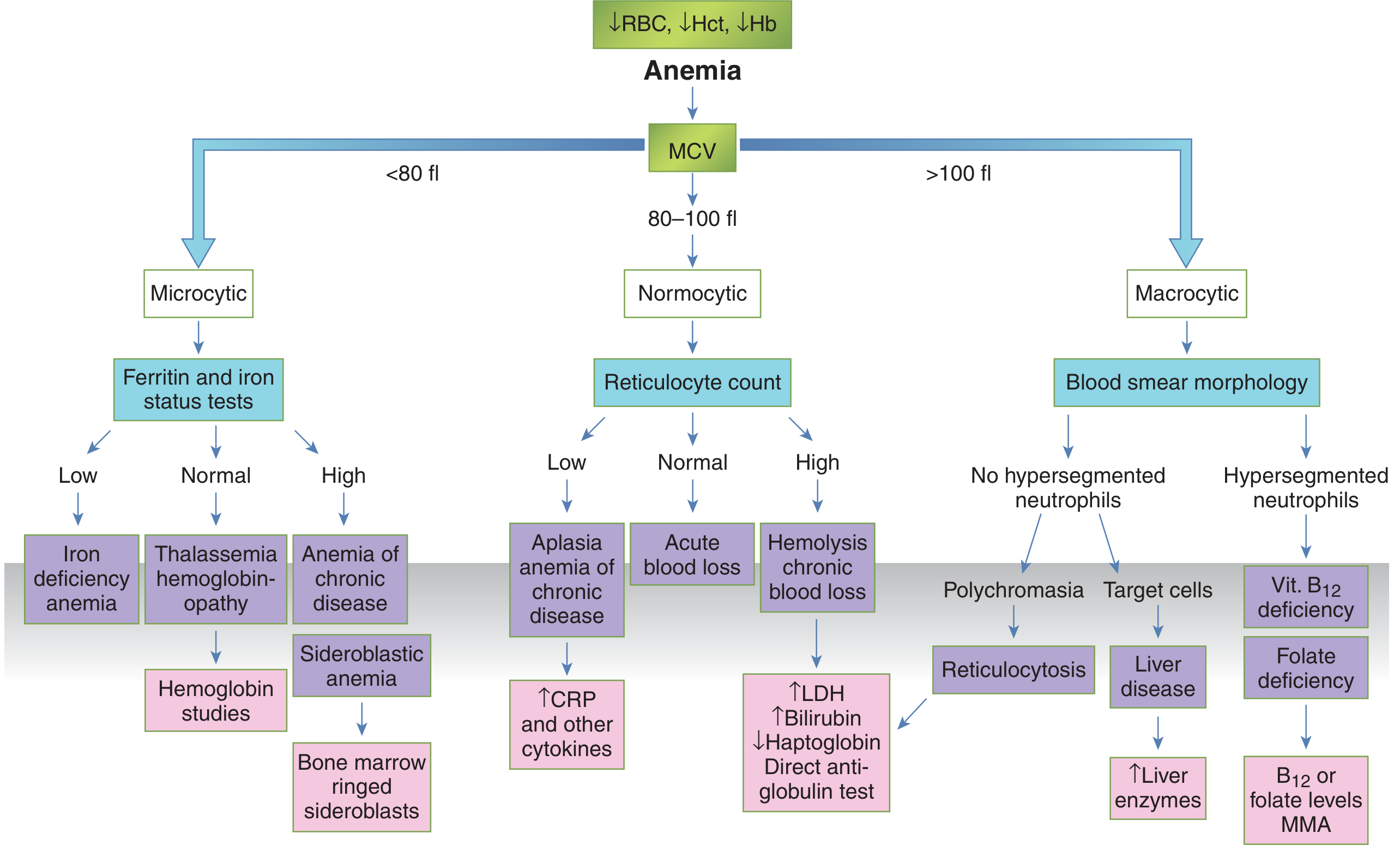
STEP 1: IRON STUDIES PANEL (Immediate, Most Important Next Test)
| Test | Send NOW |
|---|---|
| Serum iron | Low in IDA and ACD; normal/high in thalassemia |
| Total Iron Binding Capacity (TIBC) / Transferrin | High in IDA; low/normal in ACD and thalassemia |
| Transferrin saturation | Low (<16%) in IDA; low-normal in ACD |
| Serum ferritin | Low (<12 µg/L) = IDA; normal/high = ACD or thalassemia; >100 = ACD |
| Reticulocyte count | Low in IDA/ACD; elevated if hemolysis component |
| Serum transferrin receptor (sTfR) | Elevated in IDA, normal in ACD - the most reliable distinguisher |
| sTfR / log ferritin ratio (sTfR index) | >2 = IDA; <1 = ACD |
Interpretation Matrix
| Pattern | Serum Iron | TIBC | Ferritin | Diagnosis |
|---|---|---|---|---|
| Low iron, High TIBC, Low ferritin | ↓ | ↑ | ↓ | Iron Deficiency Anemia (IDA) |
| Low iron, Low TIBC, Normal/High ferritin | ↓ | ↓ | Normal/↑ | Anemia of Chronic Disease (ACD) |
| Normal iron, Normal TIBC, Normal ferritin | N | N | N | Thalassemia trait |
| Mixed pattern | ↓ | N | Low-normal | IDA + ACD coexisting |
Per Goldman-Cecil Medicine: "Microcytosis is less common in anemia of chronic disease, and the smear often shows features of inflammation such as increased rouleaux formation, background staining, and sometimes neutrophilia."
WHY THIS FINDING MATTERS FOR THE FUO DIAGNOSIS
Scenario A: If Iron Studies Show IDA (Low ferritin, High TIBC)
| Cause | Test |
|---|---|
| Occult GI blood loss (Crohn's disease, IBD, GI malignancy, polyp) | Stool for occult blood (FOBT) x3, colonoscopy/upper GI endoscopy - IBD especially Crohn's can present as FUO + IDA |
| Celiac disease | Anti-tissue transglutaminase IgA (anti-tTG IgA) + total IgA - celiac classically presents as IDA in young females |
| Heavy menstrual bleeding | Detailed menstrual history |
| Hookworm / GI parasites | Stool examination for ova and parasites |
| H. pylori | H. pylori antigen in stool / Urea breath test |
IBD (Inflammatory Bowel Disease) - especially Crohn's disease is a well-recognized cause of both FUO and IDA in young people. Crohn's can cause prolonged fever with no obvious localizing symptoms and a normal USG abdomen.
Scenario B: If Iron Studies Show ACD (Normal/High ferritin, Low iron, Low TIBC)
| Disease Category | Specific Diagnosis | Next Test |
|---|---|---|
| Chronic infection | TB (extrapulmonary), brucellosis, subacute endocarditis, kala-azar, HIV | IGRA, blood cultures x3, brucella serology, rK39, HIV |
| Malignancy | Hodgkin lymphoma, NHL, solid tumors | CECT chest/abdomen/pelvis, PET/CT |
| Autoimmune / inflammatory | AOSD, SLE, vasculitis (Takayasu), IBD | Glycosylated ferritin, anti-dsDNA, ANCA, colonoscopy |
| Other | Castleman disease, HLH | Biopsy, sCD25, triglycerides |
Scenario C: If Iron Studies Are Normal (Normal ferritin, Normal iron, Normal TIBC)
- Hemoglobin electrophoresis / HPLC - mandatory next test
- Beta thalassemia trait: HbA2 >3.5%
- Alpha thalassemia trait: normal electrophoresis (requires gene testing)
- Thalassemia itself does not cause fever, but coexisting infection or malignancy causes the FUO; the thalassemia is incidental
STEP 2: COMPLETE THE FUO MANDATORY PANEL (Still Not Done)
| Test | Why |
|---|---|
| Blood cultures x3 | Subacute endocarditis (classically causes ACD + FUO), brucellosis |
| IGRA (QuantiFERON-TB Gold) | Extrapulmonary TB is the top cause of ACD + FUO in India |
| HIV 4th gen Ag/Ab | HIV causes both FUO and microcytic/normocytic anemia |
| EBV VCA IgM/IgG + CMV IgM/IgG | Viral causes in this age group |
| Brucella Rose-Bengal + SAT | Brucellosis = undulant fever + ACD |
| Malaria smear + RDT | Malaria causes hemolytic anemia (usually normocytic, but check) |
| Anti-tTG IgA + total IgA | Celiac disease = IDA + FUO in young females |
| Stool occult blood x3 | GI blood loss causing IDA + possible IBD/Crohn's |
| Stool ova + parasites | Hookworm, other GI parasites |
| H. pylori stool antigen / UBT | H. pylori-associated IDA |
STEP 3: IMAGING ESCALATION
- Rules out Crohn's disease (terminal ileum thickening, skip lesions, mesenteric adenopathy - often not seen on USG)
- Detects mediastinal lymphadenopathy (Hodgkin lymphoma)
- Rules out occult malignancy, retroperitoneal lymph nodes
- Evaluates aortic wall (Takayasu in young female)
STEP 4: DISEASE-SPECIFIC INVESTIGATIONS BASED ON IRON STUDY RESULT
If ACD Pattern - Priority Investigations:
1. IGRA → TB (most likely in India)
2. Blood cultures x3 → subacute endocarditis, brucellosis
3. Glycosylated ferritin + IL-18 → AOSD (ANA-negative, ACD + FUO = classic AOSD)
4. CECT chest + abdomen → lymphoma, Crohn's, retroperitoneal disease
5. rK39 antigen → kala-azar (causes pancytopenia/ACD + FUO)
6. Anti-dsDNA + C3/C4 + ANCA → SLE/vasculitis
7. PET/CT if CT unrevealing
8. Bone marrow aspirate + trephine (miliary TB, lymphoma, kala-azar, HLH)
If IDA Pattern - Priority Investigations:
1. Anti-tTG IgA + total IgA → Celiac disease (classic IDA in young female)
2. Stool occult blood x3 + stool ova/parasites
3. H. pylori stool antigen / UBT
4. Upper GI endoscopy + duodenal biopsy (celiac, peptic ulcer, Crohn's upper GI)
5. Colonoscopy / ileoscopy (Crohn's, IBD, polyps)
6. Capsule endoscopy if endoscopy inconclusive (small bowel Crohn's, GI angiodysplasia)
7. CECT abdomen (small bowel Crohn's disease)
If Thalassemia Pattern (Normal iron studies):
1. Hemoglobin electrophoresis / HPLC → confirm thalassemia trait
2. Thalassemia does NOT cause fever - the FUO workup must continue independently
3. Proceed with IGRA, blood cultures, CT, PET/CT for FUO diagnosis
Practical Immediate Action Plan
SEND TODAY (all at once):
1.  Serum iron + TIBC + transferrin saturation
2.  Serum ferritin (repeat - context of inflammation can falsely normalize it)
3.  Soluble transferrin receptor (sTfR) + sTfR index
4.  Reticulocyte count + reticulocyte production index
5.  Hemoglobin electrophoresis / HPLC
6.  Blood cultures x3 (BACTEC, prolonged incubation)
7.  IGRA (QuantiFERON-TB Gold)
8.  HIV 4th gen Ag/Ab combo
9.  EBV VCA IgM/IgG + CMV IgM/IgG
10. Anti-tTG IgA + total IgA (celiac screen)
11. Stool occult blood x3 + stool ova & parasites
12. Brucella serology (Rose-Bengal + SAT)
13. Malaria thick/thin smear + RDT
14. Fasting triglycerides + fibrinogen + sCD25 (HLH screen)
15. Glycosylated ferritin + IL-18 (AOSD screen)

IMAGING THIS WEEK:
16. CECT Chest + Abdomen + Pelvis (IV contrast) - PRIORITY
17. Transthoracic echocardiography

IF ABOVE NEGATIVE:
18. Upper GI endoscopy + colonoscopy (especially if IDA pattern - celiac/Crohn's)
19. ¹⁸F-FDG-PET/CT
20. Bone marrow aspirate + trephine biopsy
Key Diagnostic Framework
Microcytic hypochromic anemia + FUO
            ↓
    Iron Studies Panel
    ↙         ↓         ↘
Low ferritin  Normal   High/Normal ferritin
(IDA)        ferritin  (ACD)
  ↓         (Thalassemia)    ↓
GI workup         ↓      Chronic inflammation
Celiac        Hb HPLC   → TB, Lymphoma, AOSD
Crohn's      (confirm)    Kala-azar, Endocarditis
Parasites                 SLE, IBD, Castleman's
Sources: Goldman-Cecil Medicine International Edition, Harrison's Principles of Internal Medicine 22E, Tietz Textbook of Laboratory Medicine 7th Ed, Robbins & Kumar Basic Pathology, Sleisenger and Fordtran's Gastrointestinal and Liver Disease