
tension lines and resting lines in maxillofacial surgery suturing. give me a detail answer going through literatures and articles. also explain the current last advancement in cocepts
resting skin tension lines facial surgery scar
tension lines resting skin tension lines maxillofacial surgery suturing RSTL Langer's lines current concepts 2023 2024
BEST lines biodynamic excisional skin tension facial surgery
https://escholarship.org/content/qt47x6c53r/qt47x6c53r_noSpl…
skin tension lines facial incision scar cosmesis dermatology surgery
Langer lines skin tension lines face maxillofacial
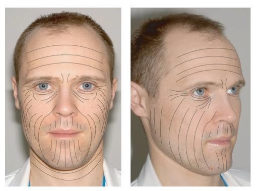
This clinical illustration consists of two photographs (frontal and three-quarter views) of a human male face with superimposed black lines representing the Lines of Langer, also known as relaxed skin tension lines (RSTLs). The diagram maps the natural orientation of skin tension across different facial regions. On the forehead, lines are predominantly horizontal and parallel. In the periorbital and malar regions, the lines exhibit a radial, curving pattern branching outward. The lower face, including the perioral area and chin, features vertical and curvilinear lines that follow the contours of the jaw and mouth, extending down the neck in horizontal bands. This anatomical mapping is essential for dermatological surgery and plastic surgery to guide surgical incisions; placing incisions parallel to these lines minimizes skin tension, optimizes wound healing, and reduces the risk of hypertrophic or keloid scarring. The visual highlights the importance of skin biomechanics in clinical outcomes.
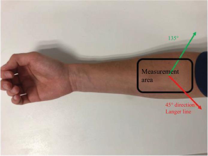
A clinical photograph of a human volar forearm, positioned for biomechanical skin testing. A black rectangular frame outlines the designated 'Measurement area' on the proximal forearm. Superimposed on this area are two directional vectors representing applied stress axes for traction testing. A red arrow is labeled '45° direction Langer line,' indicating a path following the natural skin tension lines. An orthogonal green arrow is labeled '135°,' representing a direction perpendicular to the Langer lines. The skin within the measurement area appears healthy with uniform texture and no visible lesions or hair. This setup is used in dermatology and medical engineering to study the anisotropic mechanical properties of the skin, such as elasticity and stiffness, across different layers including the epidermis and dermis.
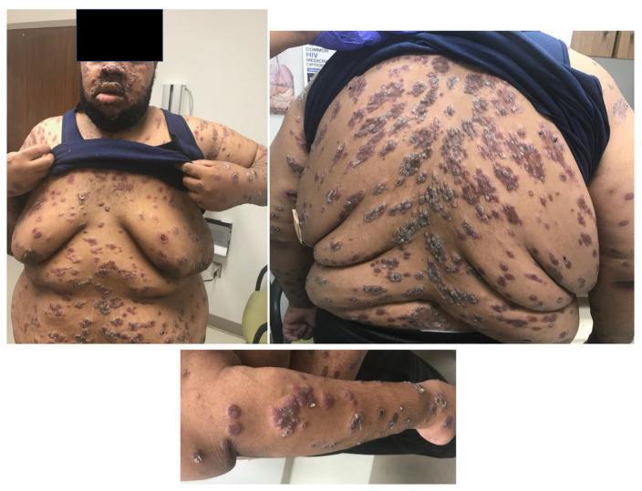
A composite of three clinical photographs demonstrating a disseminated, florid skin eruption in a patient with HIV. The images show a widespread distribution of skin lesions involving the face, trunk (chest and back), and right upper extremity. The lesions exhibit a heterogeneous morphology characterized by firm, violaceous papules and nodules intermixed with larger, crusted, and ulcerated plaques. On the back, the lesions follow a distinct 'Christmas tree' distribution, aligning with Langer's lines of skin tension, reminiscent of pityriasis rosea. Facial involvement is concentrated on the forehead and perioral areas, while the right arm displays clustered nodular and ulcerative components with visible serosanguinous crusting. The presentation is highly suggestive of malignant syphilis (lues maligna) or a severe systemic mycosis in the setting of immunosuppression. Key educational features include the varied stages of lesion evolution—from discrete papules to confluent, malodorous, ulceronodular plaques—and the specific dermatomal-like pattern on the posterior trunk.
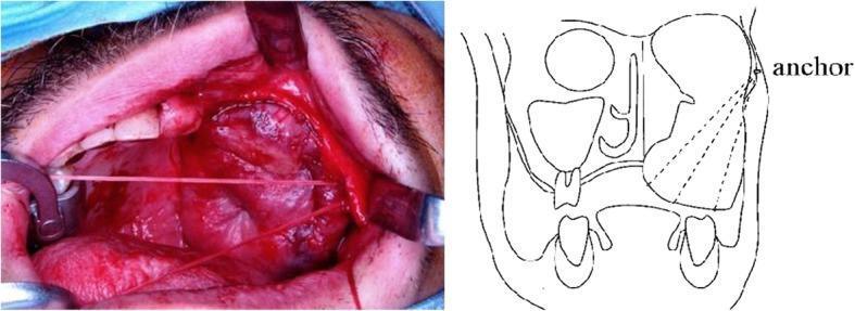
This composite educational material consists of a clinical photograph and a corresponding anatomical line drawing detailing a mid-face surgical reconstruction. The clinical photograph displays an intraoperative view of a left maxillectomy site. Key visible structures include the oral cavity, residual maxillary teeth, and a large rectus abdominis muscle flap used for reconstruction. A surgical retractor is positioned to provide exposure of the zygomatic bone area. High-tension anchor sutures are clearly visible, spanning from the zygomatic bone through the subcutaneous tissue to the skin flap. The accompanying diagram provides a schematic representation of the 'anchor' placement on the zygomatic bone, illustrating the vectors of the suture lines used to lift and stabilize the sagging reconstruction flap. This demonstrates a flap suspension technique using Mitek anchors to improve oral cavity volume and facial symmetry post-oncological resection. The clinical context involves maxillofacial surgery for secondary correction of flap ptosis to enhance prosthetic rehabilitation.
facial wound suturing technique scar outcome maxillofacial
PMID: 38926072
PMID: 40523697
Tension Lines and Resting Skin Tension Lines in Maxillofacial Surgery Suturing
Overview
1. Historical Background: Langer's Lines (Cleavage Lines)
- Derived from cadavers (often in rigor mortis), not from living, muscle-active subjects
- Reflect the passive collagen fiber orientation within the dermis
- On the face, they frequently run perpendicular to muscle action, meaning they often run at right angles to the natural wrinkle lines
- On the body (trunk, extremities), they are more congruent with functional tension lines
"Follow Langer's lines" - Pfenninger & Fowler's Procedures for Primary Care, Box 22-3 (Pearls of Suturing) - this pearl reflects the general principle but must be applied with awareness that on the face, RSTLs are the superior guide.
2. Relaxed Skin Tension Lines (RSTLs)
- Method: The skin of a relaxed, living patient is gently pinched - the wrinkles that form perpendicular to the pinch direction indicate the RSTLs (they run parallel to the crease lines created)
- RSTLs correspond to the direction of maximal skin tension at rest - the collagen and elastin fibers are most aligned in this direction
- On the face, RSTLs run perpendicular to the underlying muscles of facial expression, because these muscles pull the skin perpendicular to their long axis, creating the dynamic creases
- Forehead: Horizontal lines parallel to the eyebrows
- Glabella: Vertical/oblique lines between the eyebrows
- Periorbital: Radial ("crow's feet") lines extending laterally from the orbital rim
- Cheeks: Oblique lines running from the nose toward the ear
- Perioral: Radiating lines around the lips, perpendicular to orbicularis oris
- Neck: Horizontal lines
- RSTLs reflect living skin dynamics, not cadaveric
- On the face, RSTLs and Langer's lines often run at right angles to each other - following RSTLs aligns the incision with wrinkle lines, making the scar cosmetically invisible
- Borges demonstrated that incisions parallel to RSTLs produce narrower, less conspicuous scars because wound edges are under the least possible tension during healing
- Wound edges under less tension = less fibroblast activation, less collagen deposition = finer scar
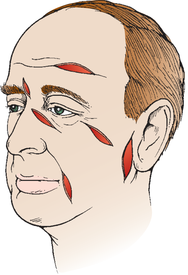
- Position the patient seated/semi-reclined, head neutral, muscles relaxed
- Ask the patient to smile (nasolabial folds, marionette lines become visible), frown, pucker lips
- Gently pinch the skin perpendicular to the suspected RSTL direction - natural parallel creases indicate RSTL direction
- Mark the incision with the long axis parallel to RSTLs
- For elliptical excisions, align the major axis along the RSTL
3. Wrinkle Lines: Kraissl and Rubin
- Photographed patients actively contracting facial muscles to map wrinkle lines
- These are dynamic lines of muscle contraction, perpendicular to the underlying muscle's long axis
- His wrinkle lines on the face largely correspond to RSTLs and are therefore often used interchangeably in facial surgery
- On the face: wrinkle lines ≈ RSTLs (both perpendicular to muscle action) - both are good surgical guides
- On the body/torso: wrinkle lines are sometimes inconsistently aligned with RSTLs, with Borges noting RSTLs are more reliable for the trunk and extremities
- The interchangeable use of "Langer's lines" and "RSTLs" is a common clinical error - particularly on the face, they can be nearly perpendicular (Pomerantz et al., Clarifying the role of skin tension lines in dermatological surgery)
4. The Biomechanics: Why Line Orientation Matters
Tension across the wound
- The skin contractile forces pull the wound edges apart
- Resulting tension stimulates fibroblasts to produce more collagen
- End result: wider, more raised, hypertrophic, or keloidal scar
- Tension acts along the wound axis, not across it
- Minimal gaping forces on wound edges
- Less mechanical stimulation of fibroblasts
- Result: narrow, flat, inconspicuous scar
Skin biomechanics
- Dermal collagen fibers are not randomly arranged; they have a preferred orientation
- The dermis behaves like a viscoelastic fiber-reinforced composite
- Young's modulus and stiffness are highest along Langer's/RSTL direction
- A cut along this direction encounters fibers parallel to the incision; fibers on either side have minimal tendency to retract or pull the wound open
5. Maxillofacial Surgery: Specific Applications
5.1 Facial Wound Closure Principles
"Facial and forehead lacerations that follow natural skin creases or lines will heal with a less noticeable scar than those that are oblique or perpendicular to the natural wrinkles of the skin."
- Orient incisions/ellipses along RSTLs - the single most impactful decision
- Conservative debridement - facial skin has excellent vascularity; even marginally perfused tissue often survives. Debride only clearly necrotic tissue to prevent sinking of scars and preserve contour
- Layered closure - traditionally favored for cosmetic repair of facial wounds. Approximate the dermis with buried absorbable sutures (SQ/subcuticular) to bring edges to within 1-2 mm of apposition before skin sutures
- Skin edge eversion - intentional slight eversion ("build pyramids, not ditches") is critical; inverting edges increases epithelial bridging time by ~300%
- Eliminate dead space - prevents seroma/hematoma; use buried inverted absorbable sutures
- Undermine extensively - double the wound width on each side to reduce tension
5.2 Region-Specific Guidance
- RSTLs run horizontally
- Horizontal lacerations/incisions heal excellently; vertical forehead incisions cross RSTLs and produce visible scars
- Coronal/bicoronal incisions used in craniofacial surgery deliberately run along the scalp to access the face
- Upper eyelid: thin, flexible skin. Use 6-0 suture or smaller. RSTLs follow the skin fold crease of the upper lid
- Lower eyelid: transconjunctival or subciliary incisions align with natural creases; do not shave eyebrows (landmarks are lost, regrowth is incomplete)
- "Crow's feet" wrinkle lines radiate from lateral canthus
- RSTLs run obliquely from nose toward the ear
- The parotid gland and facial nerve (CN VII) branches lie deep; cheek lacerations with clear discharge suggest parotid/Stensen's duct injury
- Subciliary and mid-face incisions should follow RSTL contours
- Orbicularis oris muscle runs circumferentially; RSTLs radiate perpendicular to it
- The vermilion border is the critical landmark - must be aligned precisely even 1 mm step is cosmetically unacceptable; interrupted suture at the border before closing the rest is standard
- Primary goal: cover cartilage (avascular; exposed cartilage → chondritis)
- Tension lines less relevant here; structural integrity and coverage are the priority
- RSTLs are horizontal
- Horizontal neck incisions (e.g., thyroidectomy) lie along RSTLs → excellent cosmesis
- Vertical neck incisions cross RSTLs → wider scars
6. Suturing Techniques in Context of Tension Lines
6.1 Planning the Elliptical Excision
- The major axis of the ellipse must be aligned parallel to the RSTL
- Standard ratio: 3:1 (length:width) minimum to prevent "dog-ear" formation at the tips
- Tip angle should be ≤ 30 degrees to prevent puckering
- Fusiform/spindle design with the long axis along RSTLs allows closure without bunching
6.2 Key Suturing Principles (from textbook evidence)
- Place interrupted sutures half as far apart as they are across the wound
- The more tension, the more sutures needed
- Fine sutures (5-0, 6-0) cause less scarring but require more passes; use this trade-off deliberately on the face
- Edema occurs after closure - only approximate tissues, do not strangulate
- Begin gentle wound washing at 12-24 hours; keep moist with ointment
6.3 Suture Material in Facial Wounds - Current RCT Evidence
- Resorbable sutures (Vicryl Rapide) vs. Non-resorbable (Ethilon) for adult facial lacerations
- No significant difference in aesthetic outcomes (VAS scores, Hamilton Scar Scale) between the two groups
- Patient satisfaction was high in both groups
- Early complication rates were significantly higher in the non-resorbable group at 1-week follow-up
- Cost: Resorbable sutures provided >5-fold cost saving (no removal appointment needed)
- Conclusion: Resorbable sutures are a viable, cost-effective alternative for facial laceration closure
6.4 Post-Suturing Scar Management
- Silicone gel used for ≥1 month post-suturing improves both cosmetic and symptomatic scar outcomes
- Mechanism: occlusion, hydration, and compression of healing dermis reduce fibroblast hyperactivity
- Recommendation: advise patients to use topical silicone gel for at least 1 month after suture removal
7. Current Advancements: Biodynamic Excisional Skin Tension (BEST) Lines
The Key Insight: Incisions vs. Excisions
- Incisions (simple linear cuts): During closure, collagen is mainly compressed/stretched along the long axis - historical RSTL guidance is adequate
- Excisions (removal of tissue): Creating a wound requires mobilizing and advancing tissue from multiple directions. When tissue is removed, collagen stretches AND elastin buckles simultaneously. This produces tension vectors that are fundamentally different from simple incision tension
BEST Line Derivation
- Real-time force transducers measured skin tension during excision in multiple body regions in living patients
- The direction of minimum wound closure tension was mapped across the body
- These empirical BEST lines often differ from historical lines
- On the face: BEST lines are remarkably similar to RSTLs (validating Borges' clinical approach for facial excisions) but with some regional differences
- On the scalp: Langer lines run sagittal (front-back dividing); BEST lines run coronal (side-side dividing) - less tension in coronal direction
- On the lower limb: BEST line excisions show significantly less wound closure tension than RSTL-guided excisions
- Skin tension in BEST lines shows minimal variation with age and sex - making them highly generalizable
Key Findings of the BEST Line Research
- Wrinkle lines are not suitable for excisional surgery on the body - they reflect surface crease lines but not the deep biomechanical response to tissue removal
- On the face, RSTL/BEST line correspondence is high - facial RSTLs remain clinically reliable
- Parallel relaxing incisions along BEST lines (placing small parallel incisions on either side of the excision) can further reduce wound closure tension, especially for larger excisions
- BEST lines provide an objective, consistent, patient-independent map for excisional surgery planning
8. The Confusion Problem: Clinical Implications
| Feature | Langer's Lines | RSTLs | BEST Lines |
|---|---|---|---|
| Derived from | Cadavers (rigor mortis) | Living, relaxed skin | Living patients with real-time tensiometry |
| Method | Awl puncture, ellipse orientation | Skin pinch creases | Force transducer during excision |
| Introduced | 1861 | ~1984 (Borges) | 2010s (Paul) |
| Face relevance | Poor (often ⊥ to wrinkles) | High | High (close to RSTL) |
| Body/trunk relevance | Moderate | Good for incisions | Best for excisions |
| Dynamic? | No | Partially (relaxed) | Yes (intraoperative) |
| Intended use | Incisions | Incisions + excisions | Primarily excisions |
9. Summary for Clinical Practice
-
For facial incisions and lacerations: Use RSTLs as your primary guide. Identify them preoperatively by having the patient relax and make facial expressions. Align wound closure and elliptical excisions with RSTL direction.
-
Do not use Langer's lines on the face: On the face, they frequently run perpendicular to RSTLs and are an inappropriate surgical guide.
-
For facial excisional surgery (lesion removal): RSTL guidance is appropriate - BEST lines are closely aligned with RSTLs on the face.
-
Suture materials: Current RCT evidence (TORN Face trial, 2024) supports resorbable sutures as equivalent to non-resorbable for facial lacerations, with cost and convenience advantages.
-
Post-operative scar management: Topical silicone gel for ≥1 month post-suture removal improves scar outcomes (systematic review, 2025).
-
Technical pearls (textbook-based):
- Undermine widely (double wound width on each side)
- Use buried absorbable sutures to eliminate dead space and reduce skin tension
- Evert skin edges
- Never strangulate tissue; only approximate
- Preserve subcutaneous fat in facial wounds to prevent scar sinking
- Conservative debridement given the face's superior vascularity
-
Future direction - BEST lines: As intraoperative tensiometry becomes more accessible, BEST line mapping may replace historical line systems for complex maxillofacial excisional and reconstructive surgery, providing real-time, patient-specific tension data.
- Langer K (1861) - Original cleavage lines work
- Borges AF (1984) - RSTL for facial excisions
- Kraissl CJ (1951) - Wrinkle lines for facial surgery
- Paul SP (2017). Biodynamic Excisional Skin Tension Lines. Plast Reconstr Surg Glob Open. PMID 29632788
- Basyuni S et al. (2024). TORN Face RCT - resorbable vs non-resorbable sutures. Br J Oral Maxillofac Surg. PMID 38926072
- Kendrick M & Dor A (2025). Silicone gel for facial lacerations. Emerg Med J. PMID 40523697
- Pomerantz ER et al. - Clarifying the role of skin tension lines in dermatological surgery
- Roberts & Hedges' Clinical Procedures in Emergency Medicine (Facial Wounds section)
- Pfenninger & Fowler's Procedures for Primary Care, 3rd ed. (Box 22-3, Pearls of Suturing)