Why in liver failure spironolactone is used and not other diuretics like thiazides and loop diuretics

Reading File
Searching the Web

spironolactone liver failure ascites mechanism why not loop diuretics thiazides

Finding Sources
Finding Sources
Reading File
Reading File
Finding Sources
Reading File
Finding Sources
Loading Image
I now have everything needed for a comprehensive, well-sourced answer. Here it is:

Why Spironolactone - Not Thiazides or Loop Diuretics - in Liver Failure

The short answer: the dominant mechanism of sodium retention in cirrhosis is secondary hyperaldosteronism, and spironolactone directly targets that mechanism. The others don't, and they introduce dangerous complications.

Step 1: Understand the Pathophysiology

Cirrhosis causes increased intrahepatic resistance → portal hypertension → splanchnic/systemic arteriolar vasodilation. This reduces effective arterial blood volume, which triggers the baroreceptors to activate the renin-angiotensin-aldosterone system (RAAS) and the sympathetic nervous system. The result is massive aldosterone excess from two directions:
  1. Increased production - activated RAAS drives the adrenal gland to make more aldosterone
  2. Decreased hepatic degradation - the failed liver cannot metabolize aldosterone at normal rates
Aldosterone acts on the collecting duct (principal cells) to upregulate ENaC (epithelial sodium channels) and Na+/K+-ATPase, driving avid sodium retention and potassium wasting.
This is illustrated in the pathway from Yamada's Textbook of Gastroenterology:
Site of action of therapies for ascites in cirrhosis - Yamada's Gastroenterology
Figure 98.3 - Spironolactone acts at the neurohumoral activation level, while furosemide only acts downstream at the sodium retention level. Furosemide should only be used in conjunction with spironolactone.

Step 2: Why Spironolactone Works

Spironolactone is a competitive aldosterone antagonist (mineralocorticoid receptor blocker). It directly neutralizes the primary driver of sodium retention. Because it works at the collecting duct - exactly where hyperaldosteronism is causing the problem - it is the most pathophysiologically targeted agent.
As stated in Sleisenger and Fordtran's Gastrointestinal and Liver Disease:
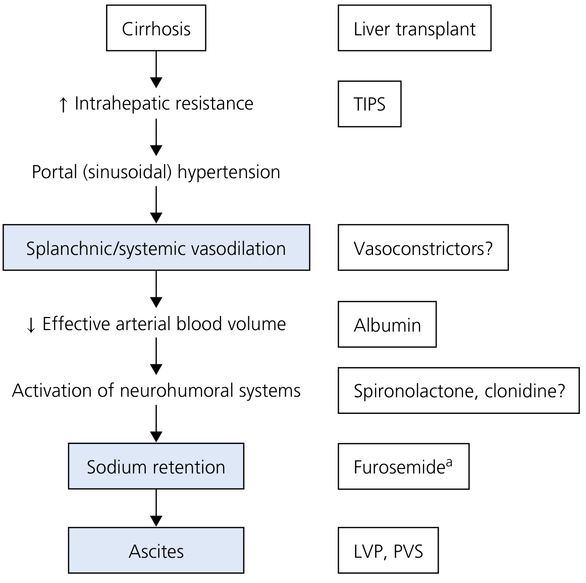
"Because hyperaldosteronism plays a key role in renal sodium retention in cirrhosis, anti-mineralocorticoids (particularly spironolactone or eplerenone) represent the first line of treatment in the management of cirrhotic ascites."

Step 3: Why Loop Diuretics (e.g., Furosemide) Fail Alone

Loop diuretics block the Na-K-2Cl cotransporter in the thick ascending limb of the loop of Henle - a segment that is proximal to where aldosterone acts. This creates a specific and critical problem in cirrhosis:
"When furosemide is used alone, sodium not reabsorbed in the loop of Henle is avidly absorbed in the distal and collecting tubules as a result of the hyperaldosteronism characteristic of most cirrhotic patients with ascites."
  • Yamada's Textbook of Gastroenterology
In other words, the loop diuretic pushes more sodium past the loop, but the downstream aldosterone-driven machinery simply absorbs it all back. The AASLD summarizes this well: RCTs consistently showed furosemide alone was significantly less effective than spironolactone in cirrhotic ascites.
Additional risks of loop diuretics in liver failure:
  • Hypokalemia - worsens hepatic encephalopathy (K+ depletion promotes ammonia production and renal ammonia generation)
  • Metabolic alkalosis - further impairs brain function in encephalopathy
  • Volume depletion - triggers hepatorenal syndrome (HRS), one of the most feared complications; only ~500 mL/day can safely be mobilized from ascites without causing circulatory dysfunction

Step 4: Why Thiazides Are Not Used

Thiazides block the NCC (Na-Cl cotransporter) in the distal convoluted tubule. The same logic applies - this is still upstream of the site where aldosterone maximally acts (the collecting duct), so the benefit is blunted. Additionally:
  • Thiazides carry a high risk of hyponatremia - already a major problem in advanced cirrhosis (dilutional hyponatremia from ADH activation)
  • Thiazides also cause hypokalemia, again risking encephalopathy
  • There is no evidence they offer any advantage over the spironolactone/furosemide combination
  • As noted by Yamada's Gastroenterology: "There is no evidence that other diuretics, such as metolazone, thiazides, or other loop diuretics such as bumetanide or torsemide, offer an advantage over spironolactone and furosemide."

Practical Clinical Protocol

SituationFirst-line
Mild-moderate ascitesSpironolactone 100 mg/day alone
Inadequate responseAdd furosemide 40 mg/day (ratio kept at 100:40 to maintain normokalemia)
Titrate up to maximumSpironolactone 400 mg + Furosemide 160 mg
Painful gynecomastiaSwitch to amiloride (another K+-sparing diuretic, though less effective)
The 100:40 spironolactone:furosemide ratio is maintained throughout titration because it preserves normal potassium - spironolactone's K+-sparing effect balances furosemide's K+-wasting effect.
Key risks to monitor regardless of regimen:
  • Renal impairment from intravascular depletion (~25% of patients)
  • Hyponatremia (~28%)
  • Hepatic encephalopathy (~26%)
  • Hyperkalemia (especially with spironolactone in renal impairment)

Summary Table

DiureticSite of actionProblem in liver failure
SpironolactoneCollecting duct (blocks aldosterone)Preferred - targets the root cause
FurosemideLoop of HenleSodium reabsorbed downstream by aldosterone; causes hypokalemia/HRS
ThiazidesDistal convoluted tubuleSame bypass problem; causes hyponatremia/hypokalemia
AmilorideCollecting duct (blocks ENaC directly)Alternative if gynecomastia; less natriuretic effect
The bottom line: spironolactone wins because it attacks the actual pathophysiological driver. Loop diuretics and thiazides act at the wrong place in the nephron and leave the hyperaldosteronism-driven sodium absorption fully intact downstream.

Sources: Yamada's Textbook of Gastroenterology (7th ed.), Sleisenger & Fordtran's Gastrointestinal and Liver Disease, Harrison's Principles of Internal Medicine 22E, AASLD guidelines on cirrhotic ascites diuretic therapy

Action of loop diuretics in the blood electrolytes levels

Finding Sources
Searching the Web

loop diuretics electrolyte effects blood Na K Mg Ca mechanism

Finding Sources
Finding Sources
Reading File
Finding Sources
Finding Sources
Finding Sources
Reading File
Loading Image
Reading File
Loading Image
I now have everything needed - two excellent diagrams from Brenner & Rector's The Kidney plus comprehensive textbook content. Here is the full answer:

Effects of Loop Diuretics on Blood Electrolytes

The Root Mechanism

Loop diuretics (furosemide, bumetanide, torsemide, ethacrynic acid) block NKCC2 - the Na-K-2Cl cotransporter on the luminal membrane of the thick ascending limb (TAL) of the loop of Henle. This single blockade creates a cascade of electrolyte disturbances, all explained mechanistically below.
TAL cell showing loop diuretic blocking NKCC2 and abolishing the 10mV lumen-positive potential - Brenner & Rector's The Kidney
Fig. 50.3 - Loop diuretics bind NKCC2 from the luminal surface, preventing Na⁺, K⁺, and 2Cl⁻ entry into the cell. This also abolishes the +10mV lumen-positive transepithelial voltage, which eliminates the electrical driving force for paracellular Ca²⁺ and Mg²⁺ reabsorption.

1. Hyponatremia (↓ Na⁺)

Mechanism:
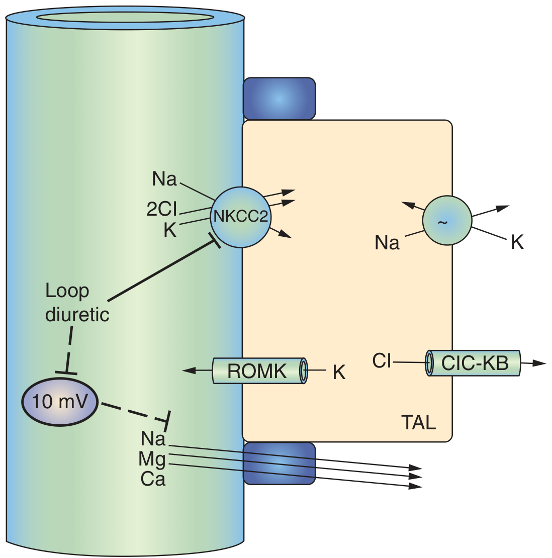
  • NKCC2 blockade directly prevents Na⁺ reabsorption in the TAL, which accounts for ~25% of all filtered sodium
  • The increased distal sodium delivery overwhelms downstream reabsorption capacity
  • Volume depletion from Na⁺ loss stimulates ADH release (non-osmotic), causing water retention - this dilutes serum Na⁺ further
  • Secondary RAAS activation causes more renin/aldosterone release, driving more Na⁺ retention (partially compensatory), but at the expense of K⁺
Net blood effect: ↓ Na⁺ (hyponatremia)

2. Hypokalemia (↓ K⁺) - the most clinically important electrolyte effect

This is a two-step mechanism:
Step 1 - Increased tubular flow to the collecting duct:
  • NKCC2 blockade means more Na⁺ is delivered to the principal cells of the collecting duct
  • The Na⁺/K⁺-ATPase in principal cells reabsorbs this extra Na⁺ by exchanging it for K⁺ secretion into the lumen - K⁺ is lost in urine
Step 2 - Secondary hyperaldosteronism:
  • Loop diuretics stimulate renin secretion directly via the macula densa (by blocking NaCl entry into macula densa cells, reducing TGF-mediated feedback)
  • As Brenner & Rector states: "Loop diuretics also stimulate renin secretion, both in the short term and long term... a major component is from direct effects on the macula densa"
  • More renin → more angiotensin II → more aldosterone → aldosterone further amplifies K⁺ secretion at the collecting duct
Net blood effect: ↓ K⁺ (hypokalemia) - which can trigger cardiac arrhythmias, muscle weakness, and (in liver disease) precipitates hepatic encephalopathy

3. Hypochloremia (↓ Cl⁻)

  • NKCC2 directly transports 2 Cl⁻ per cycle - blocking it massively increases urinary Cl⁻ loss
  • Cl⁻ is lost in urine alongside Na⁺ and K⁺
Net blood effect: ↓ Cl⁻ (hypochloremia)

4. Metabolic Alkalosis (↑ HCO₃⁻, ↑ pH)

Three mechanisms work together:
  1. Volume contraction alkalosis - loss of Cl⁻-rich fluid contracts the ECF volume; a fixed amount of HCO₃⁻ is now dissolved in a smaller volume → "contraction alkalosis"
  2. Hypokalemia drives alkalosis - K⁺ depletion causes H⁺ to shift into cells in exchange for K⁺, lowering extracellular [H⁺] → alkalosis
  3. Aldosterone effect - secondary hyperaldosteronism drives H⁺ secretion by intercalated cells of the collecting duct, generating more HCO₃⁻
Net blood effect: metabolic alkalosis (note: acetazolamide is used to treat this when problematic)

5. Hypomagnesemia (↓ Mg²⁺)

This is mediated through the transepithelial voltage mechanism:
Normally, the NKCC2-driven activity in the TAL generates a +10 mV lumen-positive transepithelial voltage. Mg²⁺ (a divalent cation) is reabsorbed passively via the paracellular pathway, driven by this positive luminal charge. As Comprehensive Clinical Nephrology states:
"Loop diuretics eliminate this potential difference and can therefore increase calcium and magnesium excretion."
When loop diuretics abolish the +10mV voltage → no electrical gradient → Mg²⁺ stays in the lumen → lost in urine
Net blood effect: ↓ Mg²⁺ (hypomagnesemia) - can cause tetany, arrhythmias, worsens hypokalemia (because hypomagnesemia impairs Na/K-ATPase, causing renal K⁺ wasting)

6. Hypocalcemia (↓ Ca²⁺) - key distinction from thiazides

Same voltage-dependent mechanism as magnesium:
  • Ca²⁺ (~20% of filtered load) is reabsorbed passively via paracellular route in the TAL, driven by the +10 mV transepithelial voltage
  • Loop diuretics abolish this voltage → Ca²⁺ lost in urine
This diagram from Brenner & Rector's The Kidney contrasts loop vs. thiazide (DCT) diuretics beautifully:
Diuretic effects on Ca and Mg - comparison of loop vs DCT diuretics - Brenner & Rector's The Kidney
Fig. 50.4 - Under loop diuretics (middle panel): ↑ Na, ↑ Cl, ↑ Ca, ↑ Mg in urine (meaning ↓ in blood). Under DCT (thiazide) diuretics (right panel): ↑ Na, ↑ Cl, ↑ Mg in urine, but ↓ Ca in urine (meaning ↑ in blood). This is the fundamental difference.
This is why:
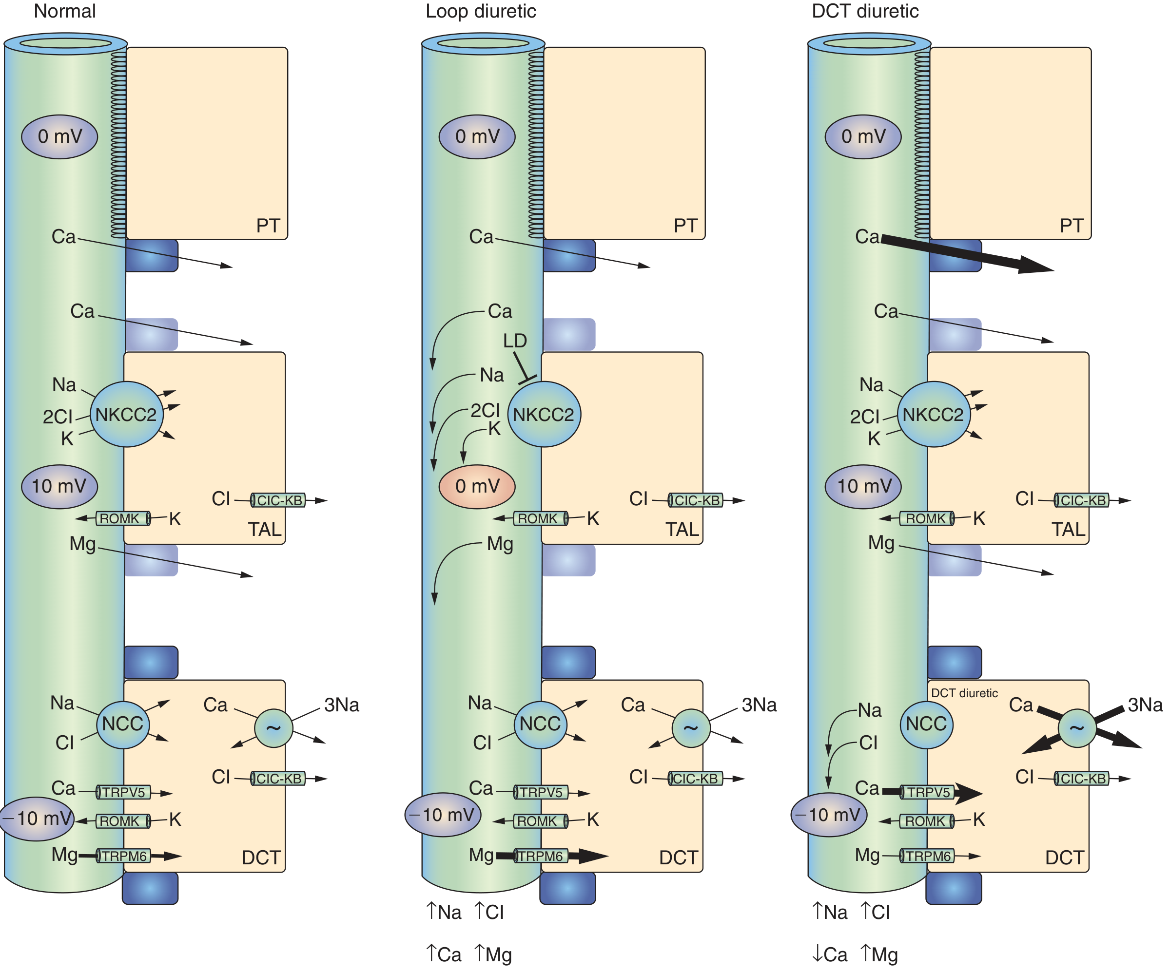
  • Loop diuretics → hypocalcemia → used clinically to treat hypercalcemia
  • Thiazides → hypercalcemia → preferred in patients with osteoporosis or calcium stones
Net blood effect: ↓ Ca²⁺ (hypocalcemia)

7. Hyperuricemia (↑ Uric acid) - bonus effect

  • Acutely, loop diuretics increase uric acid excretion
  • Chronically, volume depletion enhances proximal tubular uric acid reabsorption (via OAT transporters), raising serum uric acid
  • Can precipitate gout in susceptible patients

Summary Table

ElectrolyteBlood LevelKey Mechanism
Na⁺↓ (hyponatremia)Direct TAL blockade + ADH-driven water retention
K⁺↓ (hypokalemia)↑ distal Na⁺ delivery + secondary hyperaldosteronism
Cl⁻↓ (hypochloremia)NKCC2 directly transports 2Cl⁻
HCO₃⁻/pH↑ (metabolic alkalosis)Volume contraction + hypokalemia + aldosterone
Mg²⁺↓ (hypomagnesemia)Loss of +10mV transepithelial voltage in TAL
Ca²⁺↓ (hypocalcemia)Loss of +10mV transepithelial voltage in TAL
Uric acid↑ (hyperuricemia)Chronic proximal tubular reabsorption via volume contraction

Clinical pearls:
  • Hypokalemia and hypomagnesemia are interlinked - you often cannot correct K⁺ without first repleting Mg²⁺, because low Mg²⁺ impairs renal K⁺ retention
  • The metabolic alkalosis from loop diuretics is Cl⁻-responsive (corrects with volume and KCl repletion)
  • Furosemide's calciuric effect is exploited therapeutically in hypercalcemic crisis (IV furosemide + IV saline drives urinary Ca²⁺ loss)

Sources: Brenner & Rector's The Kidney (2-Volume Set), Comprehensive Clinical Nephrology 7th Ed., Goldman-Cecil Medicine, Harrison's Principles of Internal Medicine 22E

How does metformin causes B12 deficiency

Finding Sources
Searching the Web

metformin vitamin B12 deficiency mechanism calcium intrinsic factor ileum

Finding Sources
Finding Sources
Reading File
Reading File
Finding Sources
Reading File
Searching PubMed

metformin vitamin B12 deficiency mechanism

I have all the content needed for a thorough, well-sourced answer. Here it is:

How Metformin Causes Vitamin B12 Deficiency

To understand the mechanism, you first need to know the normal B12 absorption pathway - because metformin interferes with a specific, calcium-dependent step in it.

Normal B12 Absorption (Brief Review)

  1. Stomach - Dietary B12 (cobalamin) is released from food proteins by pepsin and gastric acid, then binds to a salivary protein called haptocorrin (R-protein)
  2. Duodenum - Pancreatic proteases release B12 from haptocorrin; free B12 then binds intrinsic factor (IF), secreted by gastric parietal cells
  3. Terminal ileum - The B12-IF complex binds to a specific receptor called cubilin on the surface of ileal epithelial cells. This step is calcium-dependent - Ca²⁺ is required for the complex to dock onto the receptor
  4. Endocytosis - The B12-IF-cubilin complex is internalized; within the cell, B12 binds transcobalamin II and enters the portal circulation
  5. Liver - B12 is stored in large amounts (enough for 3-5 years), and undergoes enterohepatic recirculation via bile
(Source: Robbins Pathologic Basis of Disease; Lippincott's Biochemistry)

How Metformin Disrupts This

There are three proposed mechanisms, with the calcium-dependent one being the most widely accepted:

Mechanism 1: Calcium-Dependent Ileal Absorption Blockade (Primary Mechanism)

This is the most evidence-supported pathway.
  • Metformin is a biguanide with a hydrophobic tail that inserts into the hydrocarbon core of intestinal cell membranes
  • This insertion imparts a net positive charge to the cell membrane surface
  • The positively charged membrane repels Ca²⁺ ions at the ileal lumen-cell interface
  • Without adequate Ca²⁺, the B12-IF complex cannot bind to the cubilin receptor on ileal epithelial cells
  • The complex passes through the gut unabsorbed and is lost in stool
This is described as "calcium-dependent ileal membrane antagonism" - and critically, this effect is reversible by calcium supplementation. Clinical trials have shown that giving calcium carbonate (1200 mg/day) to metformin-treated patients partially or fully restores B12 absorption. This is the strongest evidence that Ca²⁺ displacement is the primary mechanism.
As summarized by Goldman-Cecil Medicine:
"Metformin → Vitamin B₁₂ → Impairs absorption" (drug-nutrient interaction table)

Mechanism 2: Gut Microbiome Alteration and Bacterial Overgrowth

  • Metformin is known to significantly alter the gut microbiome - it promotes growth of certain bacteria (notably Akkermansia muciniphila and Bifidobacterium) and suppresses others
  • Some of the altered bacterial species consume vitamin B12 for their own metabolic needs, reducing the amount available for absorption
  • Additionally, bacterial overgrowth in the small intestine can compete for the B12-IF complex before it reaches the terminal ileum
  • Metformin also slows intestinal transit (contributes to its GI side effects), which may prolong bacterial exposure to the B12-IF complex

Mechanism 3: Direct Structural Interference with the B12-IF Complex

  • A third proposed mechanism suggests metformin may directly bind to the B12-IF complex, altering its structure
  • A structurally modified complex would have reduced affinity for the cubilin receptor, even if calcium is available
  • This mechanism is less well characterized than Mechanism 1

Why This Matters Clinically

The B12 deficiency from metformin is:
  • Dose-dependent - higher daily doses = greater risk
  • Duration-dependent - longer treatment = greater cumulative depletion of hepatic stores
  • Seen in up to 10-30% of long-term users in various studies
  • Clinically silent early - hepatic B12 stores last 3-5 years, so deficiency can develop insidiously over years of treatment
The key danger is that diabetic patients already have peripheral neuropathy from hyperglycemia, and B12 deficiency from metformin can worsen or mimic this neuropathy (distal symmetrical polyneuropathy, autonomic neuropathy). Because both look the same clinically, the B12 contribution is often missed. The PubMed literature highlights that metformin-induced B12 deficiency "can cause or worsen distal symmetrical, autonomic and cardiac neuropathy in the patient with diabetes."
An important additional clinical note from Goldman-Cecil: serum B12 levels in the low-normal range (200-350 pg/mL) may still be insufficient at the cellular level - a raised serum methylmalonic acid (MMA) is a more sensitive marker of true cellular B12 deficiency.

Risk Factors for Greater Depletion

Risk FactorWhy It Worsens B12 Depletion
High metformin dose (>1.5 g/day)More membrane charge displacement
Duration >5 yearsHepatic stores eventually depleted
Vegetarian/vegan dietLow dietary B12 intake to begin with
Concomitant PPI or H2 blocker usePPIs reduce gastric acid → impair B12 release from food; together with metformin, up to 65% reduction in B12 absorption
Elderly patientsMore atrophic gastritis, reduced IF secretion, reduced gastric acid
Pre-existing low B12 storesLess buffer before clinical deficiency

Summary of the Mechanism

Metformin (hydrophobic tail inserts into ileal cell membrane)
        ↓
Net positive charge on membrane surface
        ↓
Ca²⁺ ions repelled from ileal lumen-cell interface
        ↓
B12-Intrinsic Factor complex cannot bind cubilin receptor
        ↓
B12-IF complex not endocytosed → lost in stool
        ↓
Reduced circulating B12 → hepatic stores slowly depleted
        ↓
Megaloblastic changes + Peripheral/autonomic neuropathy

Management

  • Routine monitoring: Check serum B12 every 1-2 years in long-term metformin users; use MMA levels if borderline
  • Calcium supplementation: 1200 mg/day calcium carbonate can partially restore absorption
  • B12 supplementation: Oral B12 1000 µg/day (works via passive diffusion, even without IF) or IM cyanocobalamin if severe deficiency
  • Do not stop metformin for this reason alone - the cardiovascular and glycemic benefits far outweigh the risk when monitored and supplemented appropriately

Sources: Robbins & Cotran Pathologic Basis of Disease, Lippincott's Illustrated Reviews: Biochemistry, Goldman-Cecil Medicine (International Ed.), Endocrinology Advisor review on metformin and B12, Clinical Diabetology - Bahl et al.

List me the various names effects in medicine ex Macklin effect

Searching the Web

famous named effects phenomena in medicine eponymous list clinical

Finding Sources
Searching the Web

named effects in medicine "Bohr effect" "Haldane effect" "Cushing reflex" "Starling law" "Bainbridge reflex" physiology eponymous effects list

Finding Sources
Finding Sources
Finding Sources
I have excellent content. Now let me compile a comprehensive, well-organized list drawing from all sources plus my knowledge:

Named Effects in Medicine - A Comprehensive List

These are organized by system/category for easier reference. Each is a named physiological, pathological, or clinical phenomenon (not just a disease name).

RESPIRATORY

Macklin Effect Air tracks from ruptured alveoli along bronchovascular sheaths into the mediastinum, causing pneumomediastinum. Seen in trauma (especially blunt chest injury), COVID-19, and during mechanical ventilation. The air follows the path of least resistance centrally. Radiologically seen as streaky air collections around the bronchi.
Bohr Effect Increased CO₂ and H⁺ (lower pH) shift the oxyhemoglobin dissociation curve to the right, reducing Hb's affinity for O₂ and facilitating O₂ unloading at metabolically active tissues. Described by Christian Bohr (1904). - Lippincott Biochemistry
Haldane Effect The reverse of the Bohr effect for CO₂ transport. Deoxygenated hemoglobin carries more CO₂ than oxygenated hemoglobin at the same PCO₂. This is because deoxy-Hb more readily accepts protons (H⁺), shifting equilibrium toward carbamino-Hb formation and bicarbonate production. Facilitates CO₂ loading in tissues and unloading in lungs. - Fishman's Pulmonary Diseases
Venturi Effect Acceleration of fluid/gas through a constricted segment causes a drop in lateral pressure (Bernoulli principle applied). Used in Venturi masks to deliver precise FiO₂ by entraining room air in a fixed ratio.
Bernoulli Effect / Principle In a flowing fluid, increased velocity = decreased pressure. Basis for the Venturi mask, and explains dynamic airway collapse during forced expiration (airway walls collapse inward at high flow rates).
Equal Pressure Point (EPP) During forced expiration, the point in the airway where intraluminal pressure equals pleural pressure. Downstream of the EPP, airways are subject to dynamic compression. Key concept in understanding airflow limitation in COPD and emphysema.

CARDIOVASCULAR

Frank-Starling Law (Starling's Law of the Heart) Stroke volume increases in proportion to end-diastolic volume (preload). Greater sarcomere stretch → greater force of contraction, up to an optimal length. The fundamental mechanism by which the heart matches output to venous return. - Goldman-Cecil Medicine, Ganong's Physiology
Anrep Effect When aortic pressure (afterload) is abruptly increased, there is an initial fall in stroke volume followed by a slow, gradual recovery of contractility over minutes. Attributed to stretch-induced release of autocrine/paracrine factors from cardiac myocytes.
Bowditch Effect (Treppe / Staircase Effect) Increasing heart rate leads to a stepwise increase in contractility (positive force-frequency relationship). More frequent action potentials → more Ca²⁺ available per beat via sodium-calcium exchanger.
Cushing Reflex (Cushing Response) ↑ Intracranial pressure → brainstem ischemia → massive sympathetic discharge → hypertension + bradycardia + irregular breathing (Cushing's triad). The hypertension is a compensatory response to maintain cerebral perfusion pressure. A clinical sign of impending herniation.
Bainbridge Reflex ↑ Right atrial pressure (volume overload) → stretches atrial mechanoreceptors → reflexive tachycardia via vagal afferents to medullary centers. Helps increase cardiac output to clear venous overload.
Bezold-Jarisch Reflex Activation of cardiac C-fiber (vagal) receptors in the inferoposterior left ventricle (by chemical stimuli or distension) triggers bradycardia, hypotension, and apnea. Seen in inferior MI, Bezold-Jarisch reflex can cause paradoxical bradycardia. Also involved in vasovagal syncope.
Windkessel Effect The aorta and large elastic arteries act as a pressure reservoir: they expand during systole (storing energy) and recoil during diastole (maintaining diastolic pressure and continuous capillary flow). Lost in arteriosclerosis, contributing to wide pulse pressure in the elderly.
Reverse Pulsus Paradoxus (Kussmaul's Sign) Paradoxical rise in JVP on inspiration (opposite to normal). Seen in constrictive pericarditis and right heart failure - the right ventricle cannot accommodate increased venous return on inspiration.
Doppler Effect Change in frequency of a wave (sound, ultrasound) when source or observer is moving. Basis for Doppler ultrasound to measure blood flow velocity and direction in echocardiography and vascular studies.

RENAL / FLUID

Gibbs-Donnan Effect The presence of non-diffusible charged proteins on one side of a membrane creates an unequal distribution of diffusible ions across the membrane, generating an electrical potential. Important for understanding plasma vs. interstitial fluid ion differences and capillary fluid exchange.
Starling Forces The four forces determining fluid movement across capillary walls: capillary hydrostatic pressure, interstitial hydrostatic pressure, capillary oncotic pressure, interstitial oncotic pressure. Governs edema formation and fluid resuscitation physiology. Named after Ernest Starling.
Tubuloglomerular Feedback (Macula Densa Effect) Increased NaCl delivery to the macula densa → release of adenosine → afferent arteriole constriction → reduced GFR. A key autoregulatory mechanism. Blocked by loop diuretics (clinically relevant for preserving GFR in diuretic therapy).

BLOOD / HEMATOLOGY

Rouleaux Formation Red blood cells stacking like coins due to elevated plasma proteins (fibrinogen, immunoglobulins). Not strictly a "named effect" but a named phenomenon. Seen in multiple myeloma, infections, inflammation.
Rapoport-Luebering Shunt (2,3-DPG Effect) In red blood cells, 2,3-bisphosphoglycerate (2,3-BPG) binds to deoxy-Hb and stabilizes the T (tense) conformation, shifting the ODC to the right → reduced O₂ affinity → better O₂ delivery. Increases in chronic hypoxia, anemia, and high altitude.

PHARMACOLOGY / TOXICOLOGY

First-Pass Effect Orally administered drugs absorbed from the gut are carried via the portal vein to the liver before reaching systemic circulation, where hepatic metabolism can substantially reduce bioavailability. Why some drugs (e.g., nitroglycerin, morphine) have very different IV vs oral doses.
Prozone Effect (Hook Effect) At very high antigen concentrations, an immunoassay gives a falsely low or negative result because antigen excess prevents antibody cross-linking needed for precipitation. Clinically important in syphilis serology (RPR may be falsely negative in secondary syphilis).
Crabtree Effect In the presence of high glucose, cells suppress oxidative phosphorylation and favor aerobic glycolysis (Warburg-like shift). Relevant in cancer cell metabolism.
Warburg Effect Cancer cells preferentially use aerobic glycolysis (fermentation) even in the presence of oxygen, rather than oxidative phosphorylation. Basis of PET scanning (tumors consume ¹⁸F-FDG avidly due to upregulated glucose transport).
Tachyphylaxis Rapid diminution of a drug's effect with repeated doses in short succession - distinct from tolerance (which develops over longer periods). Classic example: ephedrine, certain bronchodilators.

NEUROLOGICAL / BRAIN

Kindling Effect Repeated sub-threshold electrical stimulation of the brain eventually leads to spontaneous seizures. Model for epileptogenesis. Also proposed as a mechanism for recurrent mood episodes in bipolar disorder becoming progressively easier to trigger.
Spreading Depression (Leão's Spreading Depression) A wave of neuronal depolarization followed by prolonged suppression, propagating across the cortex at ~3 mm/min. Basis for the migraine aura. Described by Aristides Leão (1944).
Mass Effect Not a physiological phenomenon but a clinical radiological term - an intracranial lesion (tumor, hematoma, abscess) displaces and compresses adjacent structures. Causes midline shift, herniation syndromes.

ENDOCRINE / METABOLIC

Somogyi Effect Rebound hyperglycemia in the morning following nocturnal hypoglycemia. The hypoglycemia triggers counter-regulatory hormone release (glucagon, cortisol, adrenaline), causing morning glucose surge. Controversial - some dispute its clinical frequency.
Dawn Phenomenon Morning hyperglycemia due to physiological rises in growth hormone and cortisol in the early morning hours (not preceded by hypoglycemia). Increases insulin requirements in the early morning. Distinct from Somogyi.
Staub-Traugott Effect Successive oral glucose loads produce progressively smaller rises in blood glucose. Reflects enhanced glucose-stimulated insulin release with repeated exposure. The physiological basis for testing glucose tolerance over multiple time points.

PULMONARY / CRITICAL CARE

Oxygen Toxicity (Paul Bert Effect) Breathing high-pressure O₂ (hyperbaric) causes CNS toxicity (convulsions). Described by Paul Bert (1878). Distinct from Lorrain Smith effect (pulmonary O₂ toxicity from prolonged normobaric high FiO₂).
Lorrain Smith Effect Pulmonary oxygen toxicity from prolonged high FiO₂ at normobaric pressure → inflammation, alveolar damage, ARDS-like picture. Relevant in mechanically ventilated patients.
Euler-Liljestrand Mechanism Hypoxia in a lung region causes local pulmonary vasoconstriction (hypoxic pulmonary vasoconstriction, HPV), diverting blood away from poorly ventilated alveoli to better-ventilated ones. Optimizes V/Q matching. Inhibited by volatile anesthetics, calcium channel blockers, and vasodilators.
Pendelluft Gas oscillation between lung units during spontaneous breathing in ARDS - regional overdistension caused by uneven lung mechanics. A mechanism of patient self-inflicted lung injury (P-SILI) even with low tidal volumes.

GASTROENTEROLOGY / SURGERY

Courvoisier's Law (Sign) A palpable, non-tender gallbladder in a jaundiced patient suggests malignant biliary obstruction (pancreatic head cancer) rather than gallstones - because chronic gallstone disease fibroses the gallbladder wall, preventing it from distending.
Pringle Manoeuvre Effect Clamping the hepatoduodenal ligament (portal triad) temporarily stops hepatic blood flow, reducing bleeding during liver surgery. "Effect" refers to the ischemia-reperfusion injury that follows its release.

IMMUNOLOGY / ONCOLOGY

Abscopal Effect Local radiotherapy to a tumor causes regression of distant, non-irradiated metastases - mediated by systemic immune activation (T cells). Rare clinically, but dramatically enhanced when combined with checkpoint inhibitors (anti-PD1, anti-CTLA4). Currently a major area of research.
Bystander Effect (Radiation) Cells not directly irradiated but adjacent to irradiated cells show DNA damage and cell death due to signals (ROS, cytokines) released by irradiated neighbors. Relevant in radiation therapy and radioprotection.
Epitope Spreading In autoimmune disease, the immune response initially targeting one antigen eventually diversifies to attack additional epitopes on the same or different proteins, broadening autoimmune attack over time. Mechanism of disease progression in conditions like lupus and multiple sclerosis.

QUICK-REFERENCE SUMMARY TABLE

EffectSystemOne-Line Summary
MacklinRespiratoryAlveolar rupture → pneumomediastinum via bronchovascular sheath
BohrRespiratory↑CO₂/↑H⁺ → right shift of ODC → O₂ unloading
HaldaneRespiratoryDeoxy-Hb carries more CO₂ than oxy-Hb
Euler-LiljestrandRespiratoryHypoxia → local pulmonary vasoconstriction → V/Q matching
Frank-StarlingCardiac↑Preload → ↑Stroke volume
Bowditch/TreppeCardiac↑Heart rate → ↑Contractility
AnrepCardiac↑Afterload → slow recovery of contractility
CushingNeuro/Cardiac↑ICP → hypertension + bradycardia + irregular breathing
BainbridgeCardiac↑Atrial pressure → tachycardia
Bezold-JarischCardiacCardiac C-fiber stimulation → bradycardia + hypotension
WindkesselVascularAortic elastic recoil maintains diastolic pressure
DopplerVascularWave frequency shift used in blood flow measurement
Starling ForcesRenalFour forces governing capillary fluid exchange
WarburgOncologyCancer cells use aerobic glycolysis (↑PET uptake)
AbscopalOncologyLocal RT causes systemic tumor regression via immunity
ProzoneImmunologyAntigen excess → false negative serology
First-PassPharmacologyHepatic pre-systemic drug metabolism
TachyphylaxisPharmacologyRapid drug tolerance with repeated doses
SomogyiEndocrineNocturnal hypoglycemia → morning hyperglycemia rebound
Dawn PhenomenonEndocrineGH/cortisol surge → morning hyperglycemia
Spreading DepressionNeuroWave of depolarization across cortex → migraine aura
KindlingNeuroRepeated sub-threshold stimulation → seizures
2,3-BPG EffectHematology↑2,3-BPG → right shift ODC → better O₂ delivery in hypoxia
CourvoisierGIPalpable painless gallbladder = malignant obstruction
This list covers the major ones, but there are hundreds more (Fick principle, Henderson-Hasselbalch, Laplace's law, etc.). Let me know if you want any of these expanded in detail.

Create a study guide PDF of named effects organized by system

Reading File
Running Command
Running Command
Writing File
Running Command
Running Command
Your study guide is ready. Here's what's inside:

Named Effects in Medicine

PDF Document · PDF


What's included:
  • Cover page - title, system overview, total count (38 effects across 8 systems)
  • Table of contents - all effects listed by system with sub-entries
  • 8 colour-coded system sections, each with a distinct banner colour:
    • Respiratory (7 effects) - Macklin, Bohr, Haldane, Euler-Liljestrand, Pendelluft, Paul Bert, Bernoulli/Venturi
    • Cardiovascular (8 effects) - Frank-Starling, Bowditch, Anrep, Cushing, Bainbridge, Bezold-Jarisch, Windkessel, Fick
    • Renal & Fluid (4 effects) - Starling Forces, Tubuloglomerular Feedback, Gibbs-Donnan, Countercurrent Multiplication
    • Haematology (3 effects) - 2,3-BPG, Prozone, Rouleaux
    • Neurology (4 effects) - Leão spreading depression, Kindling, Mass effect, Doppler
    • Pharmacology & Toxicology (4 effects) - First-pass, Tachyphylaxis, Warburg, Crabtree
    • Endocrine & Metabolic (3 effects) - Somogyi, Dawn phenomenon, Staub-Traugott
    • Gastroenterology & Surgery, Immunology & Oncology
  • Each effect includes: name, eponymous credit, mechanism description, and a clinical pearl in orange
  • Final quick-reference table - all 38 effects in a single scannable table with one-line summaries

What is brisk diuretics

Finding Sources
Searching the Web

brisk diuresis medical term meaning clinical use

Finding Sources
Finding Sources
Finding Sources
Searching the Web

"brisk diuresis" clinical meaning urine output rate mL/kg/hr

Toxaemia meaning

Finding Sources
Searching the Web

toxaemia meaning definition medicine pregnancy preeclampsia sepsis

Finding Sources
Reading File
Here is a complete answer:

Toxaemia - Meaning

Toxaemia (also spelled toxemia, American English) literally comes from Greek: "toxikon" (poison) + "haima" (blood) = "poison in the blood."
The term has two distinct uses in medicine:

1. General / Older Meaning - Blood Poisoning

In its broad, original sense, toxaemia refers to a state where bacterial toxins, metabolic waste products, or other toxic substances circulate in the bloodstream, causing systemic illness. This overlaps with:
  • Septicaemia - bacteria themselves in the blood
  • Sepsis - the body's dysregulated response to infection with systemic organ effects
  • Endotoxaemia - specifically gram-negative bacterial lipopolysaccharide (LPS/endotoxin) in the blood
In modern medicine, the word "sepsis" has largely replaced the older term "toxaemia" for this meaning.

2. Obstetric Meaning - Toxaemia of Pregnancy (Pre-eclampsia)

This is by far the most common clinical use today. "Toxaemia of pregnancy" is the old name for what is now called pre-eclampsia (and in severe form, eclampsia).
The name "toxaemia" was chosen historically because doctors believed some circulating toxin from the placenta caused the condition - which turned out to be broadly correct in concept, though the actual mediators (sFlt-1, sEng, etc.) were only identified much later.

What is Pre-eclampsia?

Pre-eclampsia is a systemic syndrome specific to pregnancy, characterised by:
  • New-onset hypertension (BP ≥ 140/90 mmHg) after 20 weeks' gestation
  • Proteinuria (≥ 300 mg/24 hours, or protein:creatinine ratio ≥ 30 mg/mmol)
  • And/or signs of end-organ damage (renal, hepatic, neurological, haematological, or uteroplacental)
It affects approximately 5% of pregnancies worldwide and is a leading cause of maternal and neonatal mortality globally - responsible for over 60,000 maternal deaths per year in developing countries. - Brenner & Rector's The Kidney

The Toxaemia → Pre-eclampsia → Eclampsia Spectrum

StageDefining Features
Gestational hypertensionHigh BP after 20 weeks, no proteinuria, no organ damage
Pre-eclampsia (Toxaemia)High BP + proteinuria and/or organ damage after 20 weeks
Severe pre-eclampsiaBP ≥ 160/110, severe headache, visual disturbance, pulmonary oedema, oliguria, epigastric pain
EclampsiaPre-eclampsia + generalised tonic-clonic seizures
HELLP syndromeHaemolysis + Elevated Liver enzymes + Low Platelets - a severe variant

Why Does Pre-eclampsia Happen? (Pathophysiology in Brief)

The root cause is abnormal placentation:
  1. Shallow trophoblast invasion of spiral arteries in early pregnancy - the arteries fail to remodel into wide, low-resistance vessels
  2. Placental ischaemia results, causing the placenta to release anti-angiogenic factors into maternal circulation, especially:
    • sFlt-1 (soluble FMS-like tyrosine kinase-1) - traps VEGF and PlGF, reducing their vasodilatory effects
    • sEng (soluble endoglin) - blocks TGF-β signalling
  3. These factors cause widespread maternal endothelial dysfunction → vasoconstriction → hypertension, proteinuria (glomerular endotheliosis), oedema, and multi-organ damage
Delivery of the placenta is the only definitive cure - symptoms resolve after delivery in most cases.

Risk Factors for Pre-eclampsia (Toxaemia)

Risk FactorRelative Risk
Prior pre-eclampsia~8×
Chronic hypertension~5×
Antiphospholipid syndrome~3×
Nulliparity (first pregnancy)~2×
Multiple gestation (twins/triplets)
Diabetes, obesity, renal disease
Source: Brenner & Rector's The Kidney

Bottom Line

"Toxaemia" = an old term with two uses:
  • Broadly: toxins/bacteria in the blood (now called sepsis/septicaemia)
  • In obstetrics: the old name for pre-eclampsia - hypertension + proteinuria after 20 weeks of pregnancy, caused by placental dysfunction and maternal endothelial injury
The obstetric meaning is what most clinicians mean today when they say "toxaemia."
This is a shared conversation. Sign in to Orris to start your own chat.