
I have my university exam of 1 st year of subject physiology These are the important long answer give me answer of this question "Describe spermatogenesis. Mention the functions of Sertoli cells. Draw and label a neat diagram of sperm. and remeber it's a long question
Reading File
Now let me search the medical library for spermatogenesis and Sertoli cells content in parallel.
Finding Sources
Finding Sources
Excellent! Now let me read the key physiology textbooks in parallel - Ganong's and Costanzo (best for 1st year physiology exams), plus Sertoli cells and sperm structure.
Reading File
Reading File
Reading File
Good content so far. Let me now get more detail from Ganong's on spermatogenesis and Sertoli cell functions, plus search for sperm structure/diagram content.
Reading File
Finding Sources
Reading File
I found a sperm diagram image in Ganong's. Let me fetch that image and also get the sperm anatomy detail and Sertoli cell secretory functions from the histology text.
Loading Image
Reading File
Reading File
I have all the content I need. Now let me also get the Developing Human / Ganong's spermatogenesis section for the complete cellular steps, and the Guyton detail.
Reading File
Reading File
I now have comprehensive content from multiple authoritative textbooks. Let me compile the full exam answer.
Spermatogenesis, Functions of Sertoli Cells, and Structure of Sperm
INTRODUCTION
Spermatogenesis is the process by which male primordial germ cells (spermatogonia) are transformed into mature spermatozoa. It occurs in the seminiferous tubules of the testes and begins at puberty under hormonal control (testosterone + FSH), continuing throughout the remainder of reproductive life.
PART 1: SPERMATOGENESIS
Site
- Seminiferous tubules of the testes (constitute ~80% of testicular volume)
- Stimulated by FSH (from anterior pituitary) and testosterone (from Leydig cells)
- Begins at puberty (~13 years) and continues lifelong, decreasing in old age
Duration
- Full cycle takes approximately 64-74 days in humans
- ~128 million sperm are produced daily (2 million spermatogonia begin the process daily; each yields 64 spermatozoa)
STAGES OF SPERMATOGENESIS
Spermatogenesis can be divided into 3 phases:
Phase 1: Mitotic Phase (Proliferation of Spermatogonia)
- Spermatogonia are the stem cells lying on the basal lamina of the seminiferous tubules
- They undergo repeated mitotic divisions to maintain the stem cell pool and produce daughter cells committed to differentiation
- Type A spermatogonia - dark (reserve stem cells) and pale (actively dividing)
- Type B spermatogonia - give rise to primary spermatocytes
- These cells remain connected by cytoplasmic bridges throughout differentiation, ensuring synchronized development
Phase 2: Meiotic Phase (Reduction Division)
Step 1 - Primary spermatocyte formation:
- Type B spermatogonia enlarge and undergo DNA replication to become primary spermatocytes (the largest germ cells in the seminiferous tubule; 46 chromosomes, 4N DNA)
- They cross the blood-testis barrier into the adluminal compartment
Step 2 - First meiotic division:
- Each primary spermatocyte undergoes Meiosis I (reductive division)
- This produces two secondary spermatocytes (haploid - 23 chromosomes each, 2N DNA)
- Secondary spermatocytes are approximately half the size of primary spermatocytes
Step 3 - Second meiotic division:
- Each secondary spermatocyte undergoes Meiosis II (equatorial division)
- This produces two spermatids per secondary spermatocyte
- Total = 4 haploid spermatids from each primary spermatocyte (23 chromosomes, 1N DNA)
Phase 3: Spermiogenesis (Spermatid to Spermatozoon Transformation)
This is the final phase in which the round, haploid spermatid is transformed into a mature, elongated spermatozoon. No further cell division occurs. The following changes take place:
| Event | Description |
|---|---|
| Acrosome formation | Golgi apparatus forms the acrosome vesicle, which spreads over the anterior 2/3 of the nucleus as a cap-like organelle |
| Nuclear condensation | Nucleus elongates and chromatin condenses; histones replaced by protamines |
| Flagellum (tail) formation | Centrioles migrate to opposite pole and give rise to the axoneme (9+2 microtubule arrangement) |
| Mitochondrial sheath | Mitochondria aggregate around the proximal tail to form the midpiece energy sheath |
| Cytoplasm loss | Excess cytoplasm shed as residual body (phagocytosed by Sertoli cells) |
After spermiogenesis is complete, mature spermatozoa are released into the tubular lumen - this release is called spermiation.
Spermatozoa leaving the testis are not yet motile. They acquire full motility and fertilizing capacity during passage through the epididymis - a process called capacitation (completed in the female reproductive tract).
HORMONAL REGULATION OF SPERMATOGENESIS
| Hormone | Source | Role |
|---|---|---|
| GnRH | Hypothalamus | Stimulates LH and FSH release |
| LH | Anterior pituitary | Stimulates Leydig cells to produce testosterone |
| FSH | Anterior pituitary | Acts on Sertoli cells; promotes ABP production and final spermatid maturation |
| Testosterone | Leydig cells | Essential for spermatogonia-to-spermatid development and Sertoli cell function |
| Inhibin | Sertoli cells | Negative feedback to inhibit FSH secretion |
PART 2: FUNCTIONS OF SERTOLI CELLS
Sertoli cells (also called sustentacular cells or nurse cells) are tall columnar cells resting on the basal lamina of the seminiferous tubules. They do NOT undergo mitosis after puberty. Their functions are:
1. Support and Nutrition of Developing Germ Cells
- Spermatids are isolated from the bloodstream and cannot absorb nutrients directly
- Sertoli cells provide nutrients, metabolic substrates (lactate, pyruvate), and growth factors to developing germ cells throughout their maturation
- Spermatids mature embedded in deep folds of Sertoli cell cytoplasm
2. Blood-Testis Barrier (BTB)
- Adjacent Sertoli cells form tight junctions (zonula occludens) with each other
- This creates the blood-testis barrier, dividing the seminiferous epithelium into:
- Basal compartment - contains spermatogonia and early primary spermatocytes (exposed to blood)
- Adluminal (luminal) compartment - contains meiotic and post-meiotic cells (protected)
- Functions of BTB:
- Prevents autoimmune attack against haploid germ cell antigens
- Maintains the unique ionic environment needed for spermatogenesis
- Protects developing sperm from bloodborne toxins and drugs
3. Secretion of Androgen-Binding Protein (ABP)
- Sertoli cells secrete ABP (stimulated by FSH and testosterone)
- ABP binds and concentrates testosterone within the lumen of the seminiferous tubule
- Maintains high local testosterone concentrations essential for spermatid maturation
4. Secretion of Inhibin
- Sertoli cells secrete inhibin (a glycoprotein hormone)
- Inhibin exerts negative feedback on FSH secretion from the anterior pituitary
- This is the key endocrine feedback loop controlling spermatogenesis rate
5. Secretion of Tubular Fluid
- Sertoli cells secrete an aqueous fluid into the seminiferous tubule lumen
- This fluid transports sperm from the tubule toward the epididymis
- The fluid has a specific composition: rich in androgens, estrogens, K⁺, inositol, glutamic acid
6. Secretion of Mullerian Inhibiting Substance (MIS / AMH)
- Also called Anti-Mullerian Hormone (AMH)
- Secreted during fetal development to cause regression of the Mullerian (paramesonephric) ducts
- This ensures male differentiation of the reproductive tract
7. Phagocytosis of Residual Bodies
- During spermiogenesis, excess cytoplasm is shed as residual bodies
- Sertoli cells phagocytose and digest these residual bodies
8. Aromatase Activity (Estrogen Production)
- Sertoli cells contain aromatase (CYP19)
- They convert androgens to estrogens
- The rete testis fluid is rich in estrogen, which promotes fluid reabsorption and sperm concentration
9. Additional Secretions
- Transferrin - transports iron to developing sperm
- Ceruloplasmin - copper-transporting protein
- Plasminogen activator - converts plasminogen to plasmin (involved in remodeling)
- GDNF (Glial cell line-derived neurotrophic factor) - regulates spermatogonial self-renewal
- SCF (Stem cell factor) - supports germ cell survival
PART 3: DIAGRAM OF SPERM (SPERMATOZOON)
Below is the textbook diagram of a human spermatozoon from Ganong's Review of Medical Physiology:
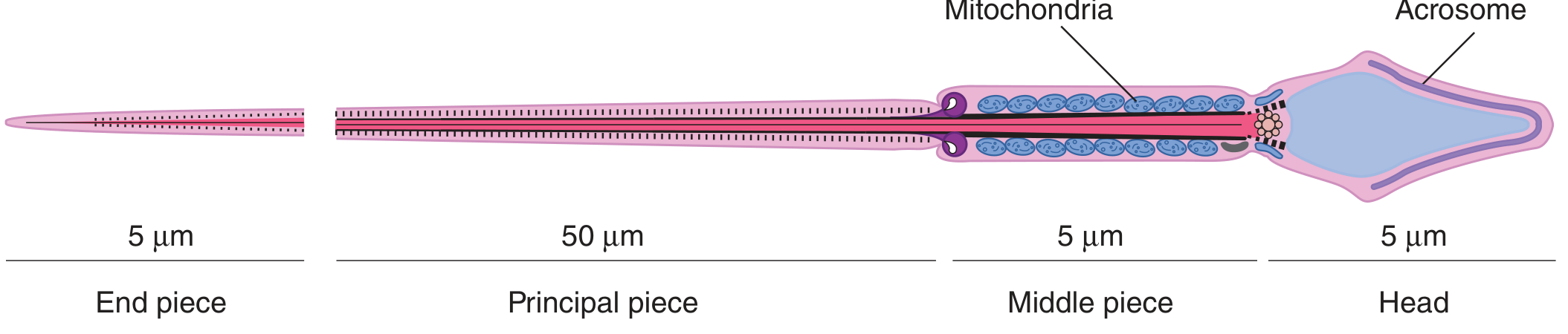
Figure: Human spermatozoon, profile view - Ganong's Review of Medical Physiology
Labeled Diagram Description (for drawing in exam):
                    ACROSOME (cap)
                  ┌──────────────┐
                  │  NUCLEUS     │  ← HEAD (~4.5 μm)
                  │  (condensed  │
                  │  chromatin)  │
                  └──────┬───────┘
                         │ NECK (connecting piece + proximal centriole)
                  ═══════╪══════════ ← MIDDLE PIECE (~5-7 μm)
                  ║ MITOCHONDRIAL SHEATH ║
                  ║ (helical, around axoneme) ║
                  ═══════╪══════════ ← ANNULUS
                         │
                  ───────┼─────────── PRINCIPAL PIECE (~50 μm)
                  (fibrous sheath + axoneme + outer dense fibers)
                         │
                  ───────┼─────────── END PIECE (~5 μm)
                         ▼           (axoneme only, no sheath)
Parts of Sperm and Their Functions:
| Part | Structure | Function |
|---|---|---|
| Head | Oval, ~4.5 μm × 3 μm | Contains nucleus with haploid DNA |
| Acrosome | Cap over anterior 2/3 of head | Contains hydrolytic enzymes (hyaluronidase, acrosin); used to penetrate zona pellucida of oocyte |
| Neck | Short, between head & tail | Connecting piece; contains proximal centriole |
| Middle piece | ~5-7 μm; proximal tail | Contains mitochondrial sheath (helically arranged) - provides ATP for motility |
| Principal piece | ~50 μm; main tail | Contains fibrous sheath + axoneme (9+2 microtubule arrangement); responsible for flagellar motility |
| End piece | ~5 μm; distal tip | Contains only axoneme; tail terminates here |
| Axoneme | 9+2 microtubule arrangement | The motor apparatus of the tail (9 outer doublets + 2 central microtubules + dynein arms) |
SUMMARY TABLE
| Phase | Cells Produced | Ploidy |
|---|---|---|
| Spermatogonium (Type B) | Primary spermatocyte | Diploid (2n, 4C) |
| Meiosis I | 2 Secondary spermatocytes | Haploid (n, 2C) |
| Meiosis II | 4 Spermatids | Haploid (n, 1C) |
| Spermiogenesis | 4 Spermatozoa | Haploid (n, 1C) |
One spermatogonium → 4 spermatozoa (compare with oogenesis: one oocyte → 1 ovum + 3 polar bodies)
Sources: Ganong's Review of Medical Physiology (26th ed.) | Costanzo Physiology (7th ed.) | Guyton & Hall Medical Physiology | The Developing Human - Clinically Oriented Embryology | Histology: A Text and Atlas (Wojciech Pawlowski, 8th ed.)
I have my university exam of 1 st year of subject physiology These are the important long answer give me answer of this question "Describe the mechanism of secretion of insulin. Mention the functions of insulin. Add a note on Diabetes Mellitus." and remeber it's a long question
Finding Sources
Finding Sources
Finding Sources
Finding Sources
Reading File
Finding Sources
Finding Sources
Excellent! Found great sources. Now let me read the key sections in parallel - Goldman-Cecil for insulin secretion mechanism, Costanzo for insulin functions, and the diabetes mellitus section.
Reading File
Reading File
Reading File
Excellent! Now let me get insulin functions from Guyton/Ganong and DM classification/complications.
Finding Sources
Reading File
Finding Sources
Reading File
Reading File
Loading Image
I now have all the content needed for a comprehensive exam answer. Let me also quickly get the DM complications and Robbins pathology content.
Reading File
I now have all the material needed. Let me compile the complete, exam-ready long answer.
Mechanism of Secretion of Insulin, Functions of Insulin, and Diabetes Mellitus
INTRODUCTION
Insulin is a polypeptide hormone secreted by the beta (β) cells of the Islets of Langerhans in the pancreas. It is the principal anabolic and glucoregulatory hormone of the body, responsible for maintaining blood glucose within the normal range of 70-140 mg/dL.
PART 1: MECHANISM OF SECRETION OF INSULIN
A. Synthesis of Insulin
Insulin is synthesized through a sequential process in the β cells:
- Preproinsulin - synthesized on ribosomes of the rough endoplasmic reticulum (RER); contains a signal peptide
- Proinsulin - signal peptide is cleaved in the RER; proinsulin is transported to the Golgi apparatus
- Insulin + C-peptide - proinsulin is packaged into secretory granules in the Golgi, where enzymatic cleavage removes the connecting (C) peptide, yielding the active two-chain insulin molecule (A-chain + B-chain linked by 2 disulfide bonds) and equimolar quantities of C-peptide
- The secretory granules are stored in the β cell cytoplasm until release is triggered
Clinical Note: C-peptide has a longer plasma half-life than insulin and is not metabolized by the liver. It is therefore a reliable biomarker for measuring endogenous insulin secretion.
Insulin has a very short plasma half-life of ~6 minutes and is degraded primarily by insulin-degrading enzyme in the liver and kidneys.
B. Stimuli for Insulin Secretion
| Stimuli | Examples |
|---|---|
| Primary stimulus | Elevated blood glucose (most important) |
| Amino acids | Arginine, leucine (after a protein-rich meal) |
| Gastrointestinal hormones | GIP (glucose-dependent insulinotropic peptide), GLP-1, CCK, secretin |
| Autonomic nervous system | Vagal (parasympathetic) stimulation via ACh on muscarinic receptors |
| Drugs | Sulfonylureas (close K⁺ channels directly) |
| Other hormones | Glucagon, cortisol, growth hormone (pharmacological doses) |
Inhibitors of insulin secretion: somatostatin, sympathetic stimulation (α₂ adrenergic), hypoglycemia, diazoxide
C. Step-by-Step Mechanism of Glucose-Stimulated Insulin Secretion
This is the most important mechanism and follows the K⁺-ATP channel model:
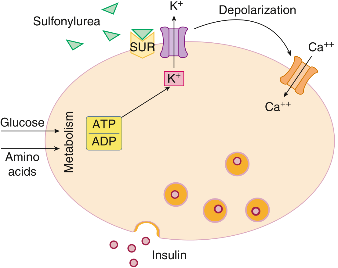
Figure: Nutrient regulation of insulin secretion - Goldman-Cecil Medicine
Steps:
Step 1 - Glucose uptake:
- Rising blood glucose after a meal is taken up by β cells via GLUT-2 transporter (high Km, insulin-independent, acts as a glucose sensor)
Step 2 - Phosphorylation by glucokinase:
- Glucose is phosphorylated to glucose-6-phosphate by glucokinase (hexokinase IV), which acts as the "glucose sensor" of the β cell
- Glucokinase is not inhibited by its product (no product inhibition), so phosphorylation is proportional to glucose concentration
Step 3 - Increased ATP/ADP ratio:
- Glucose-6-phosphate enters glycolysis and the Krebs cycle
- This leads to a rise in intracellular ATP and an increase in the ATP/ADP ratio
Step 4 - Closure of K⁺-ATP channels:
- The elevated ATP closes the ATP-sensitive potassium channels (K⁺-ATP channels / K-ATP channels)
- The K⁺-ATP channel is composed of SUR1 (sulfonylurea receptor 1) and Kir6.2 (inward rectifier K⁺ channel subunit)
- In the fasting state, these channels are open, allowing K⁺ efflux and keeping the membrane hyperpolarized
Step 5 - Membrane depolarization:
- Closure of K⁺-ATP channels prevents K⁺ efflux
- This leads to membrane depolarization of the β cell
Step 6 - Opening of voltage-gated Ca²⁺ channels:
- Depolarization opens voltage-gated L-type calcium channels
- Ca²⁺ flows into the β cell from the extracellular space
Step 7 - Calcium-mediated exocytosis:
- The increased intracellular Ca²⁺ binds to calmodulin, which activates myosin light-chain kinase
- This causes mobilization of insulin secretory granules toward the cell membrane
- Granules fuse with the plasma membrane → exocytosis of insulin (and equimolar C-peptide + small amount of proinsulin) into the portal circulation
Glucose → GLUT-2 → Glucokinase → Glucose-6-P
→ ↑ATP/ADP ratio → K-ATP channel closes
→ Membrane depolarization → Ca²⁺ channel opens
→ ↑Intracellular Ca²⁺ → Exocytosis of insulin granules
D. Biphasic Pattern of Insulin Secretion
After a glucose stimulus, insulin is released in two phases:
- First phase (rapid, 0-10 min): Release of pre-formed insulin granules already docked at the membrane
- Second phase (sustained, 10-60 min): Newly synthesized insulin granules are mobilized and released; requires ongoing glucose metabolism
In Type 2 DM, the first-phase insulin response is characteristically lost, which is an early defect in β cell function.
PART 2: FUNCTIONS OF INSULIN
Insulin is an anabolic hormone with widespread metabolic effects. Its receptor is a receptor tyrosine kinase (tetramer: 2α + 2β subunits). Insulin binding to α subunits activates the β subunit tyrosine kinase → autophosphorylation → phosphorylation of Insulin Receptor Substrates (IRS) → multiple downstream signaling cascades.
A. Effects on Carbohydrate Metabolism (ANTI-HYPERGLYCEMIC)
| Action | Mechanism |
|---|---|
| ↑ Glucose uptake into muscle and adipose | Recruits GLUT-4 transporters to cell surface |
| ↑ Glycolysis | Activates phosphofructokinase (PFK-1) |
| ↑ Glycogen synthesis | Activates glycogen synthase |
| ↓ Glycogenolysis | Inhibits glycogen phosphorylase |
| ↓ Gluconeogenesis | Inhibits PEPCK and glucose-6-phosphatase in liver |
Brain, RBCs, renal tubules, intestinal mucosa do NOT require insulin for glucose uptake (use GLUT-1, GLUT-3).
B. Effects on Fat (Lipid) Metabolism (ANTI-LIPOLYTIC)
| Action | Effect |
|---|---|
| ↑ Lipogenesis | Activates acetyl-CoA carboxylase and fatty acid synthase; promotes triglyceride synthesis |
| ↓ Lipolysis | Inhibits hormone-sensitive lipase in adipocytes |
| ↓ Ketogenesis | By reducing free fatty acid availability for the liver |
| ↑ Lipoprotein lipase activity | Promotes triglyceride uptake from blood into fat cells |
C. Effects on Protein Metabolism (ANABOLIC)
| Action | Effect |
|---|---|
| ↑ Amino acid uptake | Into muscle and other cells |
| ↑ Protein synthesis | Stimulates ribosomal activity and gene transcription |
| ↓ Protein catabolism | Inhibits proteolysis and reduces urinary nitrogen loss |
D. Effects on Potassium Balance
- Insulin activates Na⁺-K⁺-ATPase, promoting K⁺ uptake into cells
- After a meal, insulin ensures that absorbed dietary K⁺ is taken into cells → prevents hyperkalemia
- Insulin deficiency → decreased cellular K⁺ uptake → hyperkalemia
E. Growth and Mitogenic Effects
- Promotes DNA synthesis and cell proliferation
- Stimulates growth and differentiation of many cell types
- Works synergistically with IGF-1 (Insulin-like Growth Factor-1)
F. Summary Table of Insulin Actions
| Tissue | Major Actions |
|---|---|
| Liver | ↑ Glycogen synthesis, ↑ lipogenesis, ↓ glycogenolysis, ↓ gluconeogenesis, ↓ ketogenesis |
| Skeletal Muscle | ↑ Glucose uptake (GLUT-4), ↑ glycogen synthesis, ↑ protein synthesis |
| Adipose Tissue | ↑ Glucose uptake, ↑ lipogenesis, ↓ lipolysis |
| General | ↑ K⁺ uptake into cells, cell growth and division |
PART 3: NOTE ON DIABETES MELLITUS
Definition
Diabetes mellitus (DM) is a chronic metabolic disorder characterized by hyperglycemia (elevated blood glucose) resulting from defects in insulin secretion, insulin action, or both.
Classification of Diabetes Mellitus
(American Diabetes Association classification)
| Type | Key Feature |
|---|---|
| Type 1 DM | Autoimmune destruction of β cells → absolute insulin deficiency |
| Type 2 DM | Combination of insulin resistance + relative insulin deficiency |
| Gestational DM | Glucose intolerance first detected during pregnancy |
| Other specific types | MODY (monogenic), pancreatic disease, endocrinopathies, drugs |
Type 1 Diabetes Mellitus
- Accounts for 5-10% of all diabetes cases
- Most common in children and young adults (peak onset 10-14 years)
- Pathogenesis: Autoimmune "insulitis" - T-cell mediated destruction of β cells → autoantibodies (anti-GAD, anti-islet cell antibodies, anti-insulin antibodies) can be detected
- Genetic link to HLA class II genes (HLA-DR3, HLA-DR4)
- Absolute insulin deficiency
- Without insulin treatment: Diabetic ketoacidosis (DKA) develops
Features: Polyuria, polydipsia, polyphagia, weight loss, ketonuria, prone to DKA
Type 2 Diabetes Mellitus
-
Accounts for 90-95% of all diabetes cases
-
Usually adults; increasingly seen in obese children/adolescents
-
Pathogenesis - two key defects:
- Insulin resistance: Peripheral tissues (especially skeletal muscle, adipose) show decreased responsiveness to insulin → GLUT-4 recruitment is impaired
- Relative β-cell dysfunction: β cells cannot compensate adequately for the increased insulin demand → progressive decline in insulin secretion
-
Associated with obesity (80% of cases), physical inactivity, family history
-
First-phase insulin response is lost early
-
No autoimmune markers; late pathology shows amyloid deposition in islets
-
Rarely develops DKA; may develop hyperosmolar hyperglycemic state (HHS)
Diagnostic Criteria for Diabetes Mellitus
| Test | Diagnostic Value |
|---|---|
| Fasting plasma glucose (FPG) | ≥ 126 mg/dL (≥ 7.0 mmol/L) |
| 2-hour plasma glucose (OGTT) | ≥ 200 mg/dL (≥ 11.1 mmol/L) |
| HbA1c | ≥ 6.5% |
| Random plasma glucose + symptoms | ≥ 200 mg/dL |
Pre-diabetes: FPG 100-125 mg/dL (Impaired Fasting Glucose) or OGTT 140-199 mg/dL (Impaired Glucose Tolerance)
Clinical Features of Diabetes
Classic symptoms (the "3 Ps"):
- Polyuria - osmotic diuresis due to glycosuria (glucose exceeds renal threshold ~180 mg/dL)
- Polydipsia - compensatory thirst due to water loss
- Polyphagia - cellular starvation despite hyperglycemia; hyperphagia to compensate
Other features: Weight loss, fatigue, blurred vision, recurrent infections, poor wound healing
Complications of Diabetes Mellitus
Acute Complications
| Complication | Type | Key Feature |
|---|---|---|
| Diabetic Ketoacidosis (DKA) | Type 1 DM | Absolute insulin deficiency → ↑ lipolysis → ketone body production (acetone, acetoacetate, β-hydroxybutyrate) → metabolic acidosis |
| Hyperosmolar Hyperglycemic State (HHS) | Type 2 DM | Severe hyperglycemia (>600 mg/dL), hyperosmolarity, no significant ketosis |
| Hypoglycemia | Overtreatment | Blood glucose <70 mg/dL; altered consciousness, sweating, tremors |
Chronic Complications
Microvascular (due to basement membrane thickening):
- Diabetic nephropathy - Kimmelstiel-Wilson nodules in glomeruli; leading cause of chronic kidney disease
- Diabetic retinopathy - non-proliferative → proliferative; leading cause of acquired blindness in adults
- Diabetic neuropathy - peripheral (glove-and-stocking sensory loss), autonomic (gastroparesis, impotence, postural hypotension)
Macrovascular (accelerated atherosclerosis):
- Coronary artery disease - leading cause of death in diabetics
- Peripheral vascular disease - diabetic foot, gangrene
- Cerebrovascular disease - stroke
Mechanism of Vascular Complications
- Advanced Glycation End-products (AGEs) - glucose non-enzymatically glycates proteins → AGEs cross-link collagen → basement membrane thickening
- Polyol pathway activation - glucose converted to sorbitol by aldose reductase → osmotic damage to cells (nerves, lens → cataract)
- Oxidative stress - excess reactive oxygen species damage endothelium
- PKC activation - protein kinase C promotes vascular permeability and proliferation
- Hexosamine pathway - impairs insulin signaling
Management Overview
| Type | Management |
|---|---|
| Type 1 DM | Insulin replacement (basal-bolus regimen); diet and exercise |
| Type 2 DM | Lifestyle modification → Metformin (first-line) → add-on agents (sulfonylureas, GLP-1 agonists, SGLT-2 inhibitors, DPP-4 inhibitors) → insulin when needed |
| Monitoring | Self-monitoring blood glucose (SMBG); HbA1c every 3 months; target HbA1c <7% |
SUMMARY DIAGRAM (for exam drawing)
MECHANISM OF INSULIN SECRETION (Glucose-Stimulated):

Blood Glucose ↑
      ↓
GLUT-2 transporter (β cell uptake)
      ↓
Glucokinase (glucose sensor)
      ↓
Glucose-6-phosphate → Glycolysis → ↑ ATP/ADP ratio
      ↓
K-ATP channel CLOSES (SUR1 + Kir6.2)
      ↓
Membrane DEPOLARIZATION
      ↓
Voltage-gated Ca²⁺ channels OPEN
      ↓
↑ Intracellular Ca²⁺
      ↓
Insulin granule EXOCYTOSIS
      ↓
Insulin + C-peptide released into portal vein
Sources: Goldman-Cecil Medicine (International Ed.) | Lippincott's Illustrated Biochemistry (8th ed.) | Costanzo Physiology (7th ed.) | Robbins & Kumar Basic Pathology | Katzung's Basic & Clinical Pharmacology (16th ed.)
This is a shared conversation. Sign in to Orris to start your own chat.