
Where is keratin laminin and collagen in a skin layer and what are the causes in different types of epidermolysis bullosa
I'll read the medical library skill first, then research this topic in parallel.
Reading File
Now I'll run parallel searches across the library and web for skin layer proteins and epidermolysis bullosa.
Finding Sources
Finding Sources
Finding Sources
Searching the Web
epidermolysis bullosa types causes keratin collagen laminin skin layers pathogenesis
Excellent - found the key sources. Now let me read the most relevant blocks from Fitzpatrick's dermatology and the Dermatology 2-Volume set in parallel.
Reading File
Reading File
Reading File
Now let me read the main EB section from Fitzpatrick's and also the skin layer structure for keratin/laminin/collagen locations.
Reading File
Finding Sources
Finding Sources
Now let me read the main EB chapter section with the pathomechanisms and the skin layer structure from the Dermatology 5e book.
Reading File
Reading File
I have very rich data now. Let me also fetch the key diagrams showing the blister formation layers.
Loading Image
Loading Image
Loading Image
Excellent - I now have comprehensive data from authoritative dermatology textbooks plus three detailed diagrams. I have everything needed to write a thorough answer.
Keratin, Laminin, and Collagen in Skin Layers - and Epidermolysis Bullosa
Part 1: Location of Keratin, Laminin, and Collagen in Skin
The skin is anchored through a highly organized series of structures moving from the epidermis through the basement membrane zone (BMZ) to the dermis. Each of the three proteins lives at a distinct level of this architecture.
Skin Layer Architecture (from superficial to deep)
| Layer / Zone | Key Structural Proteins | Function |
|---|---|---|
| Basal keratinocytes (epidermis) | Keratin 5 (KRT5) & Keratin 14 (KRT14) | Intermediate filament cytoskeleton; provides tensile strength inside the cell |
| Hemidesmosome (intracellular plaque) | Plectin, BPAG1 (BP230), BP180 (collagen XVII) | Anchors keratin filaments to the cell membrane |
| Lamina lucida (upper BMZ) | Laminin-332 (α3, β3, γ2 subunits), integrin α6β4, anchoring filaments | Bridges hemidesmosome to the lamina densa below |
| Lamina densa (lower BMZ) | Laminin-332 (lower portion), type IV collagen, nidogen, perlecan | Dense sheet providing structural backbone of the BMZ |
| Sublamina densa / papillary dermis | Type VII collagen (anchoring fibrils), type I/III collagen fibers | Anchors the BMZ to the underlying dermis |
In plain terms:
- Keratin (KRT5/KRT14) lives inside basal epidermal cells as a cytoskeletal scaffold
- Laminin-332 spans the lamina lucida/lamina densa interface as "anchoring filaments" - it is the molecular glue between the epidermis and the BMZ
- Collagen XVII (BP180) sits in the hemidesmosome-anchoring filament complex (still in the lamina lucida zone)
- Collagen VII sits below the lamina densa in the sublamina densa zone, forming anchoring fibrils that loop around dermal collagen fibers and re-insert into the BMZ, tethering the whole structure to the dermis
- Collagen I/III are the structural fibers of the bulk dermis itself
Dermatology 2-Volume Set 5e, Table 28.6 — Localization of autoantigens and mutated proteins in EB
Part 2: Epidermolysis Bullosa (EB) - Types and Molecular Causes
EB is a group of inherited mechano-bullous disorders where mechanical trauma to the skin causes blistering. The blister plane and molecular defect define the type.
Diagram 1 - EB Simplex (intraepidermal blistering)
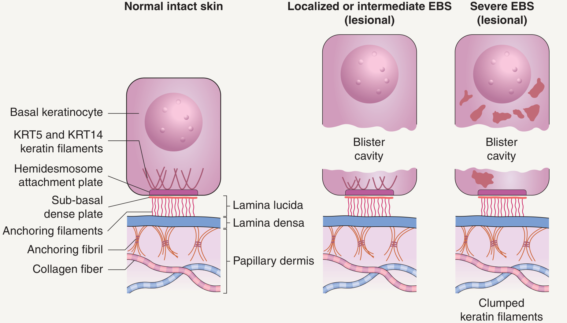
Fig. 32.1A - Blister forms within the basal keratinocyte cytoplasm. In severe EBS, keratin filaments clump into electron-dense aggregates.
Diagram 2 - Junctional EB (blistering within the BMZ)
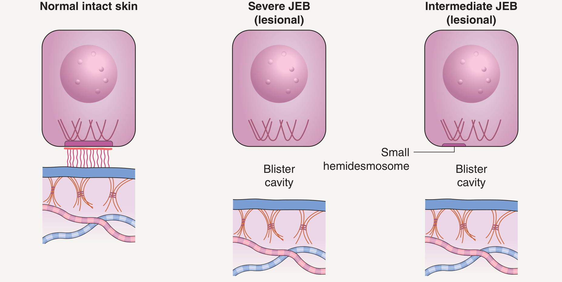
Fig. 32.1B - Blister forms within the lamina lucida. Hemidesmosomes are absent (severe) or small/rudimentary (intermediate).
Diagram 3 - Dystrophic EB (sub-BMZ blistering)
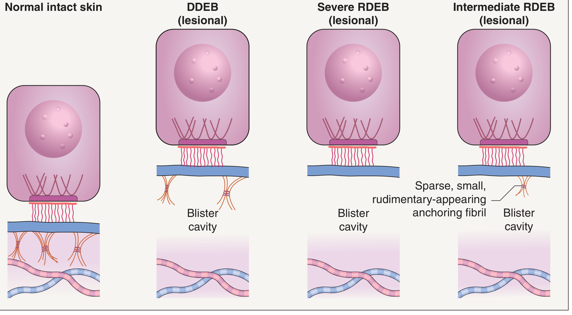
Fig. 32.1C - Blister forms below the lamina densa. Anchoring fibrils are absent (severe RDEB), sparse/rudimentary (intermediate RDEB), or structurally defective (DDEB).
Type 1: EB Simplex (EBS) - Intraepidermal
Blister level: Within the basal keratinocyte cytoplasm (cytolysis of basal cells)
Key proteins mutated: Keratin 5 and Keratin 14 (also plectin, desmoplakin, plakoglobin in rarer subtypes)
| Subtype | Gene | Protein |
|---|---|---|
| Generalized severe (Dowling-Meara) | KRT5 / KRT14 | Keratin 5 / Keratin 14 |
| Generalized intermediate (Koebner) | KRT5 / KRT14 | Keratin 5 / Keratin 14 |
| Localized (Weber-Cockayne) | KRT5 / KRT14 | Keratin 5 / Keratin 14 |
| EBS with muscular dystrophy | PLEC1 | Plectin |
| Mottled pigmentation | KRT5 | Keratin 5 |
| Acantholytic EBS | DSP, JUP | Desmoplakin, Plakoglobin |
| Plakophillin deficiency | PKP1 | Plakophillin 1 |
Mechanism: Missense mutations in KRT5 or KRT14 prevent normal keratin filament assembly. The intracellular cytoskeleton cannot withstand mechanical stress, so even minor trauma causes cytolysis of the basal cell layer. The blister roof is the upper epidermal layers; the floor is the lower portion of the basal cell. Inheritance: mostly autosomal dominant (rare recessive for plectin/BPAG1 mutations). Fitzpatrick's Dermatology, p.1045
Type 2: Junctional EB (JEB) - Within the Lamina Lucida
Blister level: Lamina lucida of the BMZ (between the hemidesmosome and lamina densa)
Key proteins mutated: Laminin-332, Collagen XVII (BPAG2/BP180), Integrin α6β4, Plectin
| Subtype | Genes | Protein |
|---|---|---|
| Generalized severe (Herlitz) | LAMB3 / LAMA3 / LAMC2 | Laminin-332 (null mutations = complete absence) |
| Generalized intermediate | LAMB3 / LAMA3 / LAMC2 / COL17A1 | Laminin-332 or Collagen XVII |
| Localized | LAMB3 / LAMA3 / LAMC2 / COL17A1 | Laminin-332 or Collagen XVII |
| EB with pyloric atresia | ITGB4 / ITGA6 / PLEC1 | Integrin β4, Integrin α6, Plectin |
| Laryngo-onycho-cutaneous | LAMA3A | Laminin α3 subunit |
| Respiratory and Renal | ITGA3 | Integrin α3 |
Mechanism: Laminin-332 is the key anchoring filament protein that bridges the hemidesmosome to the lamina densa. In Herlitz (lethal) JEB, null mutations of LAMA3/LAMB3/LAMC2 result in complete absence of laminin-332, causing total failure of the lamina lucida. Absence of laminin-332 in the airways leads to severe granulation tissue causing respiratory compromise, often limiting survival beyond 2 years. Missense mutations causing dysfunctional (not absent) laminin-332 produce milder disease (generalized atrophic benign EB). COL17A1 mutations affect the transmembrane collagen XVII, which also anchors the hemidesmosome to the BMZ. Inheritance: autosomal recessive. Dermatology 2-Volume Set 5e, p.662
Type 3: Dystrophic EB (DEB) - Sub-Lamina Densa
Blister level: Below the lamina densa, in the sublamina densa zone
Key protein mutated: Type VII Collagen (COL7A1) - the only gene involved in all DEB subtypes
| Subtype | Gene | Mechanism |
|---|---|---|
| Dominant DEB (DDEB) | COL7A1 (missense, dominant-negative) | Abnormal collagen VII incorporated into anchoring fibrils, weakening but not eliminating them |
| Recessive DEB - severe (RDEB) | COL7A1 (premature stop codon / null) | Complete or near-complete absence of anchoring fibrils |
| Recessive DEB - intermediate | COL7A1 (hypomorphic mutations) | Sparse, rudimentary anchoring fibrils present |
| Recessive DEB - localized | COL7A1 | Mildly reduced/dysfunctional anchoring fibrils |
| Recessive DEB - inversa | COL7A1 | Blistering in intertriginous areas |
Mechanism: Type VII collagen is the primary component of anchoring fibrils. The NC-1 domain of collagen VII interacts with laminin-332 in the lamina densa, while NC-2 interacts with dermal collagen fibers below, forming a looping structure that tethers the BMZ to the dermis. In DDEB, dominant-negative missense mutations produce structurally abnormal collagen VII that is incorporated into fibrils but weakens them - immunostaining for collagen VII is usually normal. In severe RDEB, null mutations eliminate anchoring fibrils entirely, causing the dermis to separate from the BMZ at the sub-lamina densa level with any friction. Severe RDEB carries a dramatically increased risk of aggressive squamous cell carcinoma (SCC) in chronic wounds. Dermatology 2-Volume Set 5e, p.662
Type 4: Kindler EB (formerly classified separately)
Blister level: Variable - can be intraepidermal, junctional, or sublaminal (mixed pattern)
Gene: FERMT1 | Protein: Kindlin-1
Mechanism: Kindlin-1 links integrins to the actin cytoskeleton. Its loss causes mixed-level blistering and progressive photosensitivity with poikiloderma. Kindler EB was re-classified as a 4th major EB type in the 2014 international consensus.
Summary Table - EB Types at a Glance
| Feature | EBS | JEB | DEB | Kindler EB |
|---|---|---|---|---|
| Blister plane | Intraepidermal (basal cell) | Lamina lucida | Sub-lamina densa | Mixed/variable |
| Key protein | Keratin 5/14 | Laminin-332 / Col XVII | Collagen VII | Kindlin-1 |
| Gene(s) | KRT5, KRT14, PLEC1 | LAMA3, LAMB3, LAMC2, COL17A1, ITGA6, ITGB4 | COL7A1 | FERMT1 |
| Inheritance | Usually AD | AR | AD or AR | AR |
| Scarring? | No | No (some atrophy) | Yes (especially RDEB) | Yes (progressive) |
| SCC risk | Low | Low | Very high (RDEB) | Moderate |
Sources: Fitzpatrick's Dermatology 9e; Dermatology 2-Volume Set 5e (Elsevier); StatPearls EB review
This is a shared conversation. Sign in to Orris to start your own chat.