
Typhoid
typhoid fever rose spots rash clinical presentation
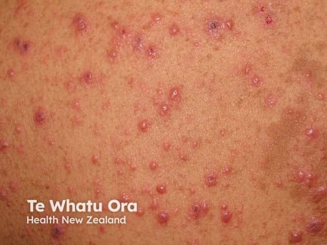
This clinical photograph shows a vesicular-papular rash on an erythematous base, distributed predominantly on the trunk with face involvement, consistent with varicella-zoster virus infection (chickenpox). The lesions appear at multiple stages, with small macules evolving to clear fluid-filled vesicles and some crusted plaques, giving the classic dew drop on a rose petal appearance. The skin is mildly erythematous around vesicles; lesions are superficial, non-palpable. No mucosal involvement is evident in this image. The distribution is generalized but has centripetal predominance. This presentation is highly suggestive of primary varicella, particularly in unvaccinated or susceptible individuals, and is contagious through vesicle fluid and respiratory secretions. Differential considerations include disseminated herpes simplex infection, disseminated herpes zoster in immunocompromised patients, or pityriasis rosea with vesicular components, though the latter is less typical. Clinically, the image supports diagnosis when accompanied by fever, malaise, pruritus, and a compatible exposure history. Reassurance, antiviral therapy consideration (e.g., acyclovir in appropriate age groups), and supportive care are relevant. The photograph is useful for medical education, telemedicine assessment, and dermatology training in recognizing staged varicella lesions, public health documentation, and patient counseling on contagion risk. This image supports clinical triage, outbreak documentation, and patient education on contagious rash in practice.
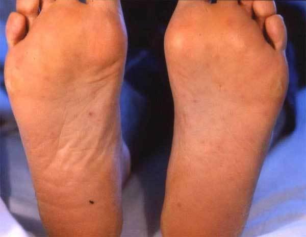
This clinical photograph displays the plantar surfaces of both feet of a 45-year-old patient, exhibiting a disseminated maculopapular rash. The lesions are characterized as non-confluent, reddish-brown macules and small papules distributed bilaterally across the soles. The rash is particularly prominent in the midfoot and heel regions. Notably, the skin integrity remains intact without evidence of ulceration, vesicles, or bullae, although some darker petechial-like spots are visible. This visual presentation is a characteristic dermatologic manifestation of rickettsial diseases, such as Mediterranean spotted fever (caused by Rickettsia conorii or R. massiliae), where involvement of the palms and soles is a key diagnostic sign. The image serves as an educational reference for infectious disease identification, specifically highlighting the distribution and morphology of cutaneous signs in tick-borne spotted fevers.
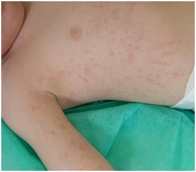
This clinical photograph displays a widespread cutaneous manifestation on a pediatric patient's torso and upper extremity. The presentation is characterized as a salmon-macular rash, featuring numerous discrete and confluent salmon-pink erythematous lesions. The lesions are primarily macular, appearing flat and flush with the skin surface, with irregular margins and varying sizes. Some areas exhibit linear or streaky patterns among the scattered spots. This specific visual finding is a hallmark clinical feature of systemic juvenile idiopathic arthritis (sJIA), often presenting during fever spikes. The image demonstrates the classic morphology and distinctive hue essential for differentiating sJIA from other pediatric exanthems or inflammatory conditions. The distribution includes the chest, abdomen, and the inner aspect of the arm, highlighting the diffuse and migratory nature of the rash in rheumatological contexts.
"typhoid fever"[MeSH Terms] AND treatment
typhoid fever Peyer patches ileum ulceration pathology histology
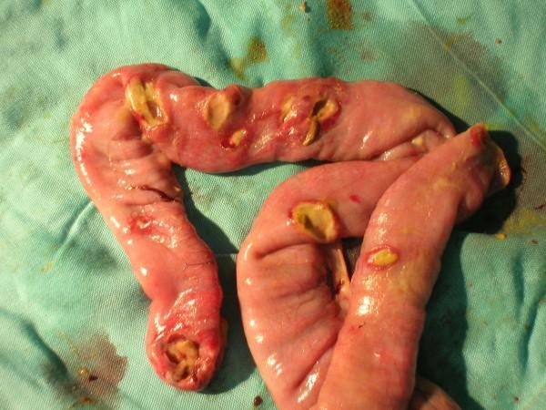
This clinical photograph shows a surgical specimen of the distal ileum placed on a green surgical drape. The anatomical structure exhibits multiple typhoid-related intestinal perforations. The ileal wall is predominantly pinkish-red, appearing moist and edematous. Approximately six to seven distinct perforations are visible, ranging in morphology from small pinholes to larger, irregular ovoid defects. The edges of these perforations are ragged and hyperemic, suggesting significant transmural inflammation. Yellowish slough or intestinal contents are visible within and surrounding several of the holes, indicative of leakage and associated peritonitis. This image illustrates a severe complication of enteric fever (Typhoid fever), specifically Peyer's patch necrosis leading to multiple intestinal perforations, which typically occur within 30 cm of the ileocecal valve. It serves as an educational reference for surgical pathology and complications of infectious gastrointestinal diseases.
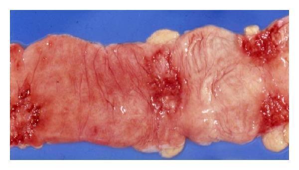
Gross pathology clinical photograph of a resected segment of the human ileum demonstrating intestinal tuberculosis. The specimen displays multiple transverse, circumferential ulcers that characteristically follow the orientation of the Peyer's patches. The ulcers exhibit distinct reddish, hemorrhagic surfaces with irregular and indurated (thickened) borders. The surrounding mucosa appears edematous and inflamed, with evidence of architectural distortion and tissue friability. These visual findings are classic for Mycobacterial infection of the gastrointestinal tract, highlighting the pathogenesis of ulcerative lesions that can lead to stricture formation or intestinal obstruction. This image serves as an educational reference for infectious disease, gastroenterology, and surgical pathology, illustrating the localized inflammatory response and mucosal destruction in abdominal tuberculosis.
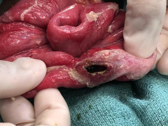
This clinical photograph is an intra-operative view depicting a typhoid intestinal perforation in the terminal ileum. The image shows a segment of small bowel held by gloved hands against a sterile surgical drape. A single, well-defined oval-shaped perforation is visible on the antimesenteric border, oriented along the longitudinal axis of the bowel. The perforation site displays dark, necrotic edges and is surrounded by an area of blanched, pale, and thickened tissue, indicating localized ischemia and inflammatory exudate. The adjacent intestinal wall appears erythematous and edematous, consistent with generalized peritonitis or severe enteritis. Small yellowish inflammatory plaques are visible on the serosal surface. This lesion is a pathognomonic finding of Typhoid Intestinal Perforation (TIP), resulting from the necrosis of Peyer's patches. It is a critical educational resource for understanding the surgical complications of Salmonella typhi infection in general surgery and infectious disease specialties.
Typhoid Fever (Enteric Fever)
Definition & Causative Agent
- Affects up to 30 million individuals worldwide per year
- Endemic in regions with poor sanitation: South Asia, Southeast Asia, Sub-Saharan Africa, and Latin America
- About 400 cases diagnosed annually in U.S. travelers returning from endemic countries
Transmission
- Fecal-oral route via contaminated food or water
- Dose-dependent: requires ingestion of 1,000 to 1 million organisms
- Chronic carriers (particularly those with gallstones) shed bacteria continuously in stool or urine and are a major reservoir
- Gallbladder colonization - especially with preexisting gallstone disease - underlies the chronic carrier state
Pathogenesis
- Ingestion - organisms penetrate the small intestinal mucosa with minimal local inflammation (explaining the absence of early intestinal symptoms)
- First bacteremia - bacilli rapidly reach lymphatics, mesenteric nodes, and bloodstream
- RES sequestration - organisms are taken up by macrophages in the reticuloendothelial system (liver, spleen, lymph nodes) where they multiply intracellularly
- Second bacteremia (symptomatic phase) - organisms re-emerge in recurrent waves after several days, disseminating widely
- Intestinal re-seeding - via direct bacteremic spread to Peyer patches in the terminal ileum, or via contaminated bile from the gallbladder
- Peyer patches in the terminal ileum enlarge into plateau-like elevations (up to 8 cm diameter)
- Mucosal shedding creates oval ulcers oriented along the long axis of the ileum - these can penetrate to cause perforation (the bowel wall becomes paper-thin)
- Typhoid nodules - small foci of parenchymal necrosis with macrophage aggregates - appear in liver, spleen, bone marrow, and lymph nodes
- Spleen: red pulp expansion from phagocyte hyperplasia
- Liver: discrete micronodular necrosis surrounded by macrophages and lymphocytes
Clinical Presentation
| Week | Features |
|---|---|
| Week 1 | Fever (stepwise rise), headache, malaise, dry cough, relative bradycardia; abdominal discomfort |
| Week 2 | High sustained fever, chills, prostration, abdominal distension; rose spots appear (pale red maculopapular lesions on chest/abdomen, especially in fair-skinned individuals); splenomegaly develops |
| Week 3 | Risk of intestinal complications (perforation, hemorrhage); delirium, typhoid state |
| Week 4 | Gradual defervescence in uncomplicated cases (or deterioration/death without treatment) |
- Relative bradycardia (pulse-temperature dissociation) - classic but may be absent
- Rose spots - transient salmon-pink maculopapular rash on trunk
- Splenomegaly
- 30% present with constipation rather than diarrhea (Tintinalli's EM)
- Leukopenia and mildly elevated liver enzymes (but lab values are often non-specific)
Complications
- Intestinal perforation (most feared - typically in terminal ileum within 30 cm of ileocecal valve)
- Intestinal hemorrhage
- Neurological: encephalopathy, meningitis, psychosis, ataxia, seizures, deafness
- Cardiovascular: myocarditis, mycotic aneurysm
- Other: pneumonia, cholecystitis, septic arthritis, DIC, renal failure, endocarditis
Diagnosis
- Blood culture - most useful in the first week; positive in ~90% of cases during the febrile phase
- Bone marrow culture - most sensitive test overall; remains positive even after antibiotic treatment
- Stool/urine culture - more useful in week 2 onward
- Rapid antigen tests - available; useful in endemic settings
- Widal test (serology) - historically used but poor sensitivity/specificity; not recommended as the sole diagnostic tool
Treatment
Antibiotics (drug of choice depends on susceptibility)
| Scenario | Preferred Agents |
|---|---|
| Susceptible strains | Ciprofloxacin 500 mg PO twice daily x 7-10 days OR Ceftriaxone 2 g IV once/twice daily x 10-14 days |
| Fluoroquinolone resistance (South/Southeast Asia) | Azithromycin 1 g daily x 5 days OR Ceftriaxone |
| Multidrug-resistant (MDR) | Ceftriaxone or azithromycin; carbapenem if XDR |
| Severe typhoid (encephalitis, shock) | Add dexamethasone (8 mg/kg x 1, then 1 mg/kg q6h x 48 hours) to parenteral antibiotics |
Resistance note: Fluoroquinolone resistance is increasing globally, especially in South and Southeast Asia (nalidixic acid-resistant strains). Ampicillin, trimethoprim-sulfamethoxazole, and chloramphenicol are now unreliable in many regions due to resistance. A 2026 systematic review (Mogasale et al., PMID 41990105) confirms the rising antimicrobial resistance burden in S. Typhi isolates in India from 1977-2024.
Supportive Care
- IV rehydration
- Blood transfusion if severe GI losses
- Surgical intervention for intestinal perforation (laparotomy, primary repair or resection)
Chronic Carrier Eradication
- Prolonged fluoroquinolone course (ciprofloxacin 750 mg twice daily x 4 weeks)
- Cholecystectomy if gallstones present
Prognosis
- Untreated mortality: 10-20%, predominantly in young children
- With appropriate treatment: mortality falls to <1%
- Relapses occur in ~10% of patients after clinical improvement (same antibiotic should be used)
Vaccination
| Vaccine | Type | Route | Age | Efficacy | Booster |
|---|---|---|---|---|---|
| Ty21a (Vivotif) | Live attenuated oral | Oral, 4 capsules alternate days | ≥6 years | ~50% at 3 years | Every 5 years |
| ViCPS (Typhim Vi) | Capsular polysaccharide | Single IM injection | ≥2 years | ~55% at 3 years | Every 2 years |
| Typhoid Conjugate Vaccine (TCV) | Vi-conjugate | IM | ≥6 months | Higher (real-world data emerging) | Being evaluated |
- Vaccine does not protect against S. Paratyphi
- Food and water precautions remain essential even with vaccination
- Indications: travelers to endemic areas, household contacts of carriers, lab workers handling S. Typhi
Microbiology
- Gram-negative, facultative anaerobe; member of Enterobacteriaceae
- Produces little or no gas from carbohydrates
- Bears the Vi (virulence) antigen on its surface - key surface marker; used in ViCPS vaccine and Widal testing
- Antigens: O (somatic), H (flagellar), Vi (capsular)
- Distinguished from other Salmonella by serotyping and genetic subtyping
- Rose spots (trunk rash)
- Oval ulcers in ileum (Peyer patches)
- Splenomegaly
- Endemic in poor sanitation areas
- Transmission fecal-oral
- Yield best from blood culture (week 1) / bone marrow
- Perforation = most feared complication
- Humans = only reservoir
- Only fluoroquinolone/cephalosporin/azithromycin reliable now (not chloramphenicol/ampicillin/TMP-SMX)
- Intracellular survival in RES macrophages
- Dexamethasone for severe/complicated disease