
Acute pancreatitis management
"acute pancreatitis"[MeSH Terms] AND management
acute pancreatitis CT scan imaging findings
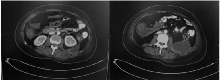
Two axial slices of a contrast-enhanced computed tomography (CT) scan of the abdomen demonstrate severe acute pancreatitis with extensive complications. The imaging shows a large, heterogeneous fluid collection in the left retroperitoneal space and left paracolic gutter, characterized by significant inflammatory fat stranding around the pancreas. Key findings include the presence of multiple air bubbles within the necrotic collections, suggestive of an emphysematous infection or secondary infected necrosis. Additionally, there is evidence of a colonic fistula involving the descending colon. The slices further reveal gallbladder stones without biliary tree dilatation. The findings are consistent with Balthazar Grade E acute pancreatitis, showcasing advanced local complications including walled-off necrosis, infection, and hollow viscus fistulization.
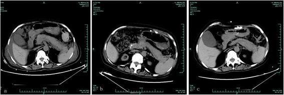
This clinical imaging series comprises three non-contrast axial CT scans (a, b, and c) of the upper abdomen, illustrating the temporal progression and resolution of acute pancreatitis (AP) in a post-transplant patient. Image 'a' demonstrates acute pancreatitis findings consistent with Balthazar Grade D, characterized by diffuse pancreatic swelling (edema) and prominent peripancreatic fluid collections/exudative effusions. No obvious necrosis is visualized in this non-contrast phase. Image 'b' represents a follow-up scan 14 days after the cessation of the suspected causative agent (tigecycline), showing a significant reduction in pancreatic volume and the resolution of surrounding fluid, returning toward a baseline morphology. Image 'c' is a further follow-up scan at 58 days, confirming the complete resolution of inflammatory changes and a normal pancreatic appearance. This series serves as an educational example of drug-induced acute pancreatitis and its radiographic recovery following the withdrawal of the offending medication. Key structures visible include the liver, spleen, kidneys, and vertebrae, providing anatomical context for the central pancreatic pathology.
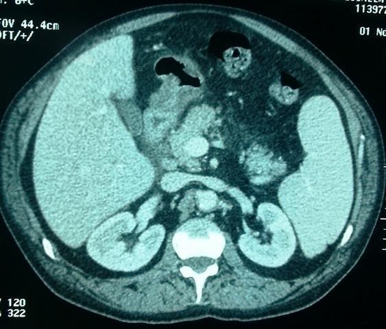
This diagnostic image is an axial contrast-enhanced abdominal computerized tomography (CT) scan focusing on the upper abdominal organs. The scan demonstrates clinical signs of acute pancreatitis. Key findings include visible infiltration and stranding of the peripancreatic fat, particularly noted as increased attenuation in the interpancréatico-duodenal region. The pancreas itself shows signs of inflammatory densification. Notably, the biliary system appears normal; the main bile duct (common bile duct) is not distended, the intra- and extra-hepatic bile ducts are fine, and the pancreatic duct (canal of Wirsung) is not dilated. Other visible landmarks include the liver, spleen, both kidneys, and a cross-section of the lumbar vertebra and aorta. This imaging is characteristic of Balthazar Grade B/C pancreatitis, illustrating the absence of significant fluid collections or biliary obstruction while highlighting localized fat-plane effacement indicative of acute inflammation.
"acute pancreatitis"[MeSH Terms] AND "fluid therapy"[MeSH Terms]
acute pancreatitis severity scoring BISAP Ranson criteria
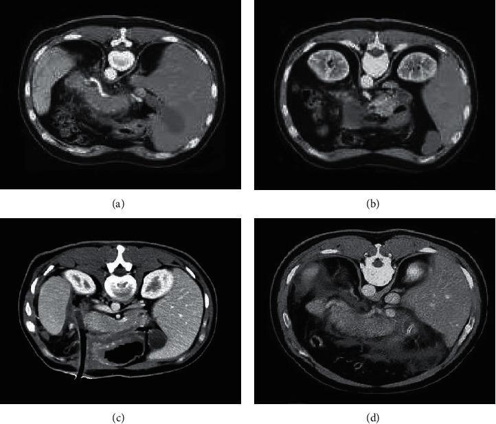
This composite diagnostic image features four axial abdominal CT scans demonstrating the spectrum of acute pancreatitis (AP) severity. Panels (a) and (b) represent mild acute pancreatitis (MAP), characterized by the pancreas showing focal or patchy low-density, non-enhancing areas. The peripancreatic fat planes remain relatively preserved with minimal fluid accumulation. In contrast, panels (c) and (d) illustrate severe acute pancreatitis (SAP). These scans reveal extensive pathology, including large, patchy peripancreatic effusion shadows and significant fluid collections in the retroperitoneal space (RPS) and anterior pararenal space (APS). The pancreatic parenchyma in SAP shows more pronounced density changes and irregular borders, reflecting higher degrees of inflammatory infiltration and potential necrosis. These images serve as educational examples for the Computed Tomography Severity Index (CTSI), highlighting the radiological differences between mild interstitial inflammation and severe exudative or necrotizing processes in the abdominal cavity.
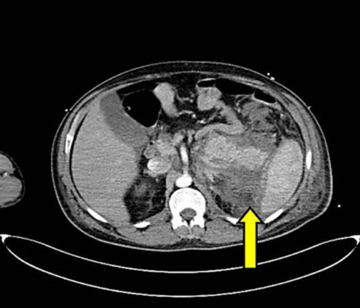
**Imaging Modality:** Contrast-enhanced computed tomography (CECT) of the abdomen. **Anatomical Region:** Axial section at the level of the upper abdomen, showing the pancreas, liver, spleen, and kidneys. **Observed Pathology:** The image demonstrates findings consistent with acute pancreatitis. There is significant inflammatory changes and edema involving the pancreatic parenchyma. A prominent, low-attenuation peripancreatic fluid collection is visible (indicated by the yellow arrow). **Characteristic Visual Features:** * **Pancreas:** Diffuse enlargement with ill-defined borders and heterogeneous enhancement. * **Fluid Collection:** An acute peripancreatic fluid collection (APFC) is noted in the anterior pararenal space, extending laterally into the splenorenal space. * **Fat Stranding:** Increased density and "dirty" appearance of the peripancreatic fat, indicating active inflammation. * **Adjacent Structures:** The collection is bordered by the tail of the pancreas and the medial aspect of the spleen. **Key Diagnostic Features:** The combination of pancreatic swelling, heterogeneous contrast enhancement, and organized peripancreatic fluid in the splenorenal space are characteristic radiologic markers for grading the severity of acute pancreatitis using the Balthazar or CT Severity Index (CTSI) criteria.
Acute Pancreatitis: Management
Diagnosis
- Typical epigastric pain (may radiate to the back)
- Serum lipase and/or amylase ≥3x upper limit of normal
- Confirmatory findings on cross-sectional imaging (CT/MRI)
- Harrison's Principles of Internal Medicine 22E, p. 2789
Severity Classification (Revised Atlanta Criteria)
| Category | Features | Mortality |
|---|---|---|
| Mild | No organ failure, no local/systemic complications | <1% |
| Moderately Severe | Transient organ failure (<48h) and/or local complications | ~10% |
| Severe | Persistent organ failure (>48h), single or multiorgan | 20-40% |
| Critical | Persistent MOF + infected necrosis | >50% |
Severity Scoring Tools
- B - BUN >25 mg/dL
- I - Impaired mental status (GCS <15)
- S - SIRS (≥2 of 4 criteria)
- A - Age >60 years
- P - Pleural effusion on imaging
- Hematocrit >44% (hemoconcentration)
- Admission azotemia / rising BUN during hospitalization
- Elevated CRP >100 mg/L
- Persistent SIRS at 24h predicts organ failure/necrosis
-
Cardiovascular: systolic BP <90 mmHg, HR >130 bpm
-
Pulmonary: PaO2 <60 mmHg
-
Renal: serum creatinine >2.0 mg/dL
-
Harrison's, p. 2791; Schwartz's Principles of Surgery 11E, p. 1471
1. Fluid Resuscitation (Most Critical Intervention)
- Initial bolus: ~10-15 mL/kg
- Maintenance: 1.5-2 mL/kg/h (recent RCT evidence favors less aggressive strategies - a 10 mL/kg bolus then 1.5 mL/kg/h showed similar outcomes to more aggressive dosing without the fluid overload risk)
- Target urine output >0.5 mL/kg/h
- Vital signs, oxygen saturation, physical exam
- Hematocrit and BUN every 8-12h
- A falling hematocrit and BUN in the first 12-24h confirms adequate resuscitation
- A rising BUN is associated with inadequate hydration AND higher in-hospital mortality
- Harrison's, p. 2790-2791
2. Analgesia
3. Nutritional Support
- Enteral nutrition (EN) is the mainstay - preferred over TPN
- EN maintains gut barrier integrity, limits bacterial translocation, is less expensive, and has fewer complications than parenteral nutrition
- Start EN within 24-72h if oral intake is not tolerated
- Nasogastric feeding is safe (NJ placement via endoscopy/fluoroscopy if NG not tolerated)
- No advantage to elemental or immune-enhancing formulas over standard polymeric formulas
- TPN only if enteral route cannot meet caloric needs
- Schwartz's, p. 1473-1474; Harrison's, p. 2792
4. Triage and Monitoring
- All patients with suspected acute pancreatitis should be admitted
- Mild (low BISAP, normal Hct/BUN, responds to fluids): Regular ward
- Persistent SIRS at 24h or comorbidities: Step-down/HDU
- Higher BISAP + elevated Hct + elevated BUN + organ failure/hypotension/respiratory failure: ICU
- Invasive monitoring of vitals, CVP, urine output, blood gases
- Supplemental oxygen
- Frequent labs: LFTs, renal function, clotting, calcium, glucose
- Supportive therapy for organ failure (inotropes, ventilatory support, hemofiltration)
5. Antibiotics
- Confirmed infected necrosis - empiric broad-spectrum therapy (carbapenems, e.g., imipenem, or ciprofloxacin + metronidazole) pending culture; duration guided by microbiology
- Ascending cholangitis - start promptly
- Concomitant respiratory or urinary infection
- Bailey & Love's Surgery 28E, p. 1472; Schwartz's, p. 1474
6. Etiology-Specific Management
Gallstone Pancreatitis (Biliary)
- Abdominal ultrasound is the initial imaging of choice (evaluates gallbladder, CBD)
- ERCP within 24-72h for:
- Ascending cholangitis (urgent, within 24h)
- Predicted severe biliary pancreatitis with evidence of biliary obstruction
- Laparoscopic cholecystectomy before discharge (or within 2-4 weeks) to prevent recurrence
Alcoholic Pancreatitis
- Alcohol cessation counseling
- Thiamine supplementation
Hypertriglyceridemia (TG >1000 mg/dL)
- IV insulin infusion, extended fasting 24-36h
- Adjuncts: heparin or plasmapheresis (limited evidence)
- Long-term: lipid-lowering agents, weight loss, avoid lipid-elevating drugs
Hypercalcemia
- Treat underlying hyperparathyroidism or malignancy
Drug-induced
- Discontinue offending agent
Post-ERCP pancreatitis (prevention)
- Pancreatic duct stenting and/or rectal indomethacin reduce risk
7. Imaging
- Organ failure
- Clinical deterioration
- Signs of sepsis
- Diagnostic uncertainty
- Interstitial edematous pancreatitis (contrast enhancement preserved)
- Necrotizing pancreatitis (lack of parenchymal enhancement)
- Acute peripancreatic fluid collections (APFC)
- Acute necrotic collections (ANC)
- Pseudocysts (>4 weeks, defined wall, fluid only)
- Walled-off necrosis (WON) (>4 weeks, defined wall, solid+fluid)
- Harrison's, p. 2789-2792; Schwartz's, p. 1474
8. Management of Local Complications
Pancreatic Necrosis
- Antibiotics with good pancreatic penetration (carbapenems, fluoroquinolones)
- Drainage/debridement - step-up approach is now preferred:
- Percutaneous catheter drainage (PCD) first
- Endoscopic (EUS-guided transmural) drainage of WON
- Minimally invasive retroperitoneal pancreatic necrosectomy (MIRPN) or video-assisted retroperitoneal debridement (VARD)
- Open necrosectomy - reserved for failure of minimally invasive approaches
Pseudocysts
- Most resolve spontaneously
- Intervene only if symptomatic, infected, or enlarging
- Endoscopic (EUS-guided) cystogastrostomy or cystoduodenostomy preferred over surgical drainage
Vascular Complications
- Pseudoaneurysm: angiographic embolization
- Splenic/portal vein thrombosis: anticoagulation in selected cases
Summary Table: Early Management of Severe Acute Pancreatitis
| Action | Details |
|---|---|
| Admission | HDU/ICU |
| Analgesia | IV opioids |
| Fluid resuscitation | LR solution; 10 mL/kg bolus, then 1.5 mL/kg/h; target UO >0.5 mL/kg/h |
| Oxygen | Supplemental O2; mechanical ventilation if needed |
| NPO / Nutrition | NPO initially; EN within 24-72h if severe |
| Monitoring | Vitals, CVP, UO, blood gases, HCT/BUN q8-12h, glucose, calcium |
| Antibiotics | Only for cholangitis, confirmed infected necrosis, or secondary infections |
| Imaging | CT if deteriorating, organ failure, or sepsis signs |
| ERCP | Within 24-72h if biliary obstruction/cholangitis |
| Cholecystectomy | Before discharge or within 2-4 weeks for gallstone pancreatitis |
- Harrison's Principles of Internal Medicine 22E (2025), Chapter 359, pp. 2789-2792
- Schwartz's Principles of Surgery 11E, Chapter 33, pp. 1471-1474
- Bailey & Love's Short Practice of Surgery 28E, Table 72.4, p. 1472