
/ Hypophysis cerebri, Histology of Suprarenal gland
I'll start by reading the medical library skill, then search for both topics in parallel.
Searching sources...
Reading File
Now I'll search both topics in parallel:
Finding Sources
Finding Sources
Finding Sources
Reading File
Good - both topics are in Junqueira's block3. Let me read the pituitary and adrenal sections in parallel:
Reading File
Finding Sources
Reading File
Finding Sources
Reading File
Finding Sources
Reading File
Searching Images
pituitary gland hypophysis histology adenohypophysis neurohypophysis
Excellent - I now have the adrenal cortex data. Let me read the adrenal medulla and neurohypophysis sections:
Reading File
Reading File
Reading File
Reading File
I now have comprehensive data from Junqueira's. Let me also read the adenohypophysis cell types section:
Reading File
Reading File
I now have all the information needed for a comprehensive answer. Let me compile it:
Histology of Hypophysis Cerebri (Pituitary Gland)
Source: Junqueira's Basic Histology - Text and Atlas, 17e
Overview & Embryology
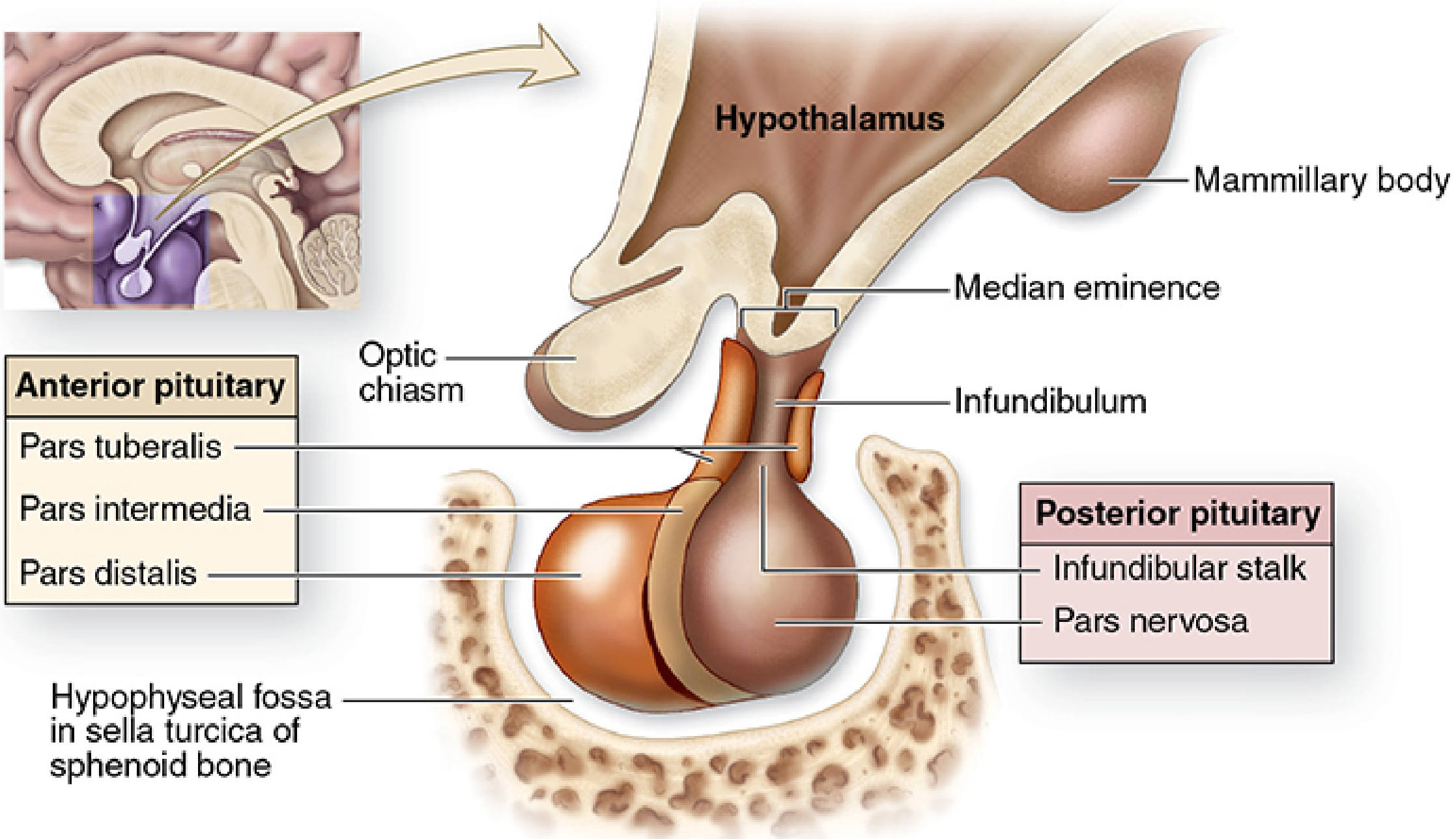
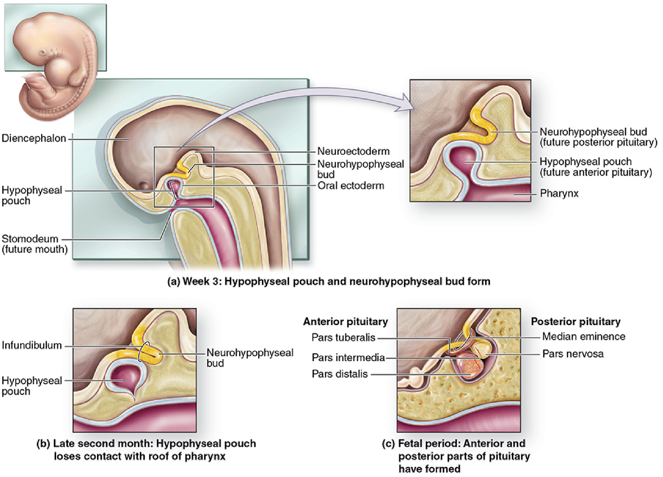
The pituitary gland (hypophysis) weighs ~0.5 g and measures ~10 × 13 × 6 mm. It sits in the sella turcica of the sphenoid bone, below the brain. It has a dual embryonic origin:
- Neurohypophysis - downward bud from the floor of the diencephalon (neural tissue)
- Adenohypophysis - upward outpocketing of oral ectoderm (Rathke's pouch)
Two Major Divisions
| Division | Parts | Origin |
|---|---|---|
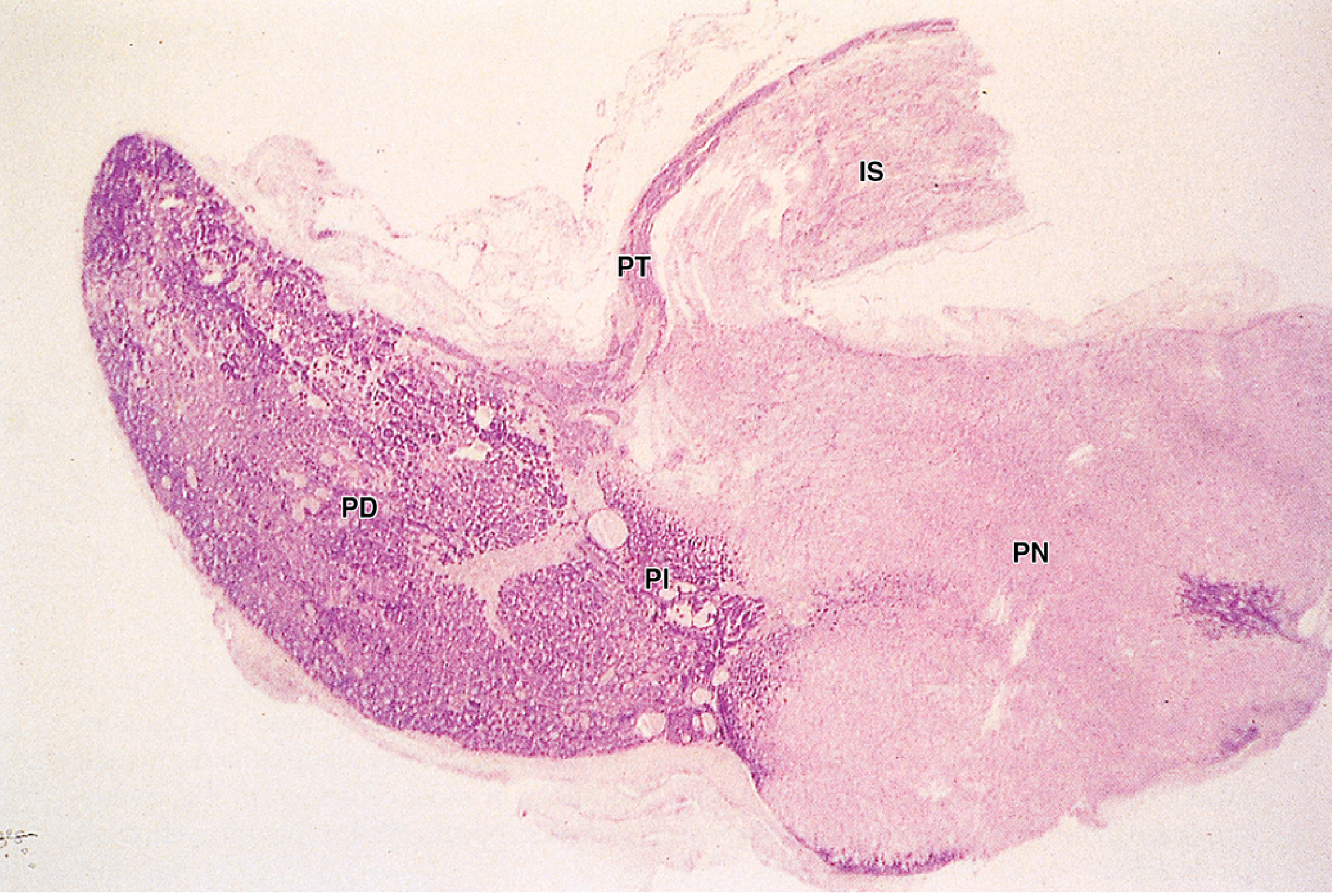
| Adenohypophysis | Pars distalis, Pars tuberalis, Pars intermedia | Oral ectoderm (Rathke's pouch) |
| Neurohypophysis | Pars nervosa, Infundibulum (stalk) | Neural tissue (diencephalon) |
1. Adenohypophysis
Blood Supply: Hypothalamic-Hypophyseal Portal System
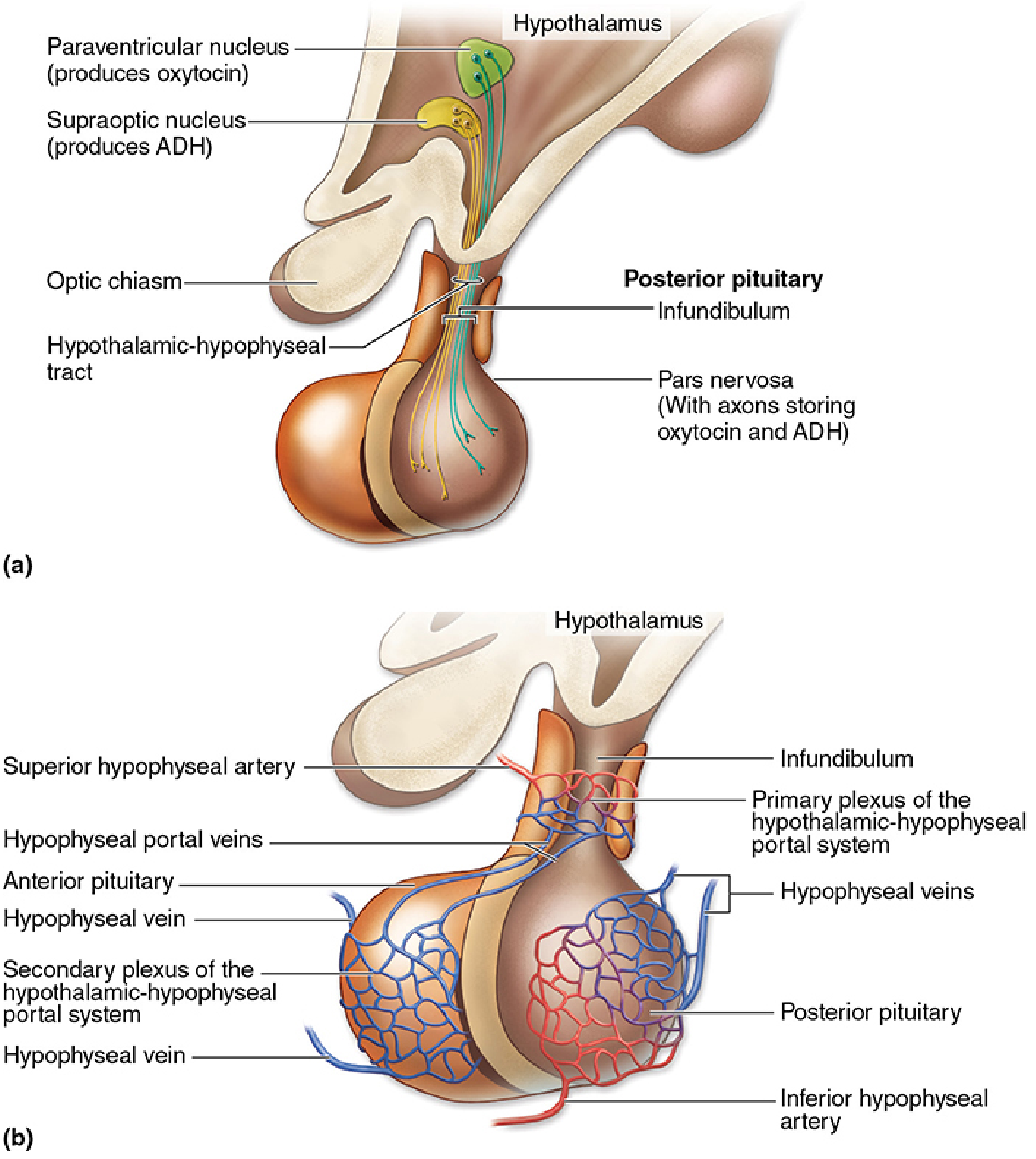
The adenohypophysis has NO direct arterial supply - it is entirely portal. Superior hypophyseal arteries form a primary capillary plexus in the median eminence/stalk, which drains into portal veins that branch as a secondary plexus throughout the pars distalis. This portal system transports hypothalamic releasing and inhibiting hormones to anterior pituitary cells.
A. Pars Distalis (75% of adenohypophysis)
Cords of secretory cells interspersed with fenestrated sinusoidal capillaries. Two major staining categories on routine H&E:
Chromophils (secretory, granule-filled cytoplasm):
- Acidophils (orange-red on H&E / Gomori trichrome) - 2 types:
- Somatotrophs (~50% of all pars distalis cells) - secrete Growth Hormone (GH/Somatotropin)
- Lactotrophs - secrete Prolactin (PRL)
- Basophils (violet on Gomori trichrome) - 3 types:
- Corticotrophs - secrete ACTH (from POMC precursor)
- Gonadotrophs - secrete FSH and LH (same cell secretes both)
- Thyrotrophs - secrete TSH (least numerous chromophils)
Chromophobes - poorly staining, granule-depleted cells (either resting chromophils or undifferentiated reserve cells)
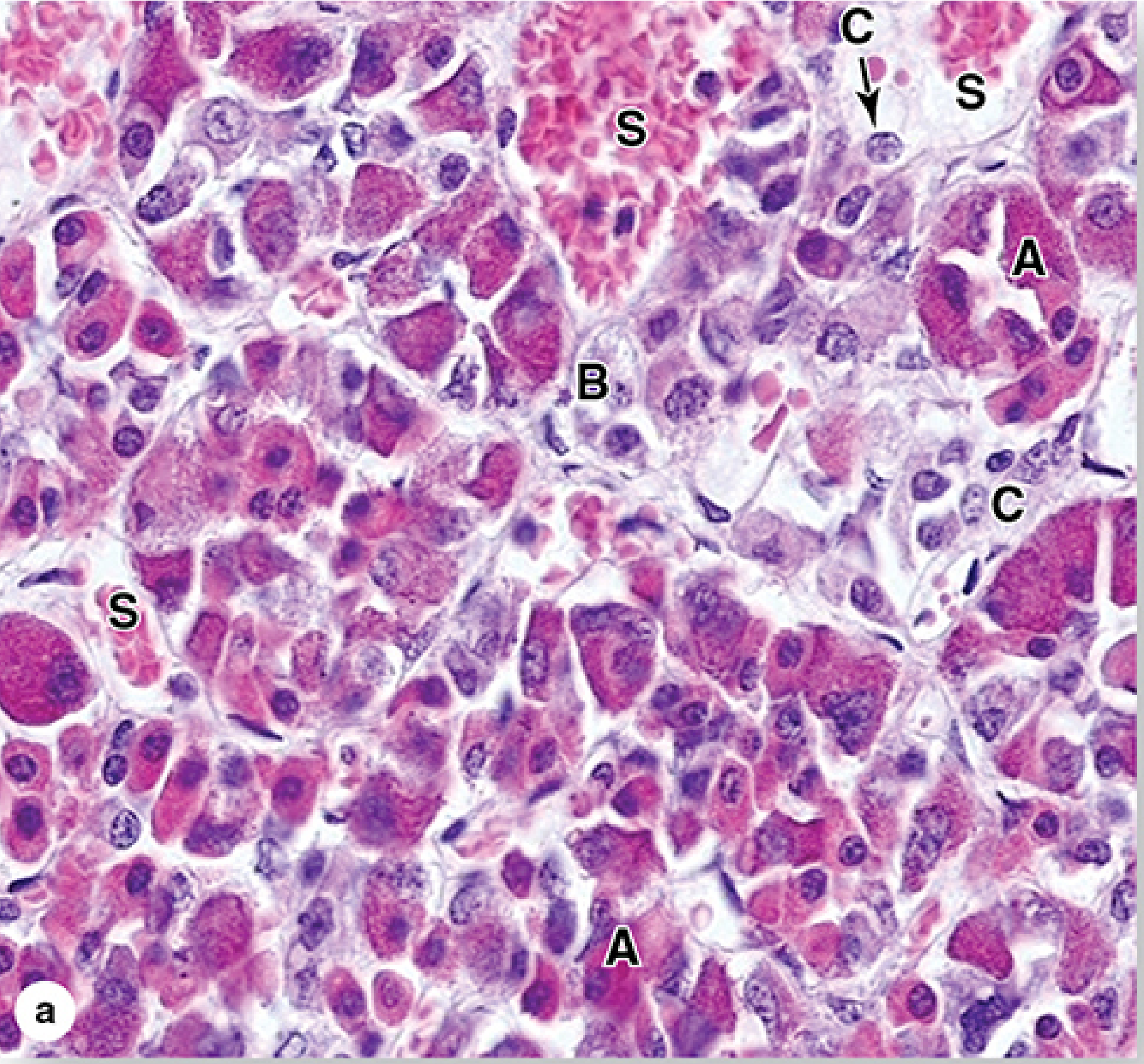
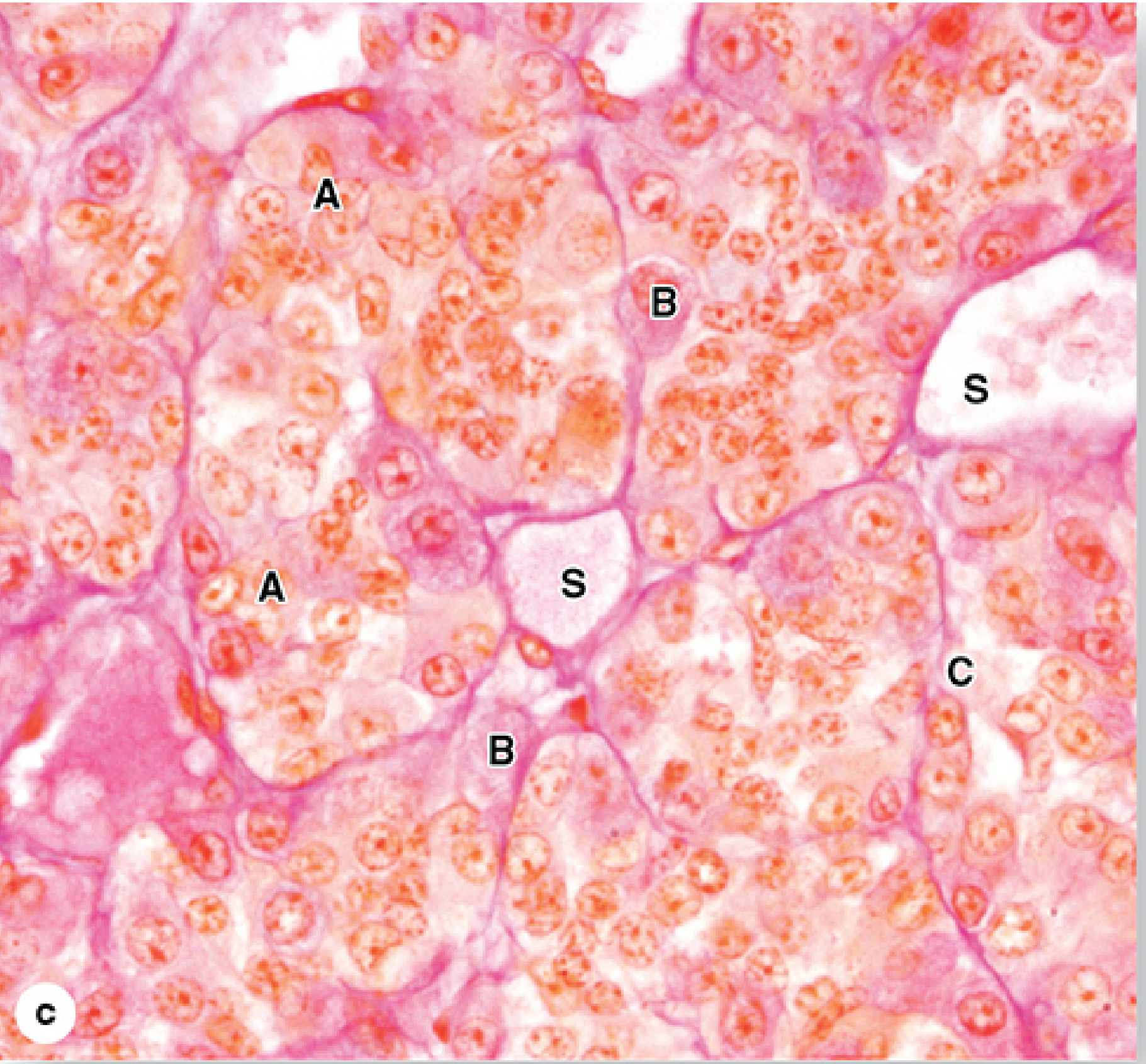
Clinical note: Benign pituitary adenomas of somatotrophs cause gigantism (before epiphyseal closure) or acromegaly (in adults).
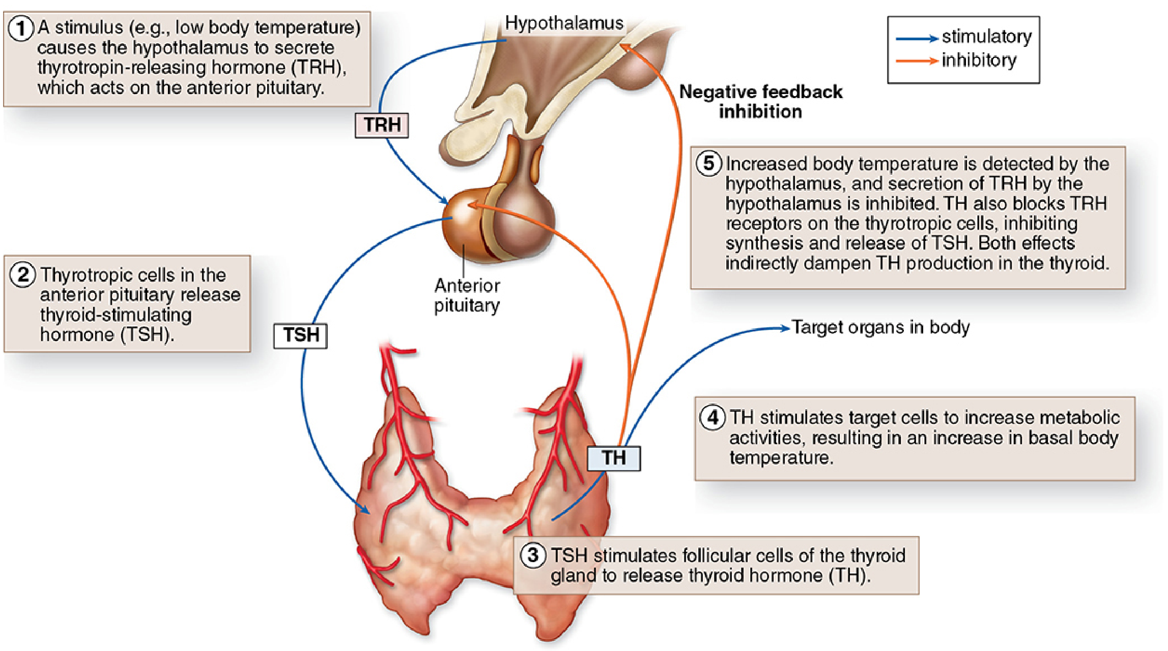
Hypothalamic Control (Table 20-2)
| Hypothalamic Hormone | Action |
|---|---|
| TRH (3 aa peptide) | Stimulates TSH release |
| GnRH (10 aa peptide) | Stimulates FSH and LH release |
| GHRH (40/44 aa polypeptide) | Stimulates GH release |
| Somatostatin (14 aa) | Inhibits GH and TSH release |
| Dopamine | Inhibits Prolactin release |
| CRH | Stimulates ACTH release |
Feedback loop: hypothalamic hormone → anterior pituitary → target organ hormone → negative feedback on both hypothalamus and pituitary.
B. Pars Tuberalis
Wraps around the infundibular stalk. Contains mainly gonadotrophs and portal blood vessels.
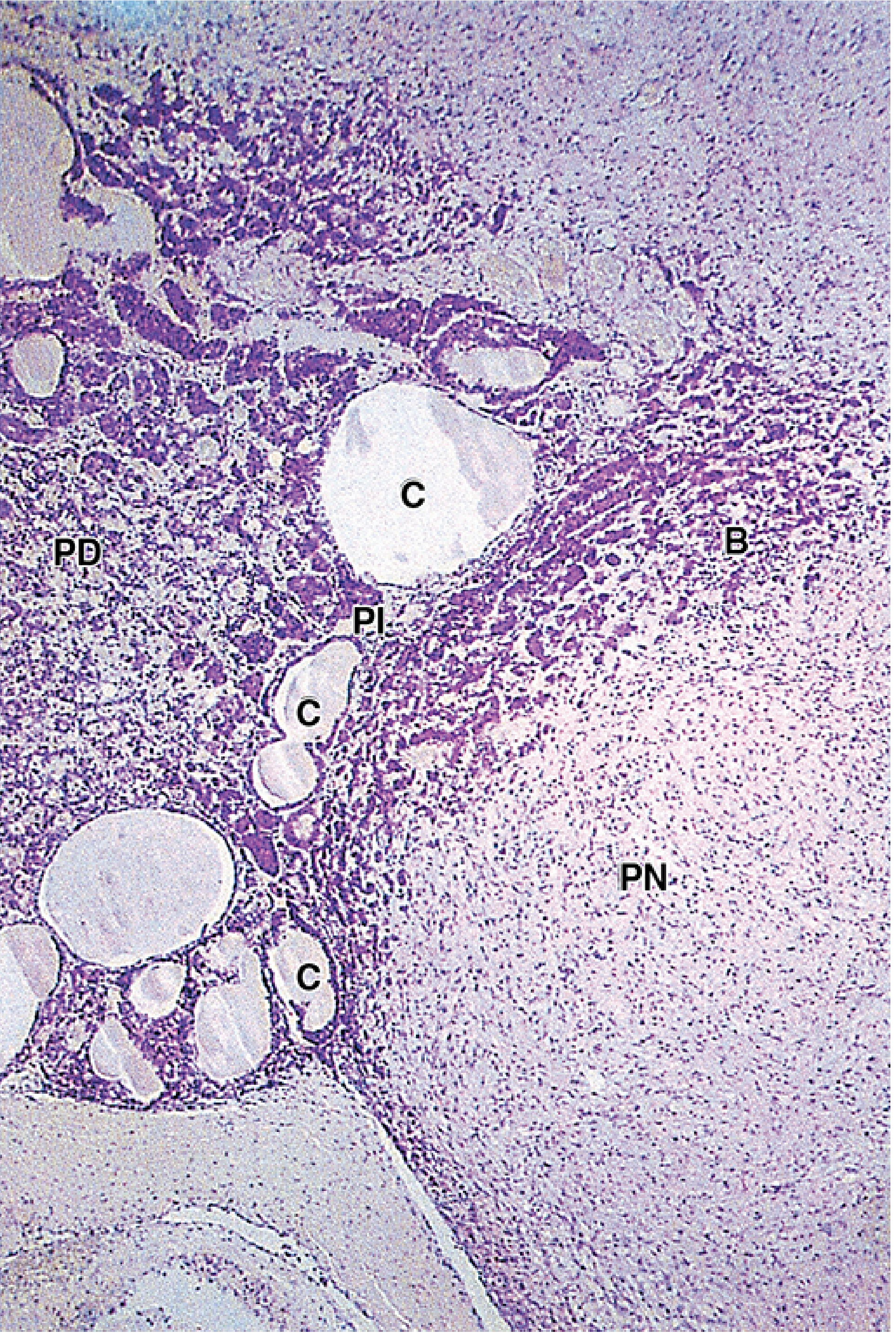
C. Pars Intermedia
Narrow zone between pars distalis and pars nervosa. Contains:
- Corticotrophs (producing MSH, γ-LPH, β-endorphin from POMC, cleaved differently than pars distalis)
- Colloid-filled Rathke's cleft cysts (remnants of hypophyseal pouch lumen)
- Chromophobes
- Best developed in fetal life; function in adults is unclear
2. Neurohypophysis (Posterior Pituitary)
The pars nervosa does NOT contain the neurons that produce its hormones. It consists of:
- ~100,000 unmyelinated axons from supraoptic (ADH) and paraventricular (oxytocin) nuclei of the hypothalamus, running down via the hypothalamic-hypophyseal tract
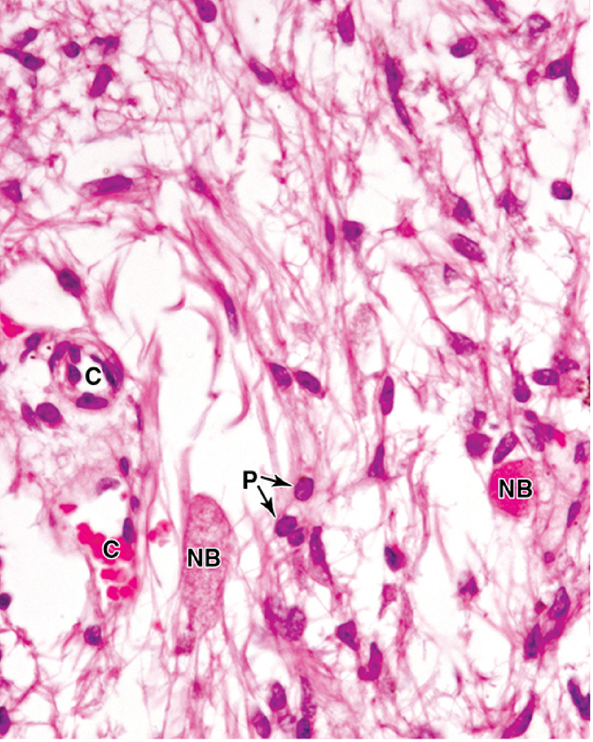
- Pituicytes - modified glial cells resembling astrocytes; the most abundant cell type
- Herring bodies (neurosecretory bodies) - axonal dilatations visible by light microscopy as faintly eosinophilic swellings; contain membrane-bound granules storing ADH or oxytocin bound to carrier proteins neurophysin II and neurophysin I, respectively
- Fenestrated capillaries for hormone uptake
Hormones released:
| Hormone | Stimulus | Action |
|---|---|---|
| ADH (Vasopressin) | Increased blood osmolality | Increases water permeability of renal collecting ducts |
| Oxytocin | Suckling, parturition | Uterine contraction; mammary myoepithelial contraction (milk ejection) |
Clinical note: Damage to posterior pituitary (trauma, tumor compression, gene mutation) → reduced ADH → Diabetes insipidus (polyuria + polydipsia).
Histology of Suprarenal (Adrenal) Gland
Source: Junqueira's Basic Histology - Text and Atlas, 17e
Overview
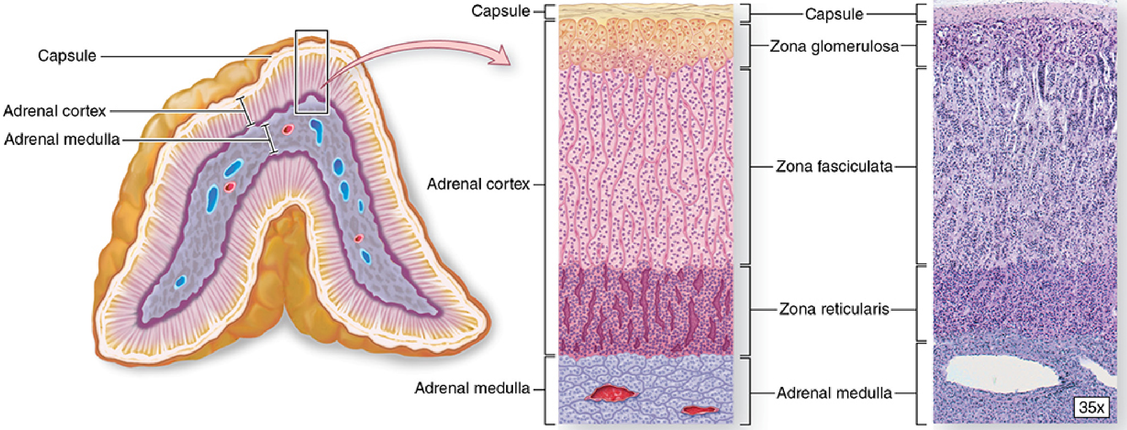
The paired adrenal (suprarenal) glands lie at the superior poles of the kidneys, each ~4-6 cm long, 1-2 cm wide, ~8 g total weight. Each is enclosed in a dense connective tissue capsule with thin trabeculae projecting inward. The stroma is reticular fibers.
Each gland has two concentric regions of completely different embryologic origin:
| Region | Color (gross) | Embryologic Origin |
|---|---|---|
| Cortex (outer) | Yellowish | Embryonic mesoderm (intermediate mesoderm / coelomic epithelium) |
| Medulla (inner) | Reddish-brown | Neural crest cells |
Adrenal Cortex
Cortical cells are arranged in three concentric zones, all producing steroid hormones. Steroids are not stored in granules - as small lipid-soluble molecules they diffuse freely through the plasma membrane when synthesized.
Ultrastructural features common to all steroid-secreting cells:
- Lipid droplets (cholesterol ester stores)
- Mitochondria with tubular/vesicular cristae (not lamellar) - for steroidogenesis
- Abundant smooth ER (SER)
- Autophagosomes (remove organelles between active periods)
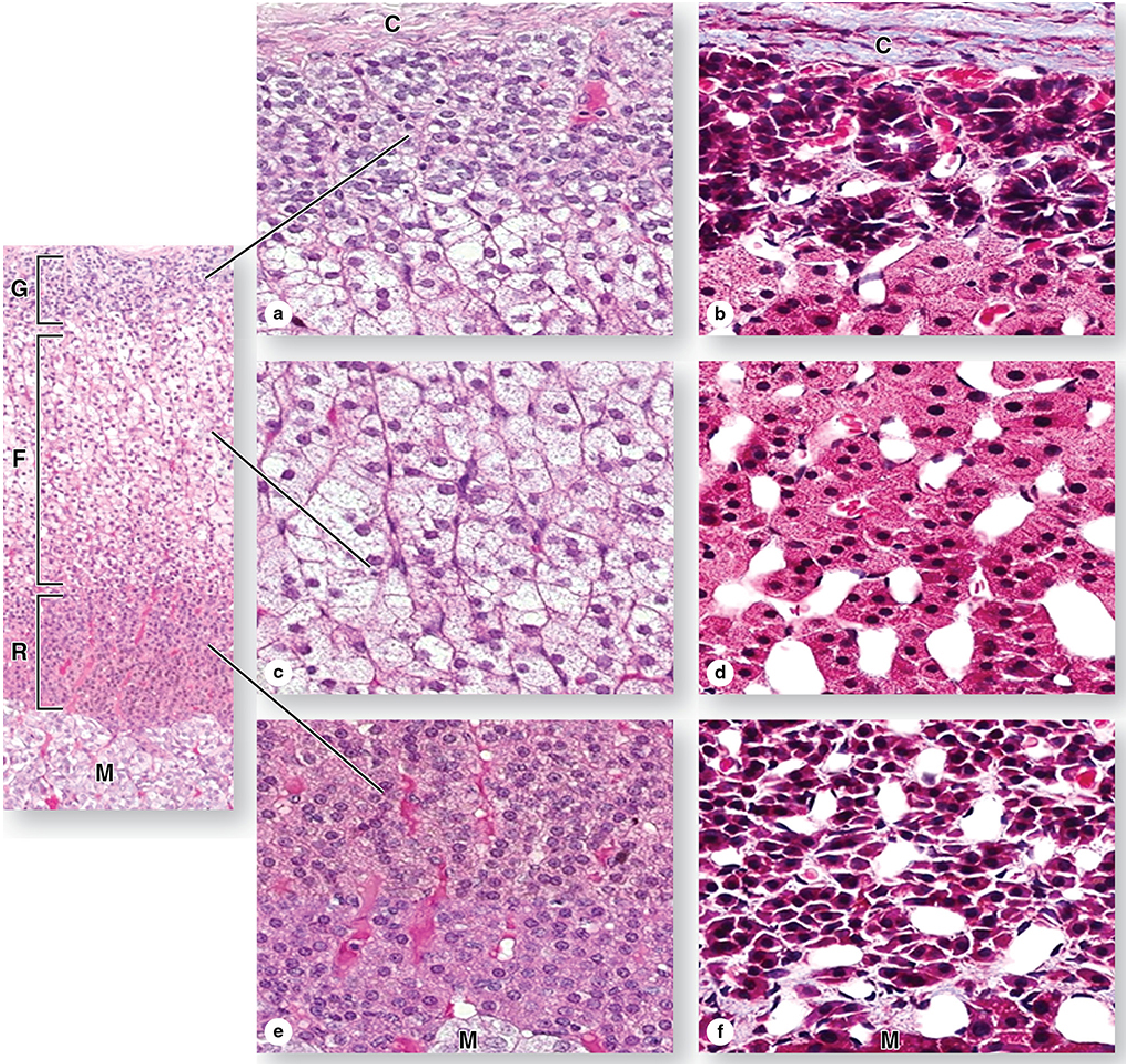
Zone 1: Zona Glomerulosa (outermost, ~15% of cortex)
- Immediately beneath the capsule
- Cells: columnar or pyramidal, arranged in rounded/arched cords or ovoid clusters
- Many capillaries
- Hormone: Mineralocorticoids → Aldosterone (principal product)
- Aldosterone regulates salt balance: stimulates Na+ reabsorption in distal convoluted tubules
- Control: primarily angiotensin II, also elevated plasma K+; only weakly by ACTH
Zone 2: Zona Fasciculata (middle, ~65-80% of cortex)
- Thickest zone
- Cells: large polyhedral cells in long straight cords (1-2 cells thick) separated by sinusoidal capillaries
- Cells are filled with lipid droplets → appear vacuolated/spongy on routine H&E ("spongiocytes")
- Hormone: Glucocorticoids → Cortisol (principal product) + small amounts of androgens
- Cortisol: stimulates gluconeogenesis, glycogen synthesis in liver; suppresses immune functions; fat mobilization; muscle proteolysis
- Control: ACTH with negative feedback
Zone 3: Zona Reticularis (innermost, ~10% of cortex)
- Cells: smaller, in irregular anastomosing network of cords with wide capillaries
- Cells are more darkly stained than zona fasciculata (fewer lipid droplets, more lipofuscin pigment)
- Hormone: mainly weak androgens (DHEA - dihydroepiandrosterone, converted to testosterone in peripheral tissues) + some cortisol
- Control: ACTH with regulatory feedback
Mnemonic for hormones: GFR = Salt, Sugar, Sex (Glomerulosa=Salt/mineralocorticoids; Fasciculata=Sugar/glucocorticoids; Reticularis=Sex/androgens)
Clinical note: Addison disease - usually autoimmune degeneration of adrenal cortex → loss of glucocorticoids, mineralocorticoids, or androgens.
Adrenal Medulla
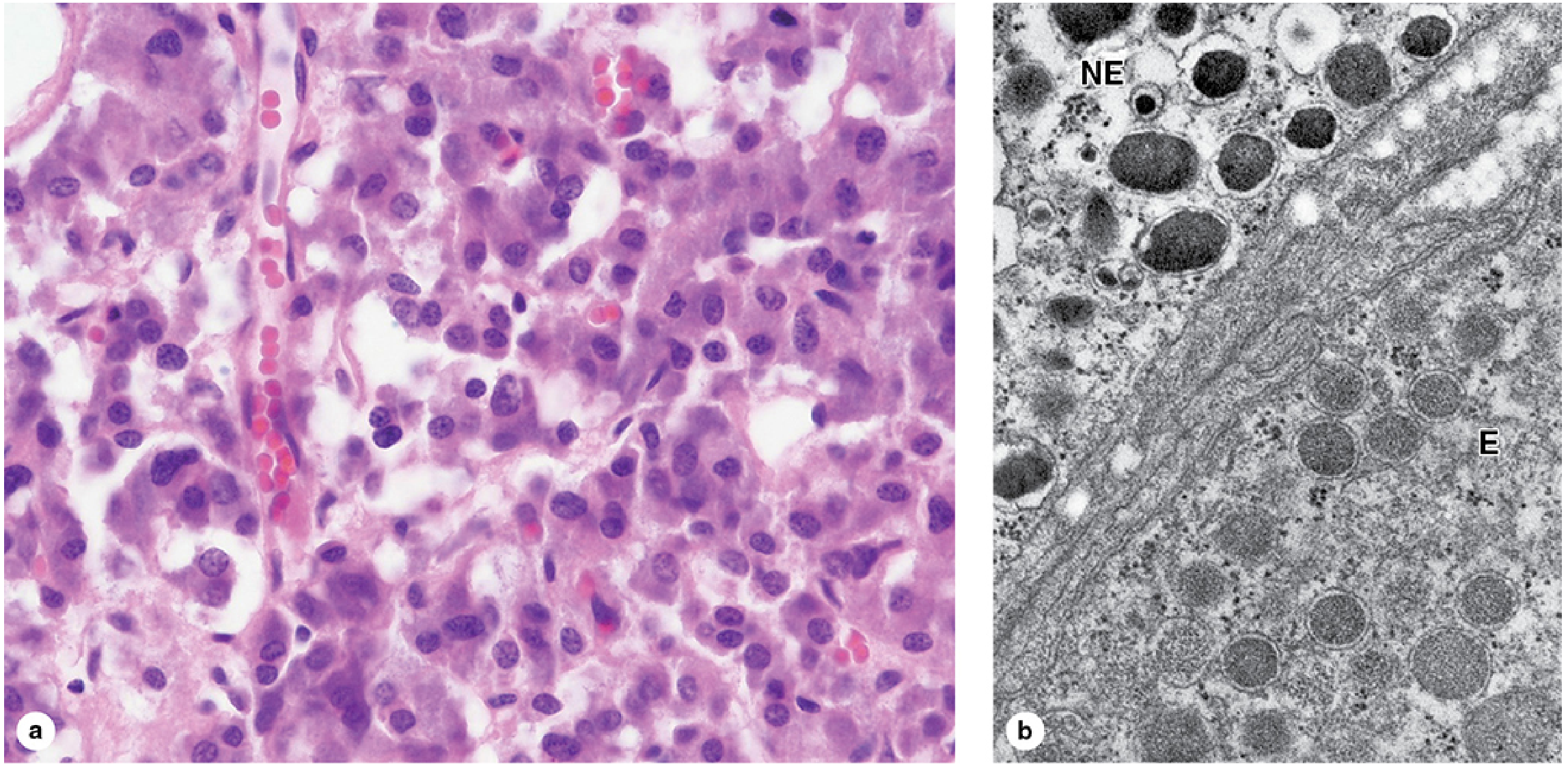
The medulla is composed of large, pale-staining polyhedral chromaffin cells arranged in cords or clumps, supported by reticular fibers with a profuse sinusoidal capillary supply. A few parasympathetic ganglion cells are also present.
Chromaffin cells:
- Derived from neural crest cells (like sympathetic postganglionic neurons)
- Considered modified sympathetic postganglionic neurons - lack axons and dendrites, specialized for secretion
- Contain electron-dense granules (150-350 nm) storing catecholamines bound to chromogranins (49 kDa proteins) with Ca2+ and ATP
- Innervated by preganglionic sympathetic neurons (direct preganglionic control, unlike other postganglionic neurons)
- Two subtypes by TEM:
- Epinephrine-secreting cells - smaller, less electron-dense granules (~80% of secretion)
- Norepinephrine-secreting cells - larger, more electron-dense granules
Key biochemistry: The conversion of norepinephrine → epinephrine occurs ONLY in adrenal medullary chromaffin cells (via PNMT enzyme, stimulated by cortisol from cortical venous blood).
Hormonal effects - Fight-or-Flight:
- Epinephrine: ↑ heart rate, dilates bronchioles, dilates arteries of cardiac/skeletal muscle
- Norepinephrine: vasoconstricts gut/skin vessels, ↑ blood flow to heart/brain/muscle
- Both: stimulate glycogenolysis → ↑ blood glucose
Clinical note: Pheochromocytoma - benign tumor of chromaffin cells; periodically secretes high catecholamines → episodic swings between hypertension and hypotension.
Summary Comparison
| Feature | Adrenal Cortex | Adrenal Medulla |
|---|---|---|
| Origin | Mesoderm | Neural crest |
| Cell type | Epithelial steroid-secreting cells | Chromaffin cells (modified neurons) |
| Hormone class | Steroids (aldosterone, cortisol, androgens) | Catecholamines (epinephrine, norepinephrine) |
| Storage | Not stored (diffuse freely) | Stored in dense-core granules |
| Control | ACTH, angiotensin II, K+ | Preganglionic sympathetic neurons |
| Organelle hallmark | Tubular mitochondrial cristae, SER, lipid droplets | Dense-core secretory granules, chromogranins |
Junqueira's Basic Histology - Text and Atlas, 17e, pp. 999-1027
This is a shared conversation. Sign in to Orris to start your own chat.