
Ectopic heartbeats
Reading File
Ectopic heartbeats are abnormal cardiac impulses that originate from sites other than the sinoatrial (SA) node - they can arise from atrial, junctional (AV nodal), or ventricular tissue.
Searching sources...
Finding Sources
Finding Sources
Searching PubMed
"ectopic beats" OR "premature contractions"
Good results. Now let me read the key sections in parallel:
Reading File
Reading File
Reading File
Excellent content retrieved. Let me now read the ventricular section from Guyton and fetch key ECG images:
Reading File
Reading File
Now let me fetch the key ECG images and read the PVC/ventricular ectopic section:
Loading Image
Loading Image
Loading Image
Reading File
I now have comprehensive information to provide a detailed, well-illustrated answer. Let me also fetch the Braunwald ECG image for atrial ectopics:
Loading Image
I now have all the material needed for a thorough, image-rich answer.
Ectopic Heartbeats
An ectopic beat (also called an extrasystole, premature beat, or premature contraction) is a cardiac impulse that originates from any site in the heart other than the sinoatrial (SA) node. It fires before the next expected normal beat and transiently interrupts the cardiac rhythm.
Mechanisms of Ectopic Beat Formation
There are two main electrophysiological mechanisms:
1. Abnormal Automaticity (Enhanced Ectopic Focus)
Normally, myocardial cells outside the SA node do not discharge spontaneously - the SA node suppresses them by firing faster (a concept called overdrive suppression). When the SA node slows or an ectopic site becomes abnormally irritable (increased automaticity), that focus can fire and produce a premature beat. If the ectopic focus discharges once, the result is a single premature beat. If it discharges repetitively at a rate faster than the SA node, it produces a sustained tachyarrhythmia (atrial tachycardia, junctional tachycardia, or ventricular tachycardia). - Ganong's Review of Medical Physiology, p. 530
2. Reentry
A more common cause of repetitive ectopic firing. A transient conduction block in one limb of a circuit allows an impulse to travel down the unblocked path, then circle back through the previously blocked path (when it recovers), establishing a continuous circus movement. - Ganong's Review of Medical Physiology, p. 530
Causes of Ectopic Foci
-
Local areas of myocardial ischemia
-
Small calcified plaques pressing on adjacent cardiac muscle
-
Toxic irritation of the AV node, Purkinje system, or myocardium: infection, drugs, nicotine, caffeine, digitalis toxicity
-
Mechanical irritation (e.g., catheter tip touching the endocardium during cardiac catheterization)
-
Electrolyte imbalances (hypokalemia, hypomagnesemia)
-
Hypoxia, sympathetic excess, thyrotoxicosis
-
Guyton & Hall Textbook of Medical Physiology, p. 168
Classification by Site of Origin
| Type | Origin | Also Called |
|---|---|---|
| Atrial | Atrial muscle (outside SA node) | PAC, APC (atrial premature complex) |
| Junctional | AV node / AV bundle (His bundle) | AV nodal premature contraction |
| Ventricular | Ventricular myocardium / Purkinje system | PVC (premature ventricular contraction) |
1. Atrial Premature Contractions (PACs)
ECG features:
- Early P wave with an abnormal morphology (different from the sinus P wave)
- P wave is frequently inscribed within the preceding T wave, making morphology unclear
- PR interval may be shortened (if ectopic focus is near the AV node) or prolonged
- QRS is usually narrow (normal) - conducted normally through the bundle of His and ventricles
- Compensatory pause follows - the premature impulse discharged the sinus node early, resetting its timing
A nonconducted (blocked) PAC - where the ectopic atrial impulse arrives when the AV node is still refractory - is one of the most common causes of an unexpected pause on an ECG and can mimic sinus bradycardia, especially in bigeminy.
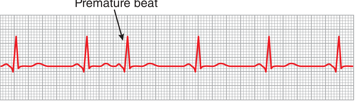
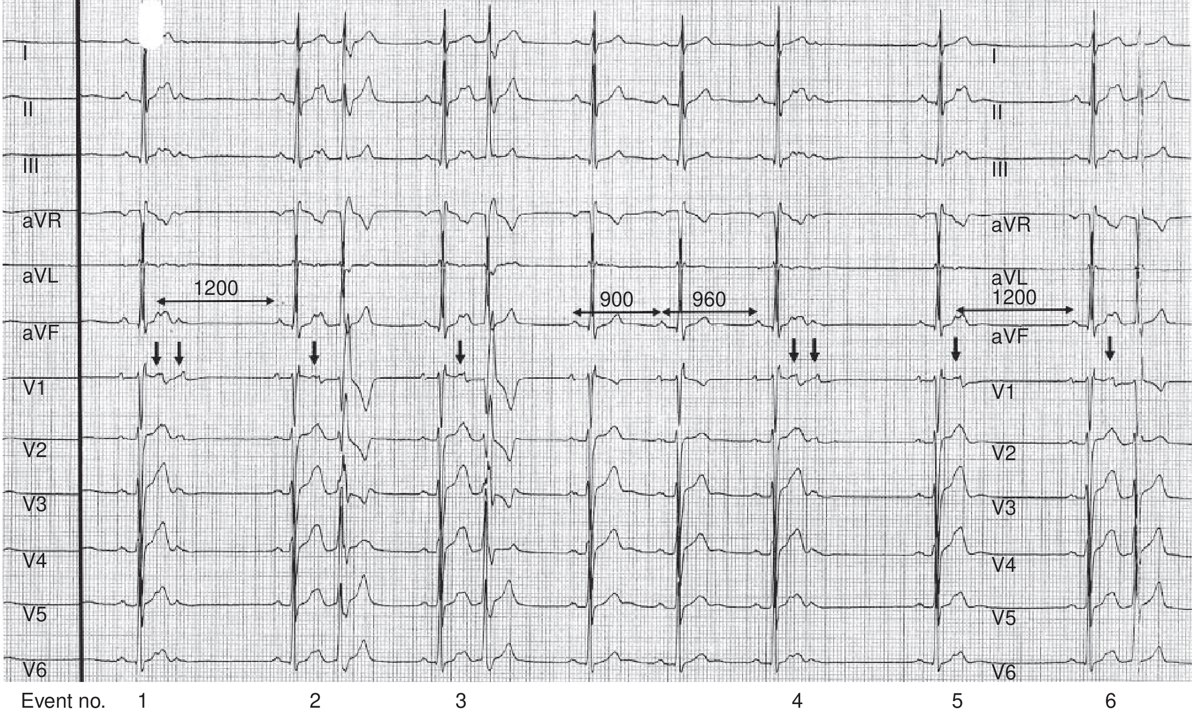
Clinical significance of PACs:
- In the vast majority, PACs are benign and require only reassurance
- However, Haïssaguerre et al.'s landmark work showed PACs from pulmonary vein sleeves can trigger atrial fibrillation (AF)
- Excess PACs (>30/hour or runs >20 beats) are associated with incident AF, stroke, and death
- Patients with excess PACs and CHADS-VASc ≥ 2 have an annual stroke risk comparable to patients with known AF
- Very frequent PACs (20-40% daily burden) may cause reversible cardiomyopathy
Braunwald's Heart Disease, p. 491
2. Junctional (AV Nodal) Premature Contractions
ECG features:
- P wave is absent or hidden - the impulse travels retrogradely into the atria and forward into the ventricles simultaneously, superimposing the P wave onto the QRS-T complex
- The QRS complex is usually narrow (normal configuration)
- The P wave, if visible, appears just before or just after the QRS and is typically inverted in inferior leads
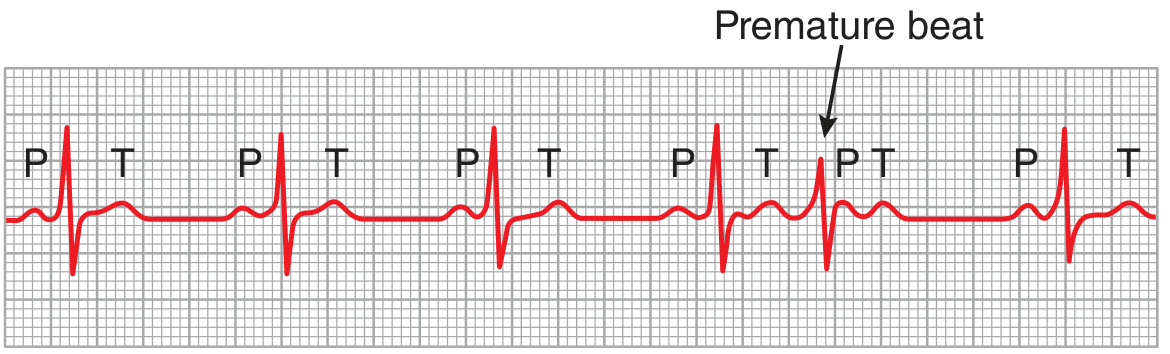
General significance and causes are similar to PACs.
3. Premature Ventricular Contractions (PVCs)
PVCs are the most clinically significant type of ectopic beat.
ECG features:
- Wide, bizarre QRS complex (usually >120 ms) - the impulse travels through the ventricular myocardium rather than the fast-conducting Purkinje system
- High voltage - in a normal beat, both ventricles depolarize nearly simultaneously so the vectors partially cancel out. In a PVC, one ventricle depolarizes ahead of the other, generating large electrical potentials
- T wave polarity opposite to the QRS - because slow muscle conduction means the first areas to depolarize are also the first to repolarize, creating discordance
- Full compensatory pause - the SA node timing is usually undisturbed; the post-PVC pause brings the next sinus beat back on schedule
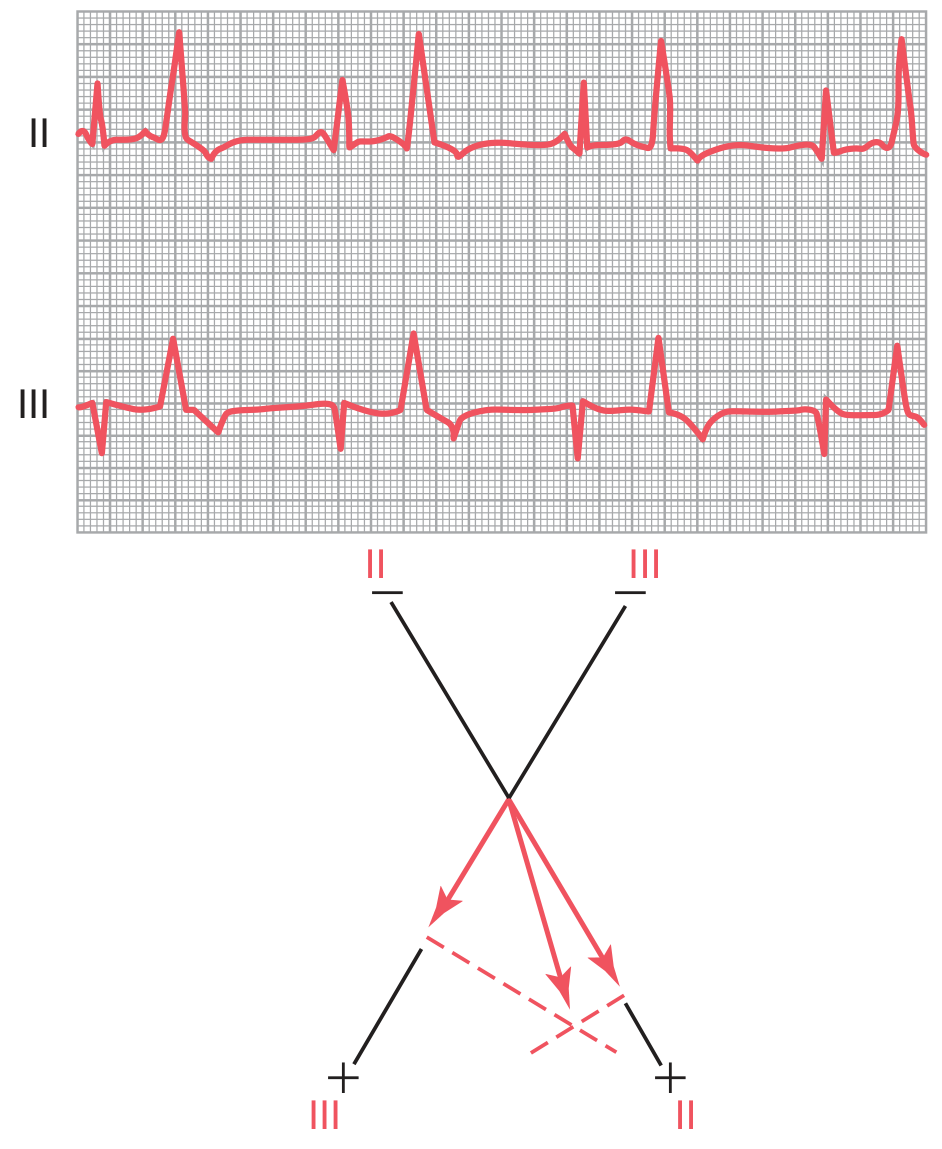
Patterns of PVCs:
| Pattern | Definition |
|---|---|
| Bigeminy | Every other beat is a PVC |
| Trigeminy | Every third beat is a PVC |
| Couplet | Two PVCs in a row |
| Triplet | Three PVCs in a row (= non-sustained VT) |
| R-on-T | PVC falls on T wave of preceding beat (dangerous) |
Pulse deficit: Because the ventricle contracts prematurely before adequate filling, stroke volume is reduced or absent. The peripheral pulse may not be felt even though the heart contracted, creating a discrepancy between apical rate and radial pulse rate.
Clinical significance of PVCs:
- Isolated, infrequent PVCs in an otherwise healthy heart are usually benign (nicotine, caffeine, fatigue, emotion)
- PVC burden >20% of beats is associated with cardiac dilation and cardiomyopathy - often reversible with treatment
- PVCs occurring in the vulnerable period (on the T wave, "R-on-T phenomenon") risk triggering ventricular fibrillation
- PVCs in the setting of ischemia, structural heart disease, or after MI carry significantly higher risk
Guyton & Hall, p. 169-170
Symptoms
Most ectopic beats are asymptomatic. When symptoms occur, patients may describe:
- A sense that the heart gave an "extra beat" or "missed a beat"
- Palpitations or fluttering in the chest or throat
- A thudding or pounding sensation (the forceful beat after the pause)
- Occasional brief dizziness or faintness
Braunwald's Heart Disease, p. 491
Diagnosis
- 12-lead ECG - detects ectopic beats occurring at the time of recording
- Ambulatory Holter monitoring (24-48 hours) - quantifies ectopic burden; essential for assessing frequency and patterns
- Event recorder / loop recorder - for infrequent symptoms; allows patient-triggered recording
- Echocardiogram - to assess for underlying structural heart disease and cardiomyopathy (especially if PVC burden is high)
Treatment
PACs: No treatment needed in most cases. If symptomatic:
- Eliminate triggers (caffeine, alcohol, sleep deprivation, stimulants)
- Beta-blocker or non-dihydropyridine calcium channel blocker (verapamil, diltiazem)
- Antiarrhythmic drugs (e.g., flecainide) in refractory cases
- Catheter ablation for high-burden, unifocal, drug-refractory symptomatic PACs
PVCs:
- Address reversible causes (ischemia, electrolyte abnormalities, thyrotoxicosis)
- Beta-blockers - first-line for symptomatic PVCs
- Antiarrhythmics (flecainide, propafenone for structurally normal heart; amiodarone, mexiletine if structural disease)
- Catheter ablation - highly effective, especially for unifocal PVCs; indicated for symptomatic or high-burden PVCs causing cardiomyopathy
Braunwald's Heart Disease, p. 491
Summary Table
| Feature | PAC | AV Junctional | PVC |
|---|---|---|---|
| P wave | Early, abnormal morphology | Hidden in QRS | Absent |
| QRS width | Narrow | Narrow | Wide (>120 ms) |
| Compensatory pause | Incomplete (usually) | Variable | Full (usually) |
| T wave | Normal | Normal | Discordant (opposite QRS) |
| Risk | AF trigger if frequent | Generally benign | VF risk if R-on-T; cardiomyopathy if >20% |
Proximal femur nailing
Reading File
Proximal femur nailing is the surgical fixation of proximal femoral fractures (subtrochanteric, intertrochanteric, basicervical) using an intramedullary nail inserted through the greater trochanter, with a cephalocervical screw locking into the femoral head/neck.
Searching sources...
Finding Sources
Finding Sources
Finding Sources
Finding Sources
Finding Sources
Finding Sources
Excellent - I have Rockwood & Green and Campbell's. Let me read the key sections in parallel now:
Reading File
Finding Sources
Finding Sources
Reading File
Reading File
Reading File
Reading File
Excellent content. Let me now read the cephalomedullary nail technique section fully and fetch key images in parallel:
Reading File
Loading Image
Loading Image
Good - I have the TAD diagram. Now let me get the cephalomedullary nail technique steps and fetch key surgical images:
Reading File
Loading Image
Loading Image
Loading Image
I now have comprehensive data from Rockwood & Green and Miller's Review. Let me compile the full answer:
Proximal Femur Nailing (Cephalomedullary Nailing)
Proximal femur nailing - also called cephalomedullary (CM) nailing - is the fixation of proximal femoral fractures using an intramedullary nail inserted via the greater trochanter, with a cephalocervical lag screw locking into the femoral head/neck. It combines the mechanical advantage of intramedullary load sharing with rotational control of the head-neck fragment.
Indications
CM nailing is the implant of choice for:
| Fracture Type | Notes |
|---|---|
| Unstable intertrochanteric fractures | Reverse oblique, comminuted, absent lateral wall (<20.5 mm) |
| All intertrochanteric fractures | Valid option; mandatory for unstable patterns |
| Subtrochanteric fractures | Long nail preferred; extends to distal interlocking |
| Basicervical femoral neck fractures | Cephalomedullary construct controls rotation |
| Pathological fractures of proximal femur | |
| Ipsilateral femoral neck + shaft fractures | Reconstruction nail |
For stable intertrochanteric fractures (intact lateral wall >20.5 mm), a Sliding Hip Screw (SHS/DHS) is also acceptable and cheaper - but a CM nail is still a valid choice for all patterns.
- Miller's Review of Orthopaedics 9th Ed, p. 918
- Rockwood and Green's Fractures in Adults 10th Ed
Implant Principles
The CM nail acts as an intramedullary buttress - it resists femoral medialization and provides less fracture collapse than extramedullary devices (SHS). The nail body sits in the medullary canal, and the lag screw slides within the nail barrel, allowing controlled fracture impaction while preventing medialization.
Key design features:
- Proximal entry angle: typically 4-6° valgus (trochanteric entry)
- Anterior bow: nail bow must match the anterior bow of the femur to avoid anterior cortex perforation distally
- Lag screw: single-screw design (e.g., PFNA, Gamma nail, TFN) or dual-screw design (e.g., InterTAN)
- Blade vs. screw: Cephalad blade has more medial migration risk than a cephalad screw
Surgical Setup and Positioning
Table: Fracture table is essential - closed reduction without it is not recommended.
Position: Supine. The injured leg is placed in traction in a traction boot. The uninjured leg is either:
- Flexed and abducted in a lithotomy stirrup (C-arm goes between legs)
- Extended in the "scissor" position (C-arm from opposite side) - preferred when contralateral hip motion is limited
A transparent plastic isolation drape suspended from a rail above the patient is preferred to separate the sterile field from the image intensifier.
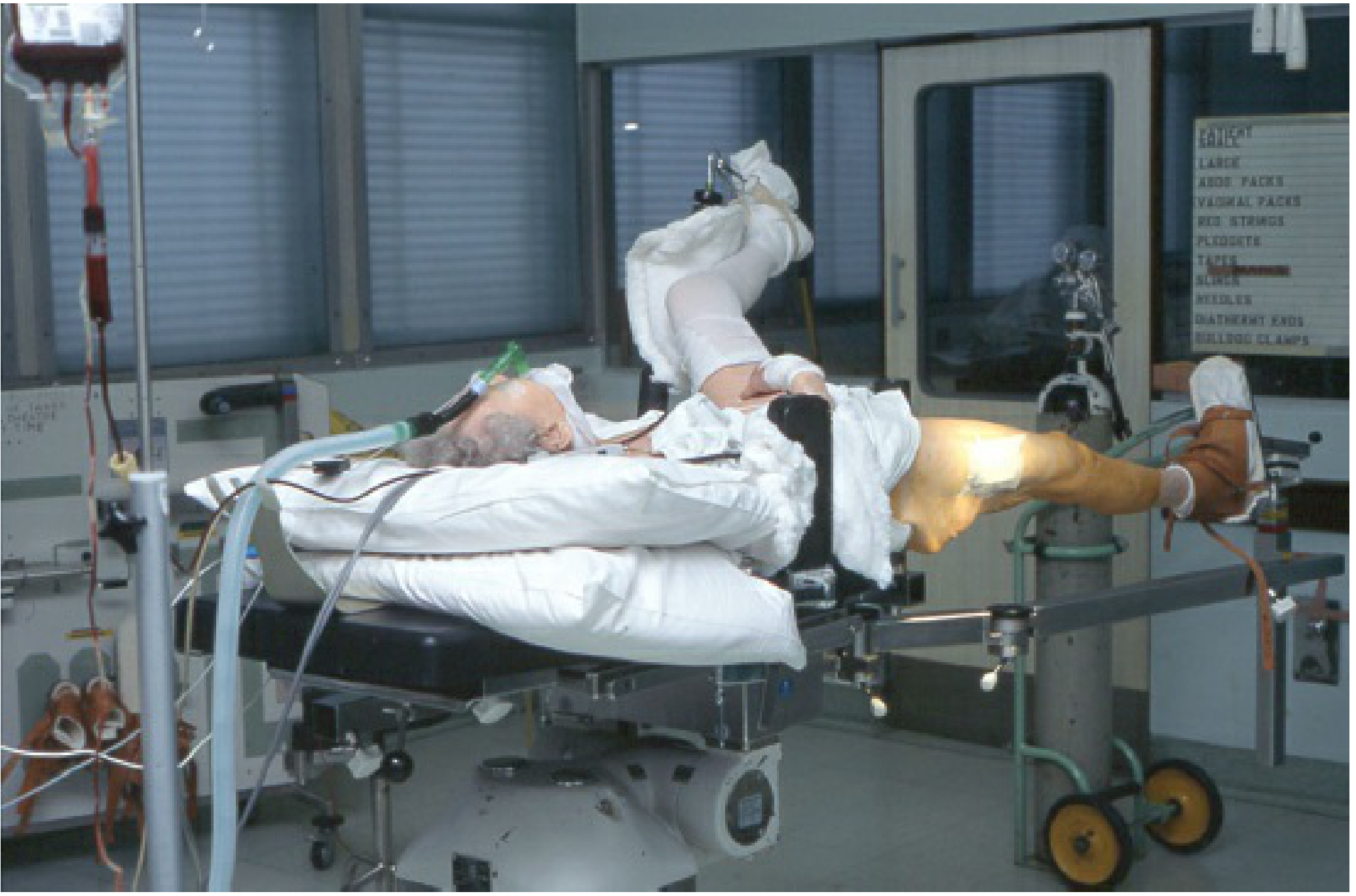
Fracture Reduction - The Most Critical Step
The two most important intraoperative steps are: (1) fracture reduction and (2) guidewire placement in the femoral head.
AP view:
- Reduce to anatomical or slight valgus - valgus is associated with the lowest risk of implant cut-out and less limb shortening
- A slight medial cortex gap in valgus is acceptable - it closes rapidly with fracture collapse
- Varus must never be accepted - mechanically unstable, leads to progressive varus collapse and cut-out
Lateral view:
- Femoral head, neck, and trochanteric region must align in a straight line
- Sagittal sag is more common in bariatric patients, high-energy injuries, and comminuted fractures
Reduction aids when closed reduction fails:
- Posterior reduction aid (attached to fracture table - provides upward pressure)
- Vertical crutch/support under the fracture site
- Bone lever or clamp placed percutaneously
- Formal open reduction (rare - mainly for A3/reverse oblique/subtrochanteric)
- Cerclage wires after open reduction in selected cases
Rotation control: Patella should face the ceiling throughout.
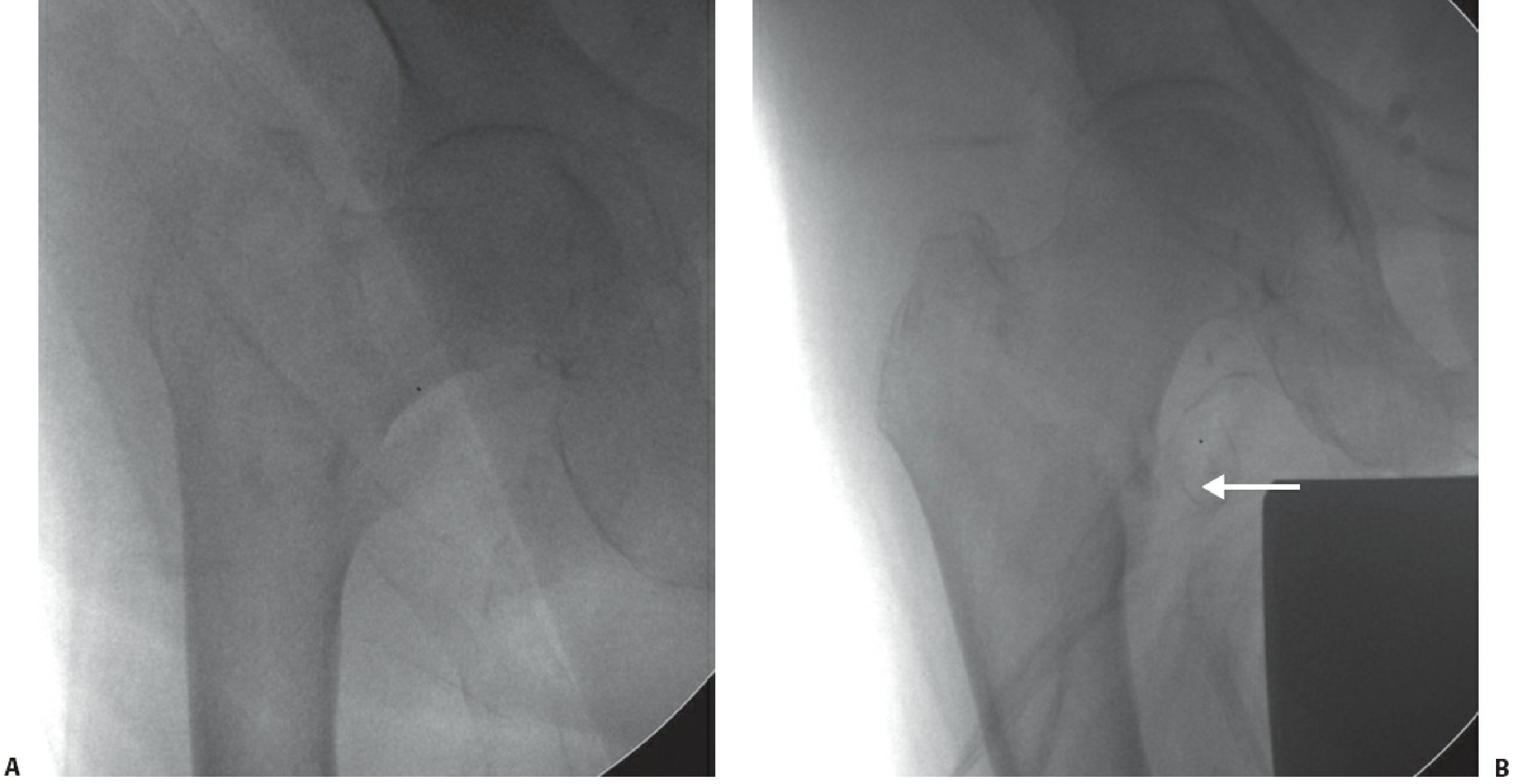
Surgical Technique - Step by Step
| Step | Detail |
|---|---|
| 1. Incision | 3-cm incision ~5 cm proximal to the tip of greater trochanter; incise fascia lata; split muscle to the trochanter |
| 2. Entry point guidewire | Insert at tip of greater trochanter - just lateral to medial aspect on AP view; centered or slightly posterior on lateral view |
| 3. Open proximal canal | Core/ream proximal 5 cm with solid channel reamer to size |
| 4. Ream medullary canal | Ream to accommodate nail diameter (if required by nail design) |
| 5. Insert nail | Advance nail under fluoroscopic guidance - confirm alignment AP and lateral |
| 6. Lag screw guidewire | Target center-center on AP (inferior half acceptable), center on lateral - to achieve TAD <25 mm |
| 7. Measure lag screw length | From guidewire measurement; advance to 5-7 mm from the joint line |
| 8. Ream femoral neck | Over the guidewire to the appropriate lag screw diameter |
| 9. Insert lag screw | Anti-rotation: place a finger on the femoral neck anteriorly to resist head rotation; use anti-rotation pin/screw |
| 10. Distal interlocking | Static lock for unstable/subtrochanteric fractures; optional for stable intertrochanteric fractures |
| 11. Closure | Fascia lata closed; absorbable subcuticular skin sutures; pressure dressing |
- Rockwood and Green's Fractures in Adults 10th Ed, p. 2726-2727
The Tip-Apex Distance (TAD)
The TAD is the single most important predictor of lag screw cut-out.
TAD = distance from screw tip to femoral head apex on AP + distance on lateral view (corrected for magnification)
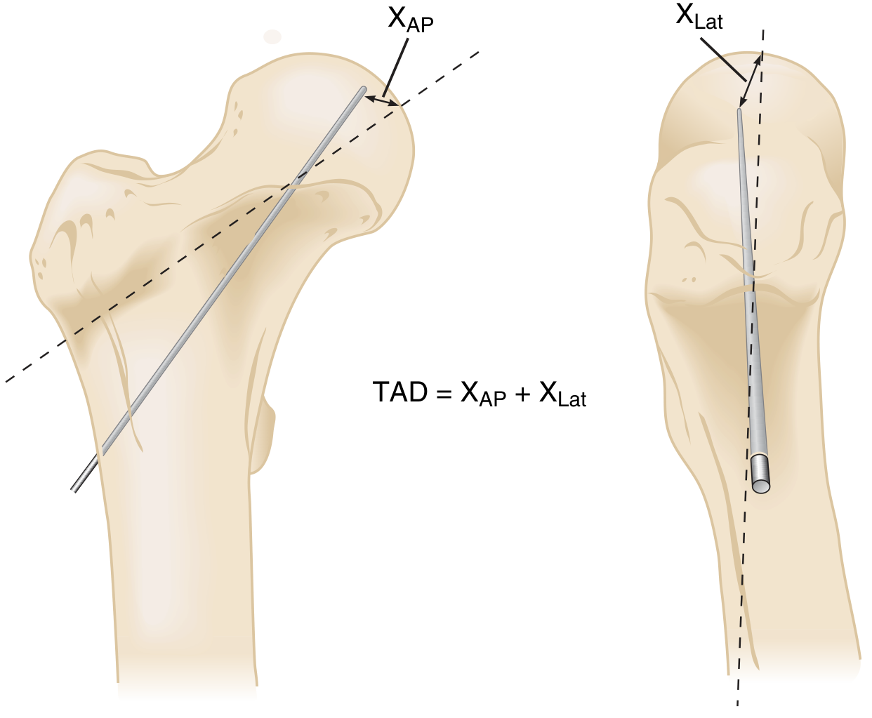
- TAD <25 mm - strongly associated with lowest cut-out rate
- TAD >25 mm - significantly higher risk of screw cut-out
- The ideal lag screw position is center-center on AP, center on lateral
Nail Length - Short vs. Long
| Short Nail | Long Nail |
|---|---|
| Standard oblique (AO 31-A1, A2) stable fractures | Reverse oblique (A3), subtrochanteric, fractures extending distally |
| Distal interlocking optional | Distal interlocking required |
| Cheaper | Reduces risk of stress fracture at nail tip |
A literature review of 1,276 patients found no statistically significant difference in periprosthetic fracture rate between short and long nails for standard intertrochanteric fractures, but long nails are preferred when the fracture pattern extends or to protect the entire femur.
Postoperative Care and Weight Bearing
- Immediate or early full weight bearing is the goal in elderly patients
- Physiotherapy and mobilization start on day 1 post-op
- Prophylactic anticoagulation for VTE prevention
- Transfer to a pressure-relieving mattress as soon as possible
Complications and Their Prevention
| Complication | Incidence | Prevention |
|---|---|---|
| Lag screw cut-out (varus collapse) | 1-5% | Valgus reduction + TAD <25 mm |
| Cut-in (craniomedial migration) | ~2% | Avoid jamming; correct positioning |
| Malunion (varus) | 5-10% | Correct reduction before fixation |
| Nonunion | 1-3% | Reduction + correct implant positioning |
| Implant breakage | 1-2% | Treat nonunion early |
| Avascular necrosis | ~1% | Avoid rotating femoral head at surgery |
| Anterior perforation distally | Rare | Match nail bow to femoral bow; avoid posterior entry |
| Superficial wound infection | 1-5% | Minimize exposure; prophylactic antibiotics |
| Deep wound infection | <1% | Strict aseptic technique |
| Peri-implant fracture at nail tip | Low | Long nail to bypass stress riser if fracture near tip |
- Rockwood and Green's Fractures in Adults 10th Ed, p. 2730
Cut-out is the most common mechanical failure. The lag screw fails to slide as the fracture collapses (jamming), acts as a fixed implant, and eventually breaches the femoral head.
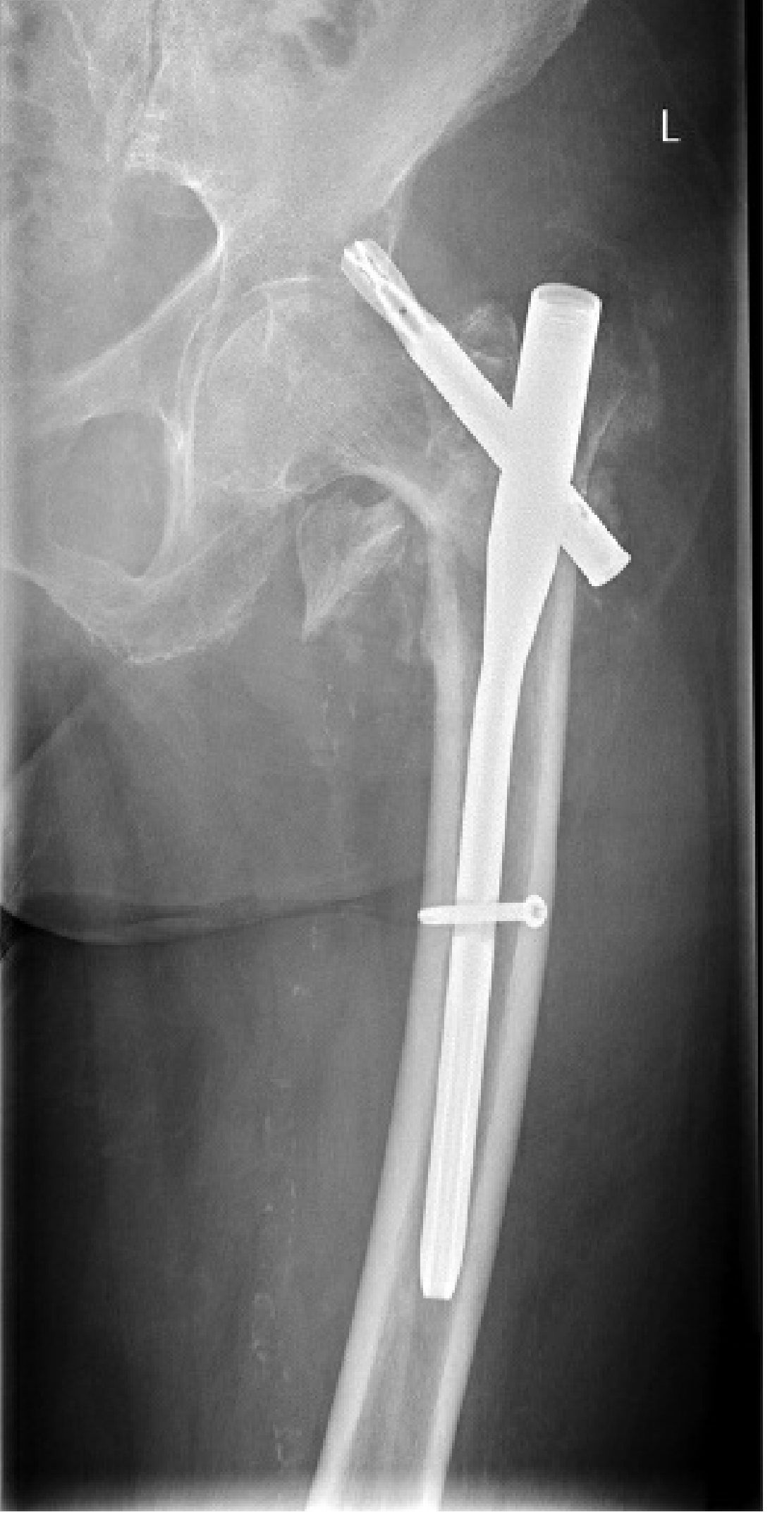
CM Nail vs. Sliding Hip Screw (SHS) - Decision Guide
| Feature | SHS (DHS) | Cephalomedullary Nail |
|---|---|---|
| Cost | Lower | Higher (long nail costliest) |
| Stable intertrochanteric (A1, A2 with lateral wall >20.5 mm) | First choice | Also valid |
| Unstable intertrochanteric (reverse oblique, lateral wall <20.5 mm) | Higher failure rate | Preferred |
| Subtrochanteric extension | Contraindicated | Required |
| Collapse/medialization | More collapse | Less collapse (IM buttress) |
| Blood loss / operative time | Similar | Similar |
| Risk of iatrogenic lateral wall fracture | Yes (if wall thin) | Lower |
- Miller's Review of Orthopaedics 9th Ed, p. 918
Vericose vein surgery
Reading File
Varicose vein surgery encompasses a range of procedures - from classical open surgery (Trendelenburg/saphenofemoral ligation + stripping) to modern minimally invasive techniques (EVLA, RFA, foam sclerotherapy, UGFS, MOCA, cyanoacrylate glue). Let me pull authoritative textbook content now.
Searching sources...
Finding Sources
Finding Sources
Finding Sources
Finding Sources
Finding Sources
Excellent. Now let me read the main textbook sections in parallel:
Reading File
Reading File
Reading File
Reading File
Reading File
Finding Sources
Now let me get the full Bailey & Love section on surgical technique and the Fitzpatrick/Dermatology content on endovenous procedures:
Finding Sources
Reading File
Reading File
Reading File
Reading File
Loading Image
Reading File
Loading Image
Reading File
Loading Image
Reading File
Loading Image
Reading File
Loading Image
Loading Image
Loading Image
Reading File
I now have comprehensive, image-rich content from Bailey & Love, Schwartz's Surgery, Harrison's, and Fitzpatrick's Dermatology. Let me compile the full answer:
Varicose Vein Surgery
Varicose veins are abnormally dilated, tortuous superficial veins resulting from venous reflux, most commonly due to incompetence at the saphenofemoral junction (SFJ) or saphenopopliteal junction (SPJ). Adult prevalence is 30-50%, increasing with age, BMI, female sex, pregnancy, and family history.
Indications for Intervention
- Persistent symptoms (aching, heaviness, throbbing, pruritus, swelling) despite conservative measures
- Complications: superficial vein thrombosis (thrombophlebitis), skin changes (lipodermatosclerosis, hyperpigmentation), venous ulceration, or bleeding
- Worsening symptoms or inadequate relief with compression hosiery
- Cosmetic concerns (patient-driven)
Conservative treatment (compression stockings 20-40 mmHg, leg elevation) is first-line and relieves symptoms in many patients.
Preoperative Assessment
Duplex ultrasound is mandatory before any intervention. It identifies:
- The site(s) of junctional incompetence (SFJ, SPJ, perforators)
- The extent and caliber of superficial axial incompetence in the GSV and SSV
- Presence or absence of deep venous obstruction or reflux
- Tortuosity and diameter of target veins (critical for selecting technique)
- Thrombus within superficial veins
- Pelvic sources of reflux
Reflux is defined as retrograde flow ≥0.5 seconds in superficial veins (≥1 second in proximal deep veins), elicited by calf compression/release or Valsalva.
Treatment Options
Modern varicose vein management uses a proximal-to-distal, highest-to-lowest pressure strategy: treat junctional/axial incompetence first, then address tributaries/telangiectasias.
Overview Table
| Technique | Mechanism | Anesthesia | Suitable For |
|---|---|---|---|
| Endovenous Laser Ablation (EVLA) | Thermal (laser energy) | Tumescent LA | GSV/SSV axial incompetence |
| Radiofrequency Ablation (RFA) | Thermal (electromagnetic current) | Tumescent LA | GSV/SSV axial incompetence |
| Ultrasound-Guided Foam Sclerotherapy (UGFS) | Chemical endothelial destruction | None/LA | Axial veins, tributaries, perforators |
| Cyanoacrylate Glue (CAC) | Non-thermal, fibrosis | Minimal/none | Axial veins |
| MOCA (Mechanochemical Ablation) | Mechanical + chemical | None (mostly) | Axial veins |
| Ambulatory Phlebectomy (Stab Avulsion) | Physical excision | Local | Branch varicosities |
| SFJ Ligation + GSV Stripping | Open surgery | GA/spinal | Large GSV (>2 cm), recurrences |
| SPJ Ligation + SSV Stripping | Open surgery | GA/spinal | SSV incompetence |
1. Endovenous Thermal Ablation
Endovenous ablation (EVLA or RFA) has largely replaced open surgery as first-line treatment in most centres. Long-term efficacy, recurrence rates, and clinical outcomes are equivalent to surgery, but with significantly less morbidity, faster recovery, less postoperative pain, wound infection, and haematoma.
Shared Technique (EVLA and RFA)
- Access: Percutaneous catheter/fibre insertion into the target vein (usually at the knee) under duplex ultrasound guidance
- Advance to 2-3 cm distal to the SFJ or SPJ termination
- Confirm position on ultrasound (longitudinal B-mode)
- Perivenous tumescent anaesthesia infiltrated under ultrasound guidance into the saphenous compartment - serves as heat sink, compresses vein onto device, protects surrounding tissues, and allows immediate ambulation
- Deliver thermal energy while slowly withdrawing the catheter/fibre
- Thermal damage destroys intima and causes collagen denaturation of the media → fibrotic occlusion
- Compression applied after procedure
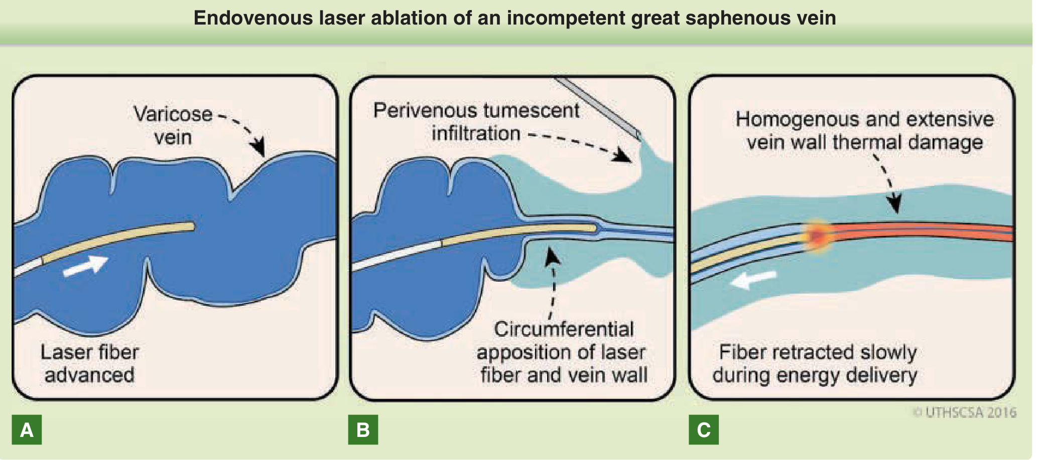
EVLA (Endovenous Laser Ablation)
- Wavelengths: 810, 940, 980 nm (haemoglobin-targeting) or 1320, 1470 nm (water-targeting - longer wavelengths cause less postoperative bruising/pain)
- Thermal damage via heat transfer from a hot fibre tip through residual intraluminal blood + photothermal absorption
- Energy delivery typically 60-80 J/cm for durable closure
- 5-year efficacy: ~90% closure rate
- EVLA requires operator knowledge of power settings and pullback speed; allows direct catheter delivery of adjuvant foam sclerotherapy via a 0.035-in lumen
RFA (Radiofrequency Ablation)
- Most popular device: ClosureFAST™ (Medtronic) - wire coil on end of catheter
- Generator heats surrounding tissue to 120°C for 20-second treatment cycles
- Catheter withdrawn by set length (3 cm or 7 cm coil) after each cycle
- More forgiving for novice users - single button press per cycle
- Automatic treatment cycle frees surgeon to concurrently perform tumescent infiltration and phlebectomy
- 2-year efficacy: 90%; 5-year: 80%
EVLA and RFA have equivocal evidence in head-to-head studies. Both achieve >95% closure rates and are suitable for the vast majority of patients with superficial axial incompetence.
- Bailey and Love's Surgery 28th Ed, p. 1055-1056
2. Ultrasound-Guided Foam Sclerotherapy (UGFS)
Mechanism: Sclerosant (typically polidocanol or sodium tetradecyl sulphate) is mixed with air or CO₂/O₂ gas to create foam (Tessari technique). Foam displaces blood, maximises endothelial contact, and causes endothelial destruction → fibrosis and occlusion.
Technique:
- Patient positioned supine with leg elevated
- Under continuous duplex guidance, foam injected into the incompetent axial vein
- When foam is visualised at the site of junctional incompetence, injection stops
- Maximum volume per session: 10-12 mL (complication risk increases with higher volumes)
- Compression applied after procedure
- 1-year occlusion: ~81%; 5-year: ~74%
Advantages of UGFS:
- No tumescent anaesthesia required (less painful procedure)
- No limitation due to vein tortuosity
- Can treat calf veins beneath damaged skin/ulcers without skin puncture
- Very low consumable cost
- Suitable as an adjunct for neovascularisation or residual varicosities
Disadvantages: Outside specialist centres, efficacy is significantly lower than thermal ablation, with higher reintervention rates and higher rates of superficial phlebitis and pigmentation.
Serious (rare) complications: Transient ischaemic attack, stroke, and visual disturbances - associated with patent foramen ovale; maximum foam volume limits mitigate risk.
- Bailey and Love's Surgery 28th Ed, p. 1061-1062
3. Non-Thermal, Non-Tumescent Techniques
Cyanoacrylate Adhesive (e.g., VenaSeal™)
- Catheter placed in target vein; glue delivered in aliquots as catheter withdrawn
- Glue causes acute inflammatory fibrosis → permanent vein occlusion
- No tumescent anaesthesia required; no compression mandatory
- 1-year occlusion: ~90%
- Adverse effects: superficial thrombophlebitis, local hypersensitivity reactions
MOCA - Mechanochemical Ablation (e.g., ClariVein™)
- Rotating wire deployed from end of catheter physically damages endothelium
- Liquid sclerosant infused simultaneously during catheter withdrawal
- No tumescent anaesthesia (some patient discomfort; occasional vein tearing)
- Early efficacy comparable to thermal methods, but higher medium/long-term recanalisation rates
- Bailey and Love's Surgery 28th Ed, p. 1062
4. Open Surgery: Saphenofemoral Ligation + GSV Stripping (Trendelenburg Operation)
Still relevant for: very large GSVs (>2 cm diameter), recurrent varicose veins requiring redo surgery, and cases not suitable for endovenous techniques. Long-term results are comparable to endovenous methods but with greater morbidity and slower recovery.
GSV (Great Saphenous Vein) - Surgical Steps
- Incision: Oblique groin incision at the level of, and lateral to, the pubic tubercle - ideally above the groin crease
- Dissection: Identify and dissect the GSV to the SFJ - clearly establish the junction before dividing (risk of inadvertent femoral vein transection)
- Tributaries: Six tributaries may be encountered near the SFJ:
- Lateral: superficial inferior epigastric vein; superficial circumflex iliac vein
- Medial: superficial external pudendal vein; deep external pudendal vein
- Distal: anterior accessory GSV; posteromedial thigh vein
- All tributaries ligated distal to their divisions
- Flush SFJ ligation performed
- Stripping: GSV stripped retrogradely to approximately the knee using a blunt-tip catheter or invagination pin stripper
- Stripping to the ankle is NOT recommended (high saphenous nerve injury risk)
- Stripping below the knee carries up to 7% saphenous nerve neuralgia risk
- Phlebectomy of tributary varicosities performed concurrently
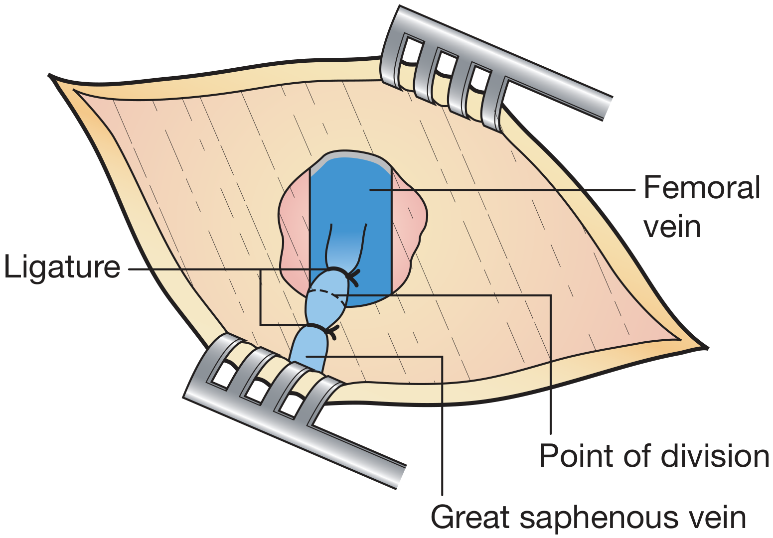
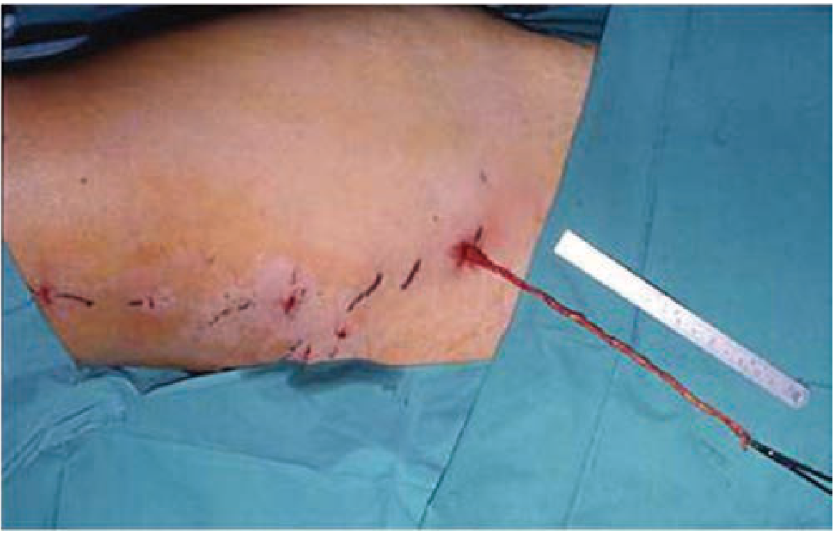
GSV stripping vs. SFJ ligation alone: Stripping results in lower recurrence rates and better quality of life than SFJ ligation alone. - Schwartz's Principles of Surgery 11th Ed, p. 1024
SSV (Small Saphenous Vein) Surgery - Saphenopopliteal Junction (SPJ) Ligation
- Preoperative duplex marking of the SPJ is mandatory - anatomy is highly variable
- Patient positioned prone
- Transverse incision over the premarked SPJ in the popliteal fossa
- Fascia divided; SSV exposed and ligated (flush ligation vs. proximal ligation - both debated)
- SSV may then be stripped or the proximal section resected
- Sural nerve injury risk with stripping; popliteal vein injury risk with flush ligation
- Sural nerve neuropraxia incidence may be as high as 20% following SSV surgery
5. Ambulatory Phlebectomy (Stab Avulsion)
An adjunct to all axial vein treatments for residual branch varicosities.
- Pre-procedure: varicosities marked with patient standing
- 2-mm stab incisions made directly over branch varicosities
- Vein dissected/hooked from subcutaneous tissue and avulsed without ligation
- Performed under local anaesthetic (tumescent)
- Bleeding controlled by leg elevation, manual compression, and tumescent effect
- Can be combined with any axial treatment in the same session
6. Perforator Vein Surgery
- SEPS (Subfascial Endoscopic Perforator Surgery): Endoscopic identification and occlusion of incompetent perforating veins - largely replaced by ablative techniques
- Duplex-guided direct ablation (thermal or foam) of incompetent perforators - preferred in patients with skin changes
- Indicated when perforators are identified as a significant source of reflux, especially in recurrent or complex cases
Sclerotherapy (Liquid)
Used for telangiectasias and small reticular veins:
| Agent | Concentration for Telangiectasias |
|---|---|
| Hypertonic saline | 11.7-23.4% |
| Sodium tetradecyl sulphate | 0.125-0.25% |
| Polidocanol | 0.5% |
After injection, elastic bandages worn continuously for 3-5 days to oppose inflamed vein walls; then compression stockings for minimum 2 weeks.
Complications
Complications of Standard Open Surgery
| Complication | Incidence | Notes |
|---|---|---|
| Wound infection (most common) | Common | Reduced by prophylactic antibiotics |
| Saphenous nerve neuralgia | Up to 7% | GSV stripping to the knee; higher to ankle |
| Sural nerve neuropraxia | Up to 20% | SSV surgery |
| Common peroneal nerve injury | Up to 4% | SSV surgery |
| VTE (DVT/PE) | ~0.5% | Individual risk assessment + prophylaxis required |
| Haematoma/ecchymosis | Common | |
| Lymphocele/lymph leak | Uncommon | Groin dissection |
| Recurrence at 5 years | Up to 50% | - Harrison's |
Complications of Endovenous Techniques
| Complication | Notes |
|---|---|
| Bruising, pain, haematoma | Less than open surgery |
| Superficial thrombophlebitis | Particularly with non-thermal techniques |
| DVT/PE | Rare; commoner with thermal ablation near SFJ |
| Skin hyperpigmentation | UGFS > thermal |
| Paresthesias | Thermal injury to adjacent saphenous nerve |
| Foam: TIA/stroke | Very rare; patent foramen ovale risk; limit foam to 10-12 mL |
Recurrent Varicose Veins
Significant clinical recurrence within 5-10 years occurs in 10-35% after open surgery; duplex-detected recurrence is up to 70%. Causes:
- Neovascularisation (most important after open surgery) - new avalvular veins bridge ligated junction to tributaries
- Reflux in residual axial vein (inadequate stripping)
- Inadequate initial surgery
- New junctional reflux
- Progression of venous disease
Recurrence is more common after SSV surgery than GSV surgery, and in patients with high BMI. Stripping of the incompetent axial vein (rather than ligation alone) reduces recurrence rates.
- Bailey and Love's Surgery 28th Ed, p. 1063-1064
Vericose vein surgery
I just covered varicose vein surgery in detail in the previous message. Here's a summary of everything that was covered — scroll up to read the full illustrated version.
Varicose Vein Surgery - Summary
Definition & Background
Dilated, tortuous superficial veins from venous reflux - usually due to incompetence at the saphenofemoral junction (SFJ) or saphenopopliteal junction (SPJ). Prevalence 30-50% in adults.
Indications
- Persistent symptoms (aching, heaviness, pruritus, swelling) despite compression
- Complications: thrombophlebitis, skin changes (lipodermatosclerosis), venous ulceration, bleeding
- Cosmetic reasons
Treatment Options (Proximal-to-Distal Strategy)
1. Endovenous Laser Ablation (EVLA)
- Laser fibre advanced to 2-3 cm from SFJ under duplex guidance
- Perivenous tumescent anaesthesia injected first (heat sink + vein compression)
- Fibre withdrawn slowly while delivering laser energy (60-80 J/cm)
- 5-year occlusion ~90%
2. Radiofrequency Ablation (RFA)
- ClosureFAST™ catheter heats tissue to 120°C in 20-second cycles
- Simpler to operate than EVLA; 7 cm coil speeds treatment
- 5-year efficacy ~80%; evidence broadly equivalent to EVLA
3. Ultrasound-Guided Foam Sclerotherapy (UGFS)
- Foam sclerosant (polidocanol/STS) injected under duplex guidance
- Maximum 10-12 mL per session
- No tumescent anaesthesia; can treat tortuous veins and veins beneath ulcers
- Lower efficacy than thermal methods; higher retreatment rates
- Rare risk: TIA/stroke (especially if patent foramen ovale present)
4. Non-Thermal Options
- Cyanoacrylate glue (VenaSeal™): ~90% 1-year occlusion; no tumescent required
- MOCA (ClariVein™): Rotating wire + liquid sclerosant; no tumescent; higher recanalisation rates long-term
5. Open Surgery: SFJ Ligation + GSV Stripping (Trendelenburg)
- Oblique groin incision above the groin crease
- Dissect GSV to SFJ; ligate all 6 tributaries; flush SFJ ligation
- Strip GSV retrogradely to the knee (not to ankle - saphenous nerve risk)
- Stripping gives lower recurrence than ligation alone
- Indicated for very large GSV (>2 cm), recurrences, or endovenous failure
6. SSV Surgery (Saphenopopliteal Junction)
- Preoperative duplex marking mandatory (variable anatomy)
- Patient prone; transverse popliteal incision over premarked SPJ
- Flush ligation or proximal ligation then strip/resect SSV
7. Ambulatory Phlebectomy (Stab Avulsion)
- 2-mm stab incisions over tributary varicosities; veins avulsed with hook
- Done under tumescent local anaesthesia; adjunct to all axial treatments
8. Perforator Ligation
- Duplex-guided foam or thermal ablation of incompetent perforators
- SEPS (subfascial endoscopic perforator surgery) largely replaced by ablative methods
Tip-Apex Sclerotherapy Concentrations
| Agent | Telangiectasias | Larger Varicosities |
|---|---|---|
| Hypertonic saline | 11.7-23.4% | 23.4% |
| Sodium tetradecyl sulphate | 0.125-0.25% | 0.5-1% |
| Polidocanol | 0.5% | 0.75-1% |
Key Complications
| Complication | Incidence | Context |
|---|---|---|
| Saphenous nerve neuralgia | Up to 7% | GSV stripping to knee |
| Sural nerve neuropraxia | Up to 20% | SSV surgery |
| Common peroneal nerve injury | Up to 4% | SSV surgery |
| VTE (DVT/PE) | ~0.5% | All open surgery |
| Wound infection | Common | Open surgery |
| Foam TIA/stroke | Very rare | UGFS, patent foramen ovale |
| Clinical recurrence at 5-10 years | 10-35% | All methods |
| Duplex-detected recurrence | Up to 70% | All methods |
Causes of recurrence: Neovascularisation (most important), residual axial vein reflux, inadequate initial surgery, new junctional reflux.
Sources: Bailey and Love's Short Practice of Surgery 28th Ed; Schwartz's Principles of Surgery 11th Ed; Harrison's Principles of Internal Medicine 22nd Ed; Fitzpatrick's Dermatology
Inguinal hernioplasty
Reading File
Inguinal hernioplasty refers to the tension-free mesh repair of inguinal hernias - both open (Lichtenstein, plug-and-patch) and laparoscopic (TEP, TAPP) techniques. It has superseded older tissue repairs (Bassini, Shouldice) as the standard of care due to lower recurrence rates.
Searching sources...
Finding Sources
Finding Sources
Finding Sources
Excellent hits from Schwartz's, Mulholland, Bailey & Love, and Sabiston. Let me read the key sections in parallel now:
Reading File
Reading File
Reading File
Reading File
Reading File
Reading File
Reading File
Reading File
Reading File
Loading Image
Loading Image
Loading Image
Reading File
Loading Image
Loading Image
I now have comprehensive, image-rich content from Schwartz's Surgery, Mulholland & Greenfield's, and Bailey & Love's. Let me compile the full answer:
Inguinal Hernioplasty
Hernioplasty specifically refers to tension-free mesh repair of inguinal hernias, as opposed to herniorrhaphy (tissue repair). It is the most commonly performed general surgical procedure worldwide. The core principle is reinforcing the defective inguinal floor with a prosthetic mesh rather than suturing together non-apposing, tension-laden tissue planes.
Anatomy Review - The Myopectineal Orifice
The myopectineal orifice (MPO) is the single large fascial opening in the lower abdominal wall through which all inguinal and femoral hernias pass. Any adequate hernia repair must cover the entire MPO:
- Hesselbach's Triangle (medial/direct hernia zone): bounded by inferior epigastric vessels laterally, inguinal ligament inferiorly, lateral border of rectus medially
- Deep inguinal ring (lateral/indirect hernia zone)
- Femoral canal (femoral hernia zone)
Key structures at risk during dissection:
- Medially: Inferior epigastric vessels, vas deferens, Cooper's ligament, bladder
- Laterally (the "triangle of doom" and "triangle of pain"): External iliac vessels, lateral femoral cutaneous nerve, femoral nerve, genitofemoral nerve
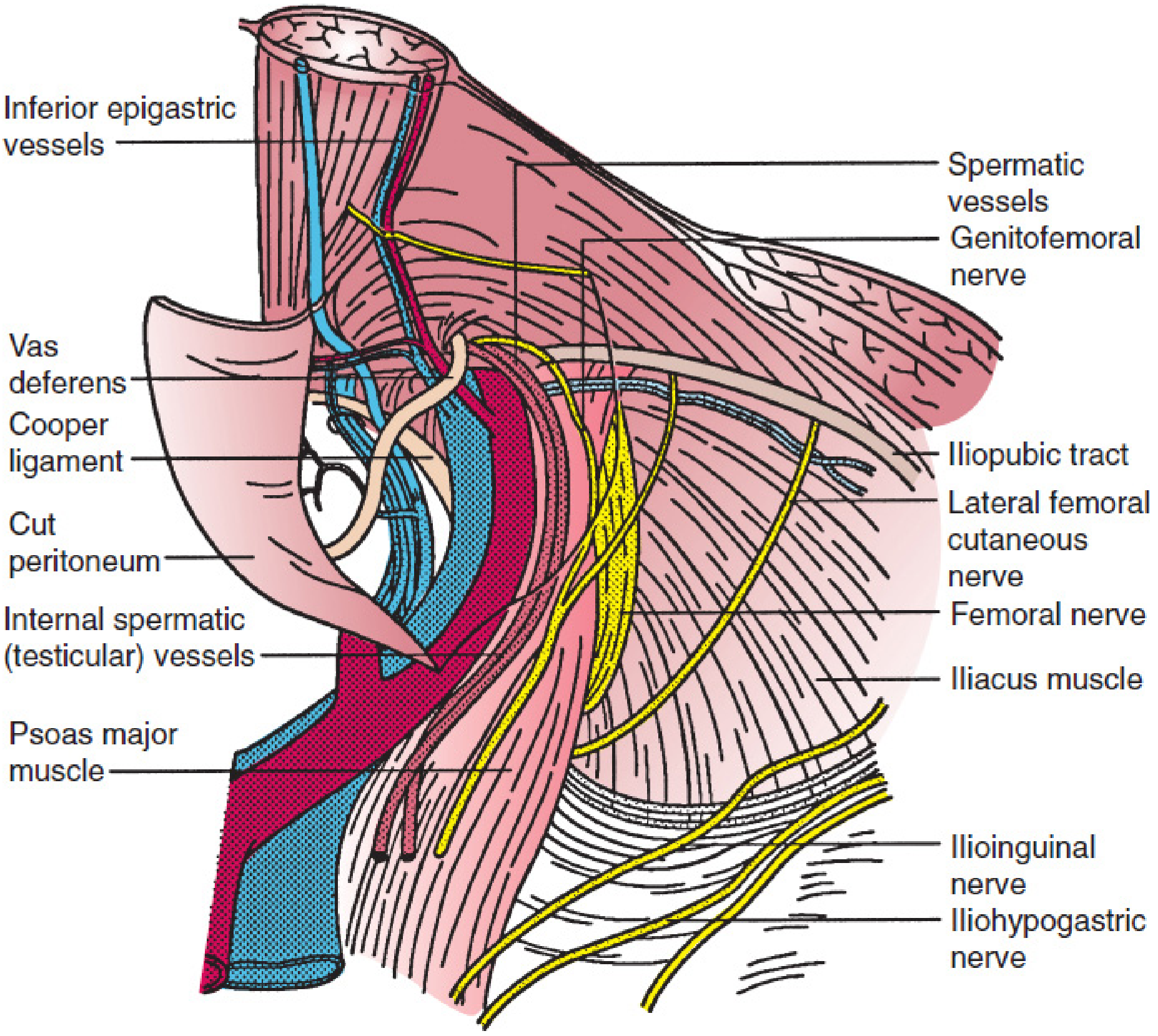
Classification of Repairs
| Category | Operations |
|---|---|
| Tissue repairs (herniorrhaphy) | Bassini, Shouldice, McVay (Cooper ligament), Desarda |
| Open tension-free mesh (hernioplasty) | Lichtenstein, Plug-and-Patch (Rutkow-Robbins), PHS (Prolene Hernia System) |
| Laparoscopic/endoscopic | TEP (totally extraperitoneal), TAPP (transabdominal preperitoneal), IPOM |
| Robotic | rTEP, rTAPP |
Part 1: Open Tension-Free Hernioplasty
A. Lichtenstein Tension-Free Repair (Gold Standard)
The Lichtenstein repair is considered the gold standard for open inguinal hernia repair, with a recurrence rate of 1% to 2% in most series - compared to 10-15% for tissue repairs in general surgical practice.
Steps:
- Incision: Skin incision 2 cm above and parallel to the inguinal ligament, from the pubic tubercle to just lateral to the deep ring
- External oblique aponeurosis (EOA): Incised in the direction of its fibres from the external ring to just beyond the internal ring; superior and inferior flaps raised
- Identify and protect nerves: Ilioinguinal nerve (runs on the cord), iliohypogastric nerve (in the superior flap), and genital branch of genitofemoral nerve (on the cord)
- Spermatic cord/round ligament: Isolated and lifted using a soft tissue retractor; cremaster divided longitudinally to expose the cord structures
- Hernia sac:
- Indirect sac: Located anteromedial to the cord; dissected free and inverted or ligated flush at the deep ring
- Direct sac: Inverted with a purse-string suture or simply reduced
- Mesh placement: "Flat iron" or keyhole-shaped mesh (typically 7.5 × 15 cm polypropylene):
- Medial edge overlaps the pubic tubercle by 1.5-2 cm (critical - prevents medial recurrence)
- Mesh extends 2-3 cm above Hesselbach's triangle superiorly
- Inferior edge fixed to the shelving edge of the inguinal ligament with a permanent monofilament suture (e.g., Prolene)
- A slit cut at the lateral end creates a narrow lower tail (1/3) and a wider upper tail (2/3); spermatic cord passes between the two tails; tails crossed and fixed - creating a new internal ring
- Mesh tails tucked under the EOA to the anterior superior iliac spine
- Wound closure: EOA closed from medial to lateral; external ring reconstructed snugly around cord; Scarpa's fascia and skin closed
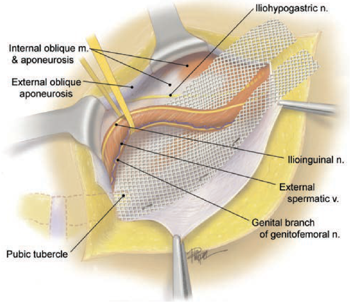
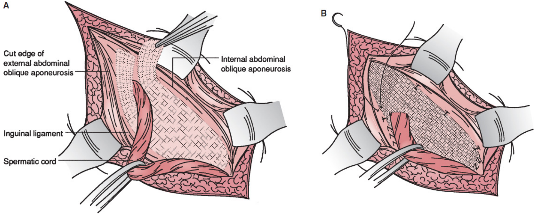
Anaesthesia: Can be performed under local, regional, or general anaesthesia - local anaesthesia suitable for most elective cases (advantage over laparoscopic which requires GA).
B. Plug-and-Patch Repair (Rutkow-Robbins / Gilbert)
A modification where a polypropylene plug is first inserted into the defect (internal ring for indirect, neck for direct hernia) and secured with interrupted sutures, then a flat mesh patch is placed over the inguinal floor as in Lichtenstein (using only 1-2 sutures or no sutures).
- Fast and easy to teach
- No demonstrated advantage over Lichtenstein in recurrence rate or chronic pain
- Disadvantages: Higher mesh burden; plug can migrate causing bowel obstruction; makes redo surgery more challenging
C. Tissue Repairs (Historical, Still Used in Select Cases)
| Repair | Technique | Notes |
|---|---|---|
| Bassini | Transversalis fascia + conjoined tendon to inguinal ligament | 10-15% recurrence in general practice |
| Shouldice | Four-layer running suture repair of transversalis fascia | ~1% recurrence at Shouldice Clinic; not reproducible elsewhere |
| McVay (Cooper Ligament) | Conjoined tendon sutured to Cooper ligament; needs relaxing incision | Covers femoral canal; indicated for femoral hernias |
| Desarda | Vascularized flap of EOA as autologous patch (mesh-free Lichtenstein) | Avoids foreign material; for patients declining mesh |
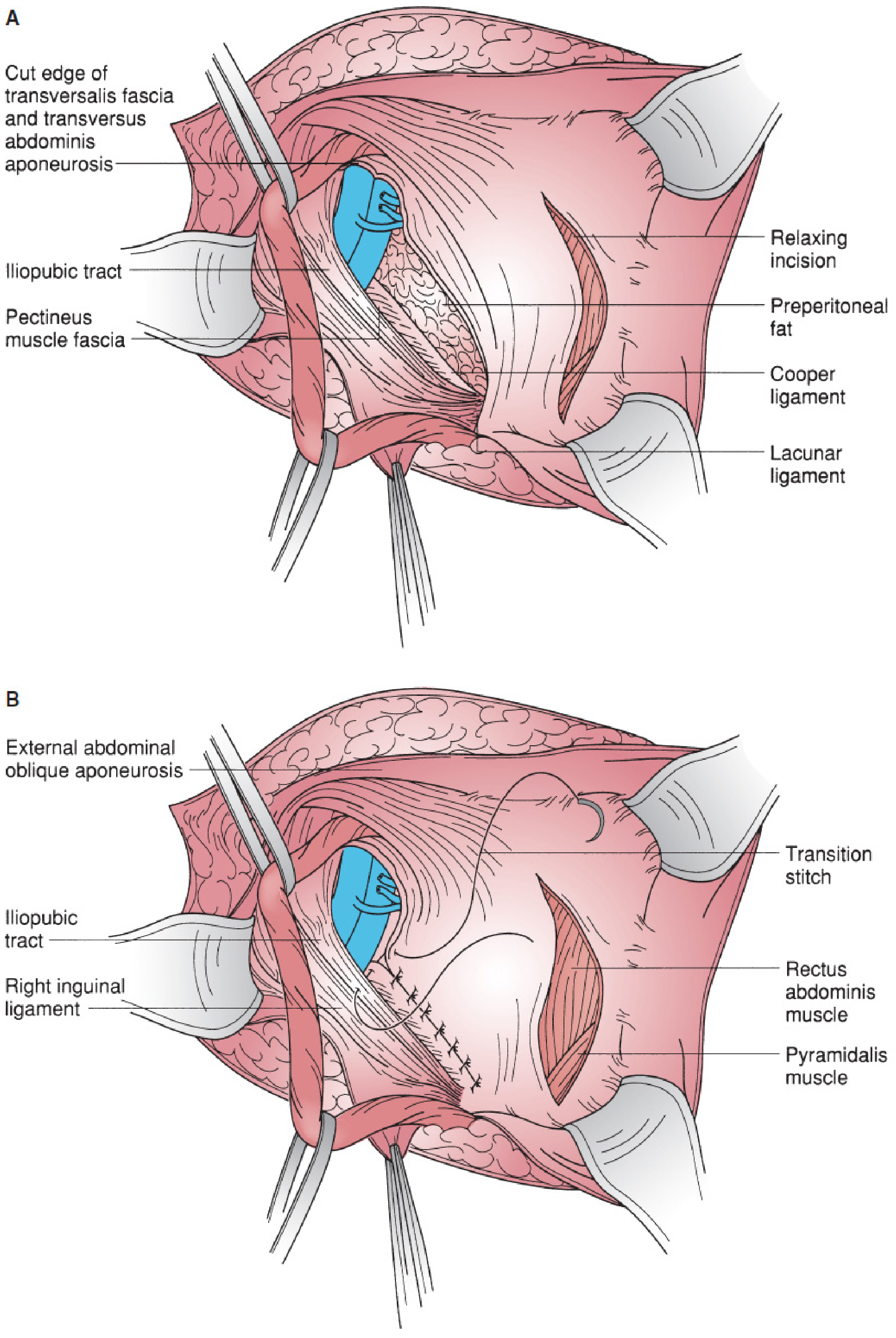
Part 2: Laparoscopic Hernioplasty
Laparoscopic repair places a large mesh (10 × 15 cm minimum) in the preperitoneal space, covering the entire myopectineal orifice - including Hesselbach's triangle, the deep ring, and the femoral canal.
Advantages over open:
- Less acute and chronic postoperative pain
- Faster return to full activity
- Lower wound complication rate (infection, seroma, haematoma)
- Superior for bilateral hernias (both repaired through same trocar sites) and recurrence after open anterior repair
- Better cosmesis
Disadvantages:
- Requires general anaesthesia (GA mandatory; patient cannot tolerate CO₂ insufflation awake)
- Longer learning curve
- Higher cost
- Risk of intra-abdominal injury (TAPP)
IEHS Grade A recommendation: TEP and TAPP are preferred alternatives to Lichtenstein for recurrent hernias after prior open anterior repair (uses a virgin preperitoneal plane).
TEP (Totally Extraperitoneal Repair)
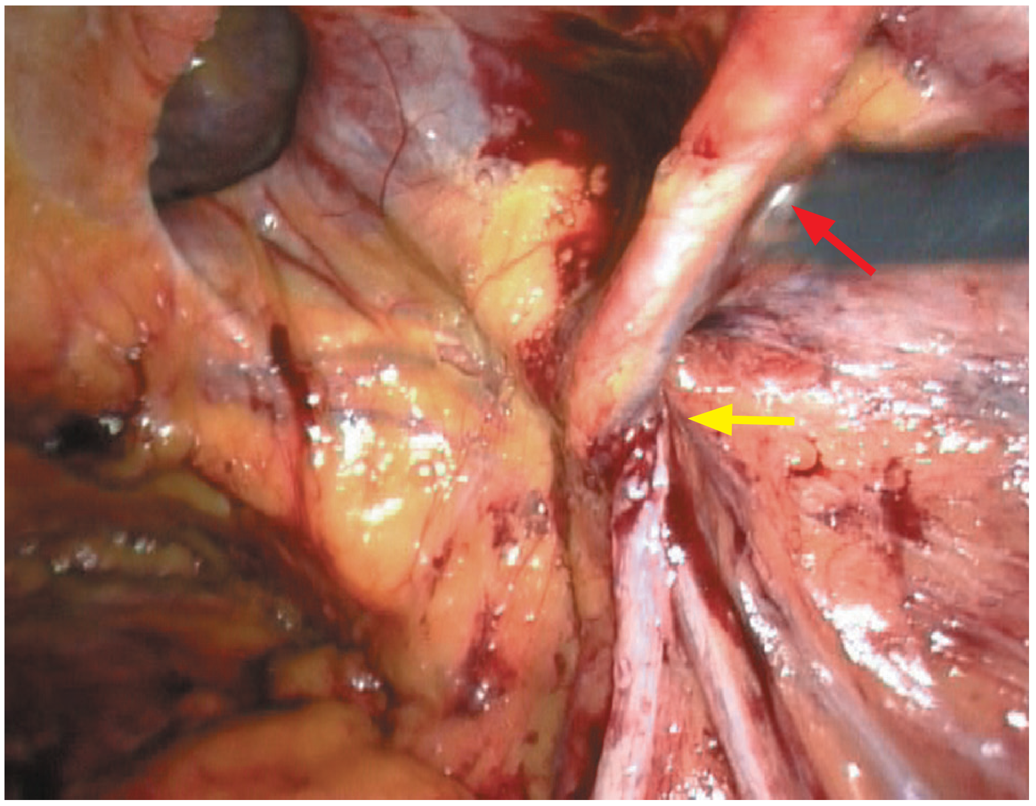
Key feature: The peritoneal cavity is never entered - entirely in the preperitoneal/extraperitoneal space.
Steps:
- Position: Supine, Trendelenburg; surgeon stands contralateral to hernia; arms tucked; screens at foot of bed
- Access: Umbilical incision; anterior rectus sheath opened ipsilaterally; rectus muscle retracted laterally; blunt finger dissection begins in space between rectus muscle and posterior rectus sheath
- Balloon dissection: Balloon dissector (or direct blunt dissection) develops the preperitoneal space under direct vision
- Trocars: 2 additional midline trocars - one ~5 cm above symphysis pubis, one midway between umbilicus and symphysis (or lateral paraumbilical trocars)
- Dissection: Complete development of retrorectus and preperitoneal space; identify Cooper ligament, inferior epigastric vessels, vas deferens, spermatic vessels
- Hernia sac: Direct sac inverted; indirect sac dissected from cord and reduced
- Mesh placement: Large mesh (≥10 × 15 cm or 14 × 11 cm) placed to cover entire MPO, extending:
- ≥2 cm above the defect superiorly
- To anterior superior iliac spine laterally
- To Cooper's ligament medially
- Fixation: Tacks, staples, or glue - or no fixation if large mesh used
- Critical: No tacks/staples placed below the iliopubic tract lateral to the internal spermatic vessels (risk of lateral femoral cutaneous nerve and femoral branch of genitofemoral nerve injury)
- Closure: No peritoneal closure needed (unlike TAPP); deflation; port site fascia of 12-mm port closed
TAPP (Transabdominal Preperitoneal Repair)
Key feature: Peritoneal cavity entered first; peritoneum incised to access the preperitoneal space.
Advantage over TEP: Intraperitoneal perspective useful for bilateral hernias, large defects, and prior lower abdominal surgery (scarring complicates TEP dissection).
Steps:
- Peritoneal access: Hasson or trocar technique; pneumoperitoneum to 15 mmHg
- Trocar placement: 12-mm umbilical trocar; two 5-mm trocars lateral and slightly inferior to umbilicus (avoiding inferior epigastric vessels)
- Peritoneal incision: Made at the medial umbilical ligament, 3-4 cm superior to the hernia defect, extended laterally to the ASIS
- For bilateral: bilateral incisions leaving a midline peritoneal bridge (avoids injuring patent urachus)
- Dissection: Preperitoneum developed inferiorly; cord skeletonized; sacs managed as above
- Mesh: 10 × 15 cm mesh covers entire MPO; fixed to Cooper's ligament medially and ASIS laterally
- Peritoneal closure: Mandatory - tacks, sutures, or glue; inferior peritoneal flap secured to transversalis fascia above mesh; prevents bowel contact with mesh
TEP vs. TAPP Comparison
| Feature | TEP | TAPP |
|---|---|---|
| Peritoneal entry | No | Yes |
| Organ injury risk | Lower | Higher |
| Port site hernia risk | Lower | Higher |
| Useful for bilateral | Yes (one dissection zone) | Yes (bilateral peritoneal incisions) |
| After prior lower abdominal surgery | Difficult | Easier |
| Peritoneal closure | Not required | Mandatory |
| Learning curve | Steeper | Easier for surgeons familiar with laparoscopy |
Robotic Repair (rTEP / rTAPP)
Robot-assisted laparoscopic inguinal hernia repair is increasing. The enhanced 3D surgical view and ergonomic benefit are advantages, but no additional patient benefit has been demonstrated over conventional laparoscopy. The cost remains hard to justify for simple primary inguinal hernia repair. - Bailey and Love's Surgery 28th Ed
Recurrence
With modern mesh-based hernioplasty, recurrence rates are <2% at specialty centres for both Lichtenstein and laparoscopic repair.
Causes of recurrence:
- Inadequate mesh overlap (medial edge not reaching pubic tubercle)
- Missed hernia at initial repair (e.g., femoral hernia after Lichtenstein - femoral canal not routinely covered)
- Mesh migration or rolled edge
- Technical failure during learning curve (especially TEP)
- Seroma, haematoma, or infection around mesh
Management of recurrence:
- After previous anterior open repair → preperitoneal repair (TEP/TAPP)
- After previous preperitoneal repair → anterior open repair (Lichtenstein)
- Rule: use the undissected space to avoid dangerous redo dissection
Chronic Postherniorrhaphy Groin Pain
With recurrence rates now below 2%, chronic groin pain persisting >3-6 months has emerged as the leading complication of inguinal hernia repair.
- Incidence: up to 10-15% of all repairs; severe pain in 2-5%
- Mechanism: nerve entrapment (ilioinguinal, iliohypogastric, genitofemoral), mesh contraction, neuromas
- Risk factors: preoperative pain, young age, open repair (higher than laparoscopic), nerve division/suture entrapment
- Prevention: Careful nerve identification and preservation intraoperatively; consideration of nerve division if nerve is at high risk of entrapment
- Treatment: physiotherapy, analgesics, nerve blocks, triple neurectomy, mesh removal
Other Complications
| Complication | Notes |
|---|---|
| Urinary retention | Most common postoperative complication; due to GA/regional anaesthesia, opioids, over-hydration, older age, prostatism; treat with catheterisation; alpha-blockers (tamsulosin, prazosin) preventive |
| Wound infection | More with open repair; prophylactic antibiotics recommended |
| Haematoma/seroma | More with laparoscopic repair; usually self-limiting |
| Ileus | More common with laparoscopic approach; self-limiting |
| Nerve injury (ilioinguinal, iliohypogastric, genitofemoral) | Open repair; causes groin/scrotal/labial dysaesthesia |
| Vas deferens injury | Rare; more common with redo/recurrent surgery |
| Testicular atrophy / ischaemia | From damage to testicular vessels; rare with primary repair |
| Mesh complications | Migration (especially plug), rejection, infection, seroma, fistula |
| Port-site hernia | TAPP; fascial defects >5 mm should be closed |
Choice of Repair - Summary Algorithm
Primary inguinal hernia
├── Fit for GA + laparoscopic experience available?
│ ├── Bilateral → TEP or TAPP preferred
│ ├── Unilateral → Lichtenstein (open) or TEP/TAPP (equivalent outcomes)
│ └── Patient prefers local anaesthesia → Lichtenstein
│
├── Recurrent after open anterior repair → TEP or TAPP (Grade A)
├── Recurrent after preperitoneal repair → Open Lichtenstein
│
└── Mesh refused / contaminated field → Shouldice or Desarda (tissue repair)
- Schwartz's Principles of Surgery 11th Ed; Mulholland & Greenfield's Surgery 7th Ed; Bailey and Love's Surgery 28th Ed
Lscs
Reading File
LSCS (Lower Segment Caesarean Section) is the most commonly performed major obstetric surgery - delivery of a baby through a transverse incision in the lower uterine segment (LUS). It accounts for ~20-30% of all deliveries globally.
Searching sources...
Finding Sources
Finding Sources
Finding Sources
Finding Sources
Finding Sources
Excellent - Creasy & Resnik's Maternal-Fetal Medicine is the primary textbook. Let me read those sections in parallel:
Reading File
Reading File
Reading File
Reading File
Reading File
Reading File
Reading File
Reading File
Reading File
Reading File
Loading Image
Loading Image
Reading File
Loading Image
I now have comprehensive content and excellent surgical images. Let me compile the full answer:
LSCS - Lower Segment Caesarean Section
LSCS (Lower Segment Caesarean Section) is the delivery of a fetus through a surgical incision in the lower uterine segment (LUS), usually via a transverse (Kerr) uterine incision through a Pfannenstiel abdominal incision. It is the most common major surgical procedure in obstetrics worldwide, accounting for 20-30% of all deliveries.
Indications
Absolute Indications (Caesarean mandatory)
- Placenta praevia (major/grade III-IV)
- Placenta accreta/increta/percreta
- Cephalopelvic disproportion (CPD) - documented radiographically
- Cord prolapse with viable fetus
- Previous classical (vertical) uterine scar (risk of uterine rupture with labour)
- Transverse lie with ruptured membranes
Relative Indications
- Dystocia (failure of progress/labour) - single largest contributor to rising caesarean rates (~30% of increase in the USA)
- Non-reassuring fetal heart rate (fetal distress)
- Breech presentation (especially footling breech)
- Previous caesarean section (repeat LSCS - though VBAC is an option in selected patients)
- Pre-eclampsia/eclampsia when induction fails or is contraindicated
- Active genital herpes simplex (protects neonate)
- Fetal anomalies (e.g., hydrocephalus, gastroschisis)
- Failed induction of labour
- Multiple pregnancy (depending on presentation)
- Maternal request (elective primary caesarean)
Four indications account for 90% of the rise in caesarean delivery rates: dystocia, repeat caesarean, breech presentation, and fetal distress. - Creasy & Resnik's Maternal-Fetal Medicine
Types of Uterine Incisions
| Incision | Also Called | Advantages | Disadvantages |
|---|---|---|---|
| Low transverse | Kerr incision | LUS is thin and less vascular; heals well; lowest dehiscence risk; used in >90% of cases | Risk of lateral extension into uterine vessels |
| Low vertical | Krönig incision | Useful for thick LUS, transverse lie, back-down breech, hydrocephalus, fibroids | Requires more bladder dissection; if extended into upper segment → repeat LSCS mandatory |
| Classical/vertical | Upper segment | Fastest; best exposure; used in extreme emergencies | High blood loss; difficult 3-layer repair; 8× higher dehiscence; mandates repeat LSCS; adhesions |
Preoperative Preparation
Informed consent covering risks: anaesthesia complications, bladder/ureter injury, bowel injury, haemorrhage requiring transfusion, infection, fetal injury, need for hysterectomy, uterine rupture in future pregnancies.
Preoperative orders:
- Nil by mouth (6 hours solids; 2 hours clear fluids for elective)
- IV access, blood group and crossmatch
- Antacid prophylaxis: Sodium citrate (non-particulate) ± H₂ blocker (ranitidine) - reduces aspiration risk under GA
- Urethral catheter (Foley) to drain bladder and monitor urine output
- Prophylactic antibiotics (reduces wound infection and endometritis rates to <3%)
- DVT prophylaxis (TED stockings ± LMWH)
- Left lateral tilt (15°) - prevents aortocaval compression by the gravid uterus
Anaesthesia
| Type | Preferred for | Considerations |
|---|---|---|
| Spinal | Most elective and urgent cases; most popular | Highest incidence of hypotension; requires maternal preloading (IV crystalloid) + uterine displacement; ephedrine ready; once induced, deliver infant promptly |
| Epidural | Labour epidural converted to surgical block | Less hypotension than spinal; onset takes 20 min - unsuitable if delivery needed within 20 min |
| Combined spinal-epidural (CSE) | When prolonged anaesthesia expected | Best of both; used for complex cases |
| General anaesthesia (GA) | True emergency (decision-to-delivery <20 min), contraindication to regional, failed regional | Rapid onset; risks: failed intubation (obstetric airway), aspiration (Mendelson syndrome - use rapid sequence induction with cricoid pressure); awareness |
Regional anaesthesia is preferred whenever possible - avoids airway risks and allows the mother to be awake for delivery.
Surgical Technique - Step by Step (Kerr/Low Transverse LSCS)
Position
Supine with left lateral tilt (15°) - either tilt the table or place a wedge under the right hip. This displaces the uterus off the inferior vena cava and aorta, maintaining maternal venous return and fetal oxygenation.
Step 1 - Skin Incision (Pfannenstiel)
Horizontal curvilinear incision 13-15 cm wide, placed 2 fingerbreadths (approximately 3 cm) above the symphysis pubis using a No. 20 blade.
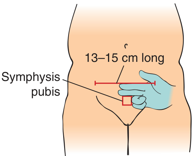
- Incision carried down through subcutaneous fat to the rectus sheath (anterior rectus fascia)
- Midline fascial incision 2 cm with scalpel, then extended laterally and superiorly in a curvilinear fashion using curved Mayo scissors
Step 2 - Fascial Layer
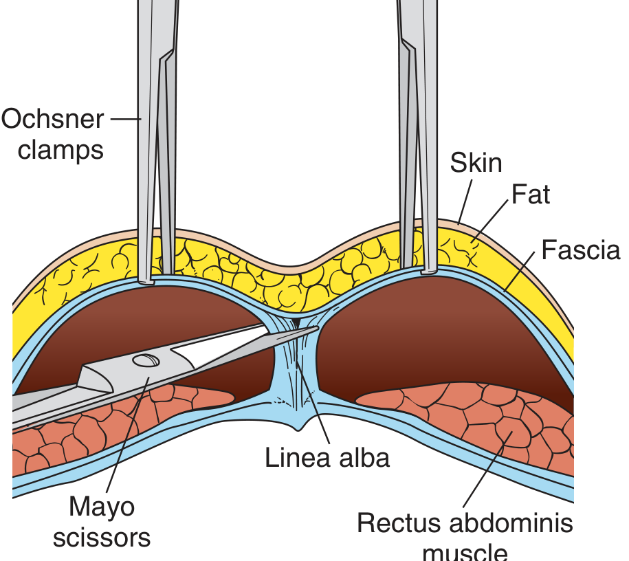
- Superior edge of fascia grasped with two Kocher clamps; fascia elevated off the underlying rectus muscle by blunt finger dissection along the midline (linea alba fibres)
- Same dissection repeated on the inferior edge of the fascia, clearing it from muscle and pubis
- In repeat LSCS: adhesions cut with Mayo scissors
Step 3 - Muscle Separation and Peritoneal Entry
- Rectus abdominis muscles separated bluntly in the midline in a vertical direction to expose the parietal peritoneum
- Peritoneum grasped with two haemostats/tissue forceps at its peak, tented upward, and a small incision made to confirm entry into the peritoneal cavity
- Peritoneal incision extended vertically to expose the lower uterine segment
- DeLee retractor (bladder blade) placed to retract bladder
Step 4 - Bladder Flap (Vesicouterine Peritoneal Flap)
- The vesicouterine serosa (peritoneum overlying the LUS) is identified, picked up with tissue forceps, and incised transversely ~2 cm above the bladder reflection
- Incision extended laterally to ~12 cm
- Bladder dissected off the lower uterine segment bluntly with fingers, pushed inferiorly
- DeLee bladder blade repositioned to include and retract the newly created bladder flap
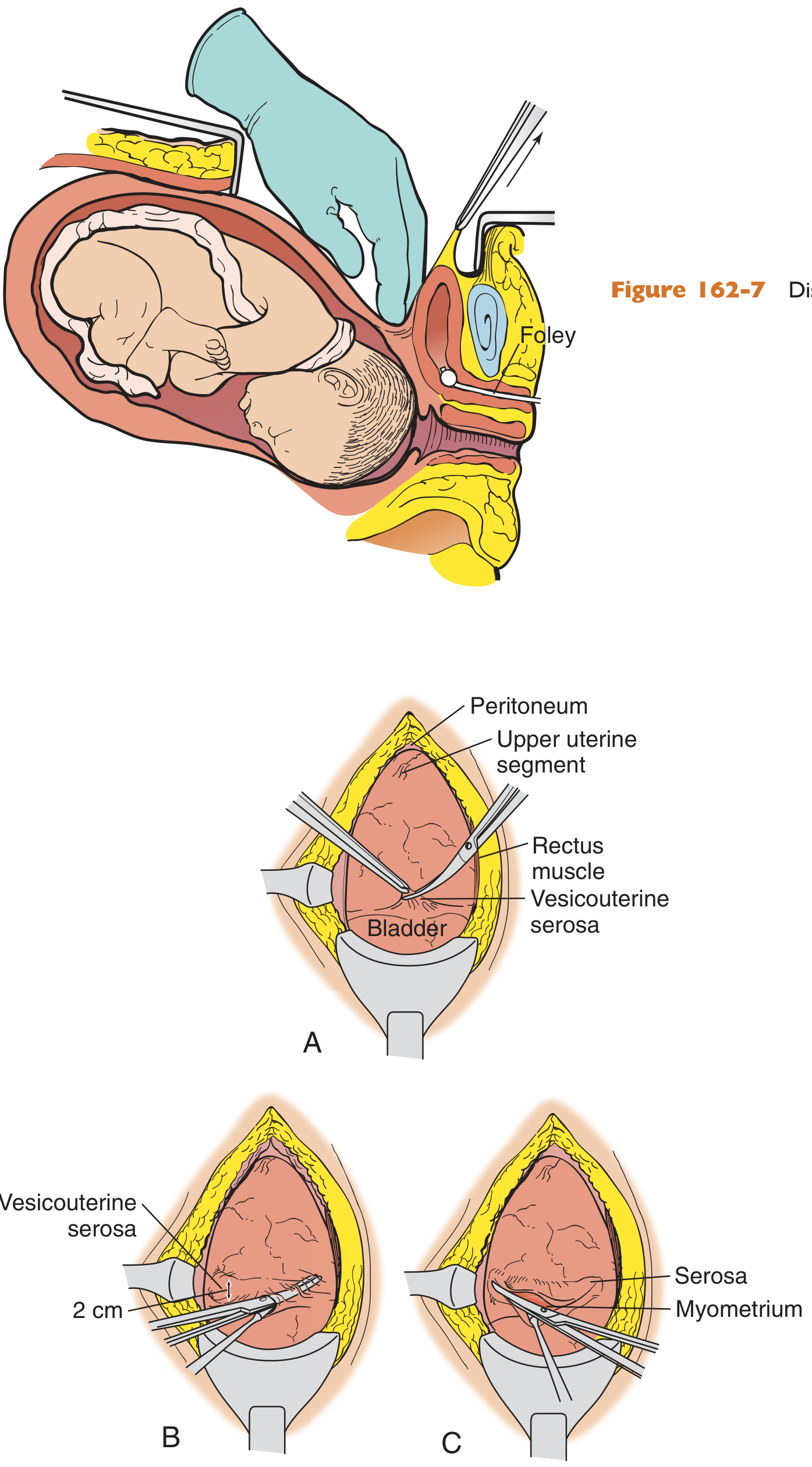
Purpose of bladder flap: Protects the bladder from the uterine incision; allows the uterine wound to be extraperitoneal → reduces risk of peritonitis.
Step 5 - Uterine Incision (Hysterotomy)
- Identify the lateral uterine vessels and confirm uterine orientation
- "Score" the LUS over the presenting fetal part with a No. 20 blade using a 2-3 cm incision, going millimetre by millimetre in depth to avoid fetal injury - announce "uterine incision" to anaesthesiologist and paediatrician
- Extend bluntly with fingers in a curvilinear direction
- Alternatively, use bandage scissors protected by two fingers placed inside the uterine opening to extend laterally for a total incision of ~10-11 cm
- Rupture membranes with an Allis clamp if intact
Step 6 - Delivery of the Baby
Cephalic presentation:
- Surgeon inserts a cupped hand under the fetal head and occiput, wrist kept straight; gently lifts upward bringing the head out of the incision
- Assistant applies gentle fundal pressure once the occiput clears the incision
- Head flexed to minimise the diameter
- If the head is deeply engaged (arrest of descent/CPD): an assistant disimpacts the head vaginally while the surgeon delivers it abdominally
If head stuck - disimpaction methods:
- Assistant pushes head up vaginally (Patwardhan's manoeuvre for deeply engaged head)
- Use of a fetal pillow or vacuum cup to elevate the head
Breech presentation:
- Feet/buttocks delivered first; then shoulders and head delivered using McRoberts-type flexion and gentle traction
Post-delivery:
- Cord clamped and cut; neonate handed to paediatrician
- Oxytocin (syntocinon) 5-10 units IV slowly given immediately after delivery of the baby to stimulate uterine contraction and minimise haemorrhage
Step 7 - Placenta and Uterine Cavity
- Controlled cord traction to deliver the placenta (preferred over manual removal - lower infection and haemorrhage rates)
- Uterine cavity cleared of clots and membranes with moist lap sponges
- If cervix is closed: cervical os dilated with dilators to allow lochia drainage
- Optional uterine externalisation for better visualization and access during closure; uterus wrapped in moist lap sponge during repair
Step 8 - Uterine Closure (Hysterotomy Repair)
- First layer (full thickness of myometrium): Running locked stitch with 0 chromic or 1 Vicryl from one angle to the other - do NOT include the endometrium in this layer
- If haemostasis is inadequate: second imbricating layer of 0 chromic over the first
- Any extensions of the incision (usually inferiorly toward the cervix) are repaired first before the main hysterotomy
- Check haemostasis; figure-of-8 stitches for bleeding points
One-layer vs. two-layer closure: Controversy exists - single-layer closure is faster but some evidence suggests two-layer closure produces a stronger scar and reduces risk of uterine rupture in subsequent pregnancies.
Step 9 - Abdominal Closure
| Layer | Suture | Notes |
|---|---|---|
| Peritoneum (parietal + vesicouterine) | Optional: 2-0 chromic | Most evidence suggests non-closure is acceptable; peritoneum reapproximates spontaneously; non-closure associated with fewer adhesions |
| Rectus sheath (fascia) | Running 1-0 Vicryl or PDS | Strong monofilament; sutures ≤1 cm apart, equally spaced |
| Subcutaneous fat | Irrigate; Bovie haemostasis | Close dead space only if pannus >2 cm (reduces seroma/infection) |
| Skin | Subcuticular 3-0 Vicryl or staples | Subcuticular preferred for cosmesis |
Step 10 - Final Checks
- Inspect uterus, tubes, and ovaries (opportunity for bilateral tubal ligation if desired)
- Irrigate pouch of Douglas with warm saline; suction clots
- Palpate colic gutters, inspect appendix and gallbladder
- Ensure blood loss accounted for; lap sponge count confirmed correct
Decision-to-Delivery Time
| Category | Target Time |
|---|---|
| Emergency (acute fetal compromise, cord prolapse) | ≤30 minutes ("30-minute rule") |
| Urgent (non-acute deterioration) | 30-75 minutes |
| Scheduled | Elective - at 39 weeks (to reduce neonatal TTN) |
VBAC (Vaginal Birth After Caesarean Section)
After one previous LSCS with a low transverse uterine scar:
- VBAC is possible and appropriate in selected patients (hospitals must have facilities for emergency CS)
- Success rates are higher if: non-recurrent indication, one prior vaginal birth, spontaneous onset of labour
- Risk of uterine scar rupture: 0.5-1% during TOLAC (trial of labour after caesarean) - serious, potentially fatal emergency
- Contraindications to TOLAC: Previous classical/vertical scar, previous uterine rupture, >2 previous caesareans, placenta praevia
Complications
Maternal Complications
Intraoperative (incidence ~2% serious complications):
| Complication | Notes |
|---|---|
| Haemorrhage/PPH | Most common serious complication; uterine atony, extensions, placenta accreta |
| Bladder injury | 1-2 per 1000 deliveries; 10× more common in CS than operative vaginal delivery |
| Ureteral injury | Rare; more with adhesions/distorted anatomy |
| Bowel injury | Associated with adhesions from prior CS |
| Anaesthesia accidents | Aspiration pneumonitis (Mendelson), failed intubation, drug reactions |
| Amniotic fluid embolism | Rare; catastrophic |
Postoperative:
| Complication | Incidence | Notes |
|---|---|---|
| Endomyometritis | 5-20% (without prophylactic antibiotics) | Polymicrobial; treat with IV clindamycin + gentamicin; add macrolide (azithromycin) for non-elective CS |
| Wound infection | <3% with prophylactic antibiotics | S. aureus 25-30%; risk in obesity, 2nd stage CS |
| DVT/PE | 1-2% DVT; PE rare | Major cause of maternal mortality; heparinisation + OAC |
| Urinary tract infection | ~11% | Largely from catheterisation; reduce by limiting catheterisation time |
| Wound haematoma/dehiscence | Uncommon | From faulty haemostasis; drain if large |
| Necrotising fasciitis | Rare, fatal | Risk factor: obesity; requires aggressive surgical debridement |
| Respiratory complications | Atelectasis common | Early physiotherapy/mobilisation |
| Ileus/adhesion formation | More with each subsequent CS | |
| Placenta accreta spectrum in future pregnancies | Increases with each CS | Major risk with anterior placenta praevia + previous CS scar |
Neonatal Complications
- Transient tachypnoea of the newborn (TTN) - more common after elective CS before 39 weeks (absence of labour reduces fetal lung fluid clearance)
- Surgical trauma (fetal lacerations - rare, ~1-2%)
- Respiratory distress syndrome (if preterm)
Reducing Morbidity - Key Principles (Creasy & Resnik)
- Anaesthesia by a skilled anaesthesiologist
- Maternal left lateral tilt; attention to blood volume
- Prophylactic antibiotics (single dose, given before incision)
- Use a transverse uterine incision whenever possible
- Await onset of labour before elective repeat CS when feasible (reduces neonatal TTN)
- Skilled neonatologist present for resuscitation
- Prompt delivery of infant after spinal/epidural - uteroplacental hypoperfusion begins even in a normotensive patient
- Creasy & Resnik's Maternal-Fetal Medicine; Pfenninger & Fowler's Procedures for Primary Care
Lscs
I covered LSCS in full detail in the previous message. Here's a concise structured summary — scroll up for the complete illustrated version.
LSCS (Lower Segment Caesarean Section) - Summary
Definition
Delivery of a fetus through a transverse incision in the lower uterine segment (LUS) via a Pfannenstiel abdominal incision. Accounts for 20-30% of all deliveries worldwide.
Indications
Absolute
- Placenta praevia (major)
- Placenta accreta/increta/percreta
- CPD (cephalopelvic disproportion)
- Cord prolapse (viable fetus)
- Previous classical uterine scar
- Transverse lie with ruptured membranes
Relative
- Dystocia / failure to progress (30% of increased CS rates)
- Non-reassuring fetal heart rate (fetal distress)
- Breech presentation
- Repeat LSCS
- Pre-eclampsia / failed induction
- Active genital herpes
- Maternal request
Types of Uterine Incisions
| Type | Details |
|---|---|
| Low transverse (Kerr) | >90% of all CS; thin LUS; best healing; lowest rupture risk |
| Low vertical (Krönig) | Thick LUS, transverse lie, hydrocephalus, fibroids |
| Classical (vertical) | Emergency only; 8× higher rupture risk; mandates future repeat CS |
Preoperative Preparation
- Informed consent
- IV access, blood group & crossmatch
- Antacid: Sodium citrate ± ranitidine (GA aspiration prophylaxis)
- Foley catheter to drain bladder
- Prophylactic antibiotics (single dose before incision)
- DVT prophylaxis
- Left lateral tilt 15° (aortocaval decompression)
Anaesthesia
| Type | Notes |
|---|---|
| Spinal | Most common; highest hypotension risk; preload + ephedrine; deliver promptly |
| Epidural | Converted labour epidural; slower onset (~20 min); not for <20 min emergencies |
| CSE | Complex/prolonged cases |
| GA | True emergency only; rapid sequence induction; cricoid pressure; aspiration risk |
Operative Steps (Kerr LSCS)
| Step | Action |
|---|---|
| 1 | Left lateral tilt of table/wedge under right hip |
| 2 | Pfannenstiel incision: 13-15 cm, 2 fingerbreadths above symphysis pubis |
| 3 | Incise through subcutaneous fat → fascial incision (midline then extended laterally with Mayo scissors) |
| 4 | Elevate fascia off rectus muscles; separate rectus muscles in midline vertically |
| 5 | Open peritoneum: Tent with haemostats; small incision; extend vertically |
| 6 | Bladder flap: Incise vesicouterine serosa; bluntly dissect bladder downward; reapply DeLee retractor |
| 7 | Hysterotomy (uterine incision): Score LUS with No. 20 blade 2-3 cm; extend bluntly with fingers or bandage scissors to 10-11 cm; rupture membranes |
| 8 | Deliver baby: Cupped hand under head + occiput; lift out; assistant applies fundal pressure |
| 9 | Clamp cord; hand to paediatrician; give oxytocin 5-10 units IV |
| 10 | Deliver placenta by controlled cord traction (preferred over manual) |
| 11 | Clear uterine cavity; dilate cervical os if needed |
| 12 | Close hysterotomy: Running locked 0 chromic/Vicryl (2 layers if needed); do NOT include endometrium |
| 13 | Inspect tubes, ovaries; BTL if requested |
| 14 | Irrigate pouch of Douglas; palpate gutters/appendix |
| 15 | Close fascia: Running 1-0 Vicryl/PDS (peritoneum closure optional) |
| 16 | Irrigate subcutaneous fat; close skin: subcuticular 3-0 Vicryl or staples |
Decision-to-Delivery Time
- Emergency (cord prolapse, acute fetal compromise): ≤30 minutes
- Urgent: 30-75 minutes
- Elective: At 39 weeks (prevents neonatal TTN)
VBAC (Vaginal Birth After Caesarean)
- Possible after one previous LSCS with low transverse scar
- Uterine rupture risk during TOLAC: 0.5-1%
- Good candidates: non-recurrent indication, prior vaginal birth, spontaneous labour
- Contraindicated: Previous classical scar, >2 prior CS, prior uterine rupture
Complications
Maternal - Intraoperative
| Complication | Note |
|---|---|
| Haemorrhage/PPH | Most common serious complication |
| Bladder injury | 1-2/1000 deliveries; 10× commoner than operative vaginal delivery |
| Anaesthesia accidents | Aspiration (Mendelson), failed intubation |
| Ureteral/bowel injury | More with adhesions (repeat CS) |
Maternal - Postoperative
| Complication | Incidence | Management |
|---|---|---|
| Endomyometritis | 5-20% (without prophylaxis) | IV clindamycin + gentamicin; add azithromycin for non-elective CS |
| Wound infection | <3% (with antibiotics) | Antibiotics; drainage |
| DVT/PE | DVT 1-2%; PE rare but major mortality cause | LMWH; OAC |
| Bacteriuria/UTI | ~11% | Minimise catheterisation time |
| Wound haematoma | Uncommon | Drainage |
| Necrotising fasciitis | Rare; high mortality | Surgical debridement |
| Placenta accreta spectrum | Increasing with each CS | Anticipate in previa + scar |
Neonatal
- TTN (transient tachypnoea of newborn) - elective CS before 39 weeks
- Fetal laceration (rare)
- RDS if preterm
5 Principles to Minimise Morbidity (Creasy & Resnik)
- Skilled anaesthesiologist
- Left lateral tilt + adequate blood volume
- Prophylactic antibiotics
- Transverse uterine incision whenever possible
- Skilled neonatologist present
Sources: Creasy & Resnik's Maternal-Fetal Medicine; Pfenninger & Fowler's Procedures for Primary Care
This is a shared conversation. Sign in to Orris to start your own chat.