
Embryology of kidney
Reading File
Now let me search the medical library for kidney embryology content in parallel.
Finding Sources
Finding Sources
Finding Sources
Finding Sources
Found a dedicated chapter in Brenner and Rector's The Kidney. Let me read it fully along with a paragraph search for detailed content.
Reading File
Finding Sources
Excellent! Multiple rich sources found. Let me now read the detailed content from the main kidney embryology section and the urogenital development sections.
Reading File
Reading File
I have rich content. The first file read was truncated with a lot of detail. Let me fetch the key images and read the Campbell-Walsh section on early kidney development for additional clinical details.
Loading Image
Reading File
I now have comprehensive information from the medical library. The truncated section from block1 contained very detailed information about all three stages. I have enough to write a thorough answer. Let me compile the full response now.
Embryology of the Kidney
The kidney derives from the intermediate mesoderm of the urogenital ridge, a structure along the posterior wall of the developing fetal abdomen. Development proceeds through three successive, overlapping stages - each more advanced than the last - in a rostral-to-caudal sequence.
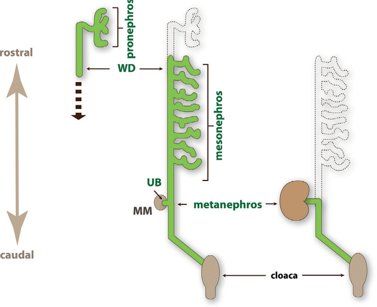
1. Three Stages of Kidney Development
Stage 1 - Pronephros (Week 3-4)
- The most primitive and rudimentary kidney structure
- Appears in the cervical region at approximately 3 weeks' gestation
- Consists of a few vestigial tubules that connect to the pronephric duct, which extends caudally
- Completely nonfunctional in humans
- Involutes fully and disappears, but its duct (which becomes the Wolffian/nephric duct) persists and is used by the next stage
Stage 2 - Mesonephros (Week 4-8)
- Develops at approximately 4 weeks' gestation in the thoracolumbar region, caudal to the pronephros
- Larger and better organized; consists of mesonephric tubules and glomeruli draining into the mesonephric (Wolffian) duct
- Provides a transient excretory function in the embryo
- Partially regresses but leaves important remnants:
- Males: mesonephric duct gives rise to the vas deferens, seminal vesicles, prostate, and part of the epididymis; mesonephric tubules form testicular efferent ductules
- Females: remnants persist as the epoophoron and paroophoron (vestigial structures)
- The mesonephric duct is integral to further urinary tract development - it gives rise to the ureteric bud
Stage 3 - Metanephros (Week 5 onward) - The Permanent Kidney
- Begins forming at approximately 5 weeks' gestation
- Arises from two distinct embryonic components interacting via inductive signaling:
- Ureteric bud (UB) - an outgrowth from the caudal mesonephric (Wolffian) duct
- Metanephric mesenchyme (MM) - a condensing blastema of intermediate mesoderm caudal to the mesonephros
- Nephrogenesis is complete by 34-36 weeks' gestation
2. Metanephric Development - The Key Events
Ureteric Bud Branching Morphogenesis
- The ureteric bud grows out from the Wolffian duct and invades the metanephric mesenchyme
- Under signaling from MM, the UB undergoes repeated dichotomous branching (~10-12 rounds)
- Each branch tip eventually forms a collecting duct; the entire branched tree forms the collecting system: collecting ducts, minor calyces, major calyces, renal pelvis, and ureter
- Key signal: GDNF (glial cell-derived neurotrophic factor) secreted by MM activates RET receptor on UB tips, driving branching
Mesenchymal-to-Epithelial Transition (MET) - Nephrogenesis
- The ureteric bud tip signals back to the MM, inducing condensation of mesenchymal cells around each branch tip (the cap mesenchyme or nephron progenitor cells)
- These cap mesenchyme cells undergo mesenchymal-to-epithelial transition (MET) to form nephrons
- Sequence of nephron formation from induced mesenchyme:
- Pretubular aggregate
- Renal vesicle
- Comma-shaped body
- S-shaped body
- Mature nephron (glomerulus through distal tubule)
- Key signals driving MET include Wnt9b and Wnt4 from the UB and stroma
Cell Lineages in the Mature Kidney
| Structure | Origin |
|---|---|
| Collecting ducts, renal pelvis, ureter | Ureteric bud (UB lineage) |
| Connecting tubule, distal tubule, loop of Henle, proximal tubule, parietal epithelial cells, podocytes | Metanephric mesenchyme (via MET) |
| Interstitial stromal cells | Metanephric mesenchyme (non-condensing stroma) |
| Renal vasculature | Vasculogenesis + angiogenesis from endothelial progenitors |
3. Ascent (Positional Migration) of the Kidney
- The metanephros initially forms in the sacral/pelvic region
- As the embryo grows and the tail end "straightens," the kidneys appear to ascend to the lumbar region (L1-L2)
- During ascent, the kidneys are vascularized by a succession of transient aortic sprouts arising at progressively higher levels; the final pair forms in the upper lumbar region and becomes the permanent renal arteries
- The kidneys also rotate 90° medially so the hilum faces anteromedially
- Clinical correlate: Failure of ascent = pelvic kidney; failure of rotation = malrotated kidney; fusion of lower poles before ascent = horseshoe kidney (most common renal fusion anomaly), which gets caught on the inferior mesenteric artery and stops ascending
4. Nephron Endowment
- Humans average 1 million nephrons per kidney (range: 0.2 to 2.7 million)
- All nephrons are formed by 34-36 weeks of gestation - no new nephrons form after birth
- Low nephron endowment (from prematurity, IUGR, low birth weight) is a risk factor for hypertension and chronic kidney disease in adult life (the Barker/Brenner hypothesis)
5. Key Molecular Players
| Gene/Molecule | Role |
|---|---|
| GDNF | Secreted by MM; drives UB outgrowth via RET receptor |
| RET / GFRα1 | Receptor complex on UB tip; essential for branching |
| WT1 (Wilms Tumor 1) | Transcription factor in MM; required for metanephric mesenchyme survival |
| Pax2 | Expressed in UB and MM; required for initial kidney development |
| Wnt9b | UB-derived signal; initiates MET in cap mesenchyme |
| Wnt4 | Drives MET in pretubular aggregates |
| Six2 | Maintains cap mesenchyme in self-renewing progenitor state |
| FOXD1 | Stromal cell transcription factor; regulates UB branching and vascular development |
6. Congenital Anomalies (CAKUT - Congenital Anomalies of the Kidney and Urinary Tract)
Dysregulated kidney embryology underlies a spectrum of anomalies collectively called CAKUT, the most common cause of end-stage renal disease (ESRD) in children:
| Anomaly | Embryological Basis |
|---|---|
| Renal agenesis | Failure of ureteric bud to form or failure of UB-MM induction |
| Horseshoe kidney | Fusion of lower poles before ascent; caught on inferior mesenteric artery |
| Pelvic kidney | Failure of renal ascent |
| Multicystic dysplastic kidney | Abnormal UB-metanephric blastema signaling |
| Duplex collecting system | Premature splitting of the ureteric bud |
| Posterior urethral valves | Abnormal integration of the Wolffian duct into the urogenital sinus |
| Vesicoureteral reflux | Abnormal ureteric insertion into the bladder |
Summary Timeline
| Week of Gestation | Event |
|---|---|
| Week 3 | Pronephros forms; completely regresses |
| Week 4 | Mesonephros forms; functional transiently |
| Week 5 | Metanephros begins - ureteric bud invades metanephric mesenchyme |
| Week 5-34 | Progressive nephrogenesis; UB branching and MET |
| Week 6-9 | Renal ascent from pelvis to lumbar region; renal rotation |
| Week 34-36 | Nephrogenesis complete (~1 million nephrons/kidney) |
| Birth | No further nephrons added; postnatal maturation of GFR continues to age 1-2 years |
Sources: Brenner and Rector's The Kidney, Chapter 1 (Embryology of the Kidney); Campbell-Walsh-Wein Urology, Chapter on Renal Development; Color Atlas of Human Anatomy Vol. 2
This is a shared conversation. Sign in to Orris to start your own chat.