
let's go
python3 -c " import pdfplumber pdf_path = '/tmp/workspace/attachments/1bb5e42c-d0b9-4d0c-b099-98580f48c2c9/13@. The Midbrain A-Prof. Royhaan.pdf' with pdfplumber.open(pdf_path) as pdf: print(f'Total pages: {len(pdf.pages)}') for i, page in enumerate(pdf.pages): text = page.extract_text() print(f'--- PAGE {i+1} ---') print(text) print() "
The Midbrain - A-Prof. Royhaan Folarin (33 slides)
Structure
| Section | Key Points |
|---|---|
| Location | Between pons and diencephalon, around the cerebral aqueduct |
| Tectum | Dorsal to aqueduct - superior + inferior colliculi |
| Crus cerebri | Corticospinal, corticonuclear, corticopontine fibres (somatotopic) |
| Substantia nigra | Pars compacta (dopaminergic) + pars reticulata |
| Tegmentum | CN nuclei, red nucleus, reticular formation, sensory/motor tracts, MLF |
Two Key Levels
- Inferior colliculus level - trochlear nucleus (CN IV), lateral lemniscus, SCP decussation
- Superior colliculus level - oculomotor complex (CN III), Edinger-Westphal, red nucleus, pretectal nuclei
Clinical Syndromes
| Syndrome | Lesion | Findings |
|---|---|---|
| Weber | CN III + crus cerebri | Ipsilateral CN III palsy + contralateral hemiplegia |
| Benedikt | CN III + red nucleus | Ipsilateral CN III palsy + contralateral tremor/incoordination |
| Parinaud | Dorsal midbrain/pretectal | Impaired upward gaze |
- Quiz me on the content
- Explain a specific topic in depth
- Create flashcards or notes
- Summarize for exam revision
Generate a study guide with clinical cases
mkdir -p /tmp/workspace/midbrain-study-guide && python3 << 'EOF' from docx import Document from docx.shared import Pt, Inches, RGBColor from docx.enum.text import WD_ALIGN_PARAGRAPH from docx.oxml.ns import qn from docx.oxml import OxmlElement doc = Document() # ── Page margins ────────────────────────────────────────────────────────────── for section in doc.sections: section.top_margin = Inches(1) section.bottom_margin = Inches(1) section.left_margin = Inches(1.1) section.right_margin = Inches(1.1) # ── Helper: coloured heading ────────────────────────────────────────────────── def add_heading(doc, text, level=1, colour=None): p = doc.add_heading(text, level=level) if colour: for run in p.runs: run.font.color.rgb = RGBColor(*colour) return p # ── Helper: add a shaded box paragraph ─────────────────────────────────────── def add_shaded_para(doc, text, shade_hex="E8F4FD", bold_label=None): p = doc.add_paragraph() if bold_label: r = p.add_run(bold_label + " ") r.bold = True p.add_run(text) # Apply shading via XML pPr = p._p.get_or_add_pPr() shd = OxmlElement('w:shd') shd.set(qn('w:val'), 'clear') shd.set(qn('w:color'), 'auto') shd.set(qn('w:fill'), shade_hex) pPr.append(shd) p.paragraph_format.left_indent = Inches(0.2) p.paragraph_format.right_indent = Inches(0.2) p.paragraph_format.space_before = Pt(4) p.paragraph_format.space_after = Pt(4) return p # ── Helper: table with header row ──────────────────────────────────────────── def add_table(doc, headers, rows, header_colour="1F4E79"): table = doc.add_table(rows=1+len(rows), cols=len(headers)) table.style = 'Table Grid' # Header hdr = table.rows[0] for i, h in enumerate(headers): cell = hdr.cells[i] cell.text = h run = cell.paragraphs[0].runs[0] run.bold = True run.font.color.rgb = RGBColor(0xFF, 0xFF, 0xFF) run.font.size = Pt(10) # background tc_pr = cell._tc.get_or_add_tcPr() shd = OxmlElement('w:shd') shd.set(qn('w:val'), 'clear') shd.set(qn('w:color'), 'auto') shd.set(qn('w:fill'), header_colour) tc_pr.append(shd) # Data rows for r_i, row_data in enumerate(rows): row = table.rows[r_i+1] for c_i, cell_text in enumerate(row_data): cell = row.cells[c_i] cell.text = cell_text cell.paragraphs[0].runs[0].font.size = Pt(10) doc.add_paragraph() # spacing after table return table # ══════════════════════════════════════════════════════════════════════════════ # TITLE PAGE # ══════════════════════════════════════════════════════════════════════════════ title = doc.add_paragraph() title.alignment = WD_ALIGN_PARAGRAPH.CENTER run = title.add_run("THE MIDBRAIN") run.bold = True run.font.size = Pt(28) run.font.color.rgb = RGBColor(0x1F, 0x4E, 0x79) subtitle = doc.add_paragraph() subtitle.alignment = WD_ALIGN_PARAGRAPH.CENTER r2 = subtitle.add_run("Internal Organization · Functional Pathways · Clinical Localization") r2.font.size = Pt(13) r2.font.color.rgb = RGBColor(0x44, 0x72, 0xC4) doc.add_paragraph() byline = doc.add_paragraph() byline.alignment = WD_ALIGN_PARAGRAPH.CENTER byline.add_run("Study Guide — Based on lecture by A-Prof. Royhaan Folarin").italic = True doc.add_page_break() # ══════════════════════════════════════════════════════════════════════════════ # SECTION 1 — OVERVIEW # ══════════════════════════════════════════════════════════════════════════════ add_heading(doc, "1. Overview of the Midbrain", level=1, colour=(0x1F, 0x4E, 0x79)) doc.add_paragraph( "The midbrain is the uppermost segment of the brainstem. It lies between the pons " "inferiorly and the diencephalon superiorly, surrounds the cerebral aqueduct, and " "sits anterior to the cerebellum." ) add_heading(doc, "Key Functions", level=2, colour=(0x1F, 0x4E, 0x79)) for fn in [ "Motor-pathway transmission (corticospinal, corticonuclear, corticopontine)", "Eye-movement control (CN III, CN IV, MLF)", "Visual and auditory reflexes (superior and inferior colliculi)", "Movement modulation (substantia nigra, red nucleus)", "Arousal and autonomic integration (reticular formation, Edinger-Westphal)", ]: p = doc.add_paragraph(fn, style="List Bullet") p.runs[0].font.size = Pt(11) # ══════════════════════════════════════════════════════════════════════════════ # SECTION 2 — MAJOR SUBDIVISIONS # ══════════════════════════════════════════════════════════════════════════════ add_heading(doc, "2. Major Subdivisions", level=1, colour=(0x1F, 0x4E, 0x79)) doc.add_paragraph( "The cerebral aqueduct divides the midbrain into the tectum (dorsal) and the " "cerebral peduncles (ventral). From anterior to posterior, a transverse section shows:" ) add_table(doc, ["Structure", "Location", "Key Contents"], [ ["Crus cerebri", "Ventral", "Corticospinal, corticonuclear, corticopontine fibres"], ["Substantia nigra", "Between crus & tegmen", "Pars compacta (dopaminergic), pars reticulata"], ["Tegmentum", "Central", "CN nuclei, red nucleus, lemnisci, MLF, reticular formation"], ["Central grey", "Periaqueductal", "Surrounds cerebral aqueduct; autonomic & pain modulation"], ["Tectum", "Dorsal", "Superior colliculi (visual), inferior colliculi (auditory)"], ] ) # ── Crus cerebri detail ─────────────────────────────────────────────────────── add_heading(doc, "Fibre Arrangement in the Crus Cerebri", level=2, colour=(0x1F, 0x4E, 0x79)) add_table(doc, ["Region", "Fibres"], [ ["Medial 1/6", "Frontopontine fibres"], ["Middle 2/3", "Corticospinal (head medial → leg lateral) and corticonuclear fibres"], ["Lateral 1/6", "Temporopontine, parietopontine, occipitopontine fibres"], ] ) # ── Substantia nigra ────────────────────────────────────────────────────────── add_heading(doc, "Substantia Nigra", level=2, colour=(0x1F, 0x4E, 0x79)) doc.add_paragraph( "The substantia nigra sits between the crus cerebri and tegmentum. It contains neuromelanin " "and appears dark in unstained sections." ) add_table(doc, ["Division", "Neurotransmitter", "Projection", "Function"], [ ["Pars compacta", "Dopamine", "Striatum (nigrostriatal pathway)", "Movement modulation"], ["Pars reticulata", "GABA", "Thalamus, superior colliculus", "Major output; analogous to GPi"], ] ) add_shaded_para(doc, "Degeneration of pars compacta → reduced striatal dopamine → Parkinson disease " "(bradykinesia, rigidity, resting tremor, reduced facial expression, shuffling gait).", shade_hex="FDEBD0", bold_label="⚠ Clinical:" ) doc.add_paragraph() # ══════════════════════════════════════════════════════════════════════════════ # SECTION 3 — TWO LEVELS # ══════════════════════════════════════════════════════════════════════════════ add_heading(doc, "3. Two Key Midbrain Levels", level=1, colour=(0x1F, 0x4E, 0x79)) add_heading(doc, "A. Inferior Colliculus Level (Lower Midbrain)", level=2, colour=(0x1F, 0x4E, 0x79)) add_table(doc, ["Structure", "Notes"], [ ["Inferior colliculus", "Auditory relay & reflex; receives lateral lemniscus; projects to MGB"], ["Trochlear nucleus (CN IV)", "In central grey; fibres decussate in superior medullary velum; exit posteriorly; supply superior oblique"], ["Mesencephalic nucleus of CN V", "Proprioception from jaw muscles"], ["Superior cerebellar peduncle decussation", "Dentatorubrothalamic fibres cross here"], ["Lemnisci present", "Medial, spinal, trigeminal, lateral lemnisci"], ] ) add_heading(doc, "B. Superior Colliculus Level (Upper Midbrain)", level=2, colour=(0x1F, 0x4E, 0x79)) add_table(doc, ["Structure", "Notes"], [ ["Oculomotor nucleus (CN III)", "Ventral central grey; supplies all extraocular muscles except SO and LR, plus levator palpebrae"], ["Edinger-Westphal nucleus", "Parasympathetic; via CN III to sphincter pupillae and ciliary muscle"], ["Red nucleus", "Receives input from cortex, cerebellum, striatum; projects via rubrospinal tract (crosses in ventral tegmental decussation)"], ["Superior colliculus", "Visual orientation; receives retina + visual cortex + IC; outputs via tectospinal, tectobulbar, tectocerebellar tracts"], ["Pretectal nuclei", "Pupillary light reflex pathway; bilateral EW projections → consensual reflex"], ] ) # ══════════════════════════════════════════════════════════════════════════════ # SECTION 4 — PATHWAYS # ══════════════════════════════════════════════════════════════════════════════ add_heading(doc, "4. Major Pathways Through the Midbrain", level=1, colour=(0x1F, 0x4E, 0x79)) add_heading(doc, "Ascending Sensory Pathways", level=2, colour=(0x1F, 0x4E, 0x79)) add_table(doc, ["Pathway", "Modality", "Location in Tegmentum"], [ ["Medial lemniscus", "Discriminative touch, vibration, conscious proprioception", "Medial"], ["Spinal lemniscus", "Pain, temperature, crude touch", "Lateral"], ["Trigeminal lemniscus", "Somatic sensation from face", "Near medial lemniscus"], ["Lateral lemniscus", "Auditory information", "Lateral tegmentum"], ] ) add_heading(doc, "Medial Longitudinal Fasciculus (MLF)", level=2, colour=(0x1F, 0x4E, 0x79)) doc.add_paragraph( "The MLF is a paired paramedian fibre bundle that connects CN III, IV, VI nuclei and the " "vestibular nuclei with cervical spinal motor neurons. It coordinates:" ) for fn in ["Conjugate eye movements", "Vestibulo-ocular reflexes", "Head-eye alignment and gaze stabilization"]: doc.add_paragraph(fn, style="List Bullet") add_heading(doc, "Pupillary Light Reflex Pathway", level=2, colour=(0x1F, 0x4E, 0x79)) steps = [ "Light → Retina → Optic nerve → Optic tract", "Pretectal nuclei (midbrain)", "Bilateral projection → Edinger-Westphal nuclei (both sides)", "Preganglionic parasympathetic fibres ride CN III → ciliary ganglion", "Postganglionic fibres → sphincter pupillae → pupil constriction", ] for i, s in enumerate(steps, 1): p = doc.add_paragraph(f"{i}. {s}") p.paragraph_format.left_indent = Inches(0.3) p.runs[0].font.size = Pt(11) add_shaded_para(doc, "Bilateral EW projections explain why light in ONE eye produces BOTH direct and consensual constriction.", shade_hex="E8F8F5", bold_label="Key point:" ) doc.add_paragraph() # ══════════════════════════════════════════════════════════════════════════════ # SECTION 5 — CLINICAL LOCALIZATION TABLE # ══════════════════════════════════════════════════════════════════════════════ add_heading(doc, "5. Clinical Localization at a Glance", level=1, colour=(0x1F, 0x4E, 0x79)) add_table(doc, ["Structure Damaged", "Side of Finding", "Typical Clinical Finding"], [ ["Oculomotor fascicles (CN III)", "Ipsilateral", "CN III palsy: ptosis, mydriasis, 'down-and-out' eye"], ["Trochlear nerve (CN IV)", "Contralateral", "Superior oblique weakness; head tilt away from lesion"], ["Corticospinal fibres (crus)", "Contralateral", "Hemiplegia / hemiparesis"], ["Red nucleus", "Contralateral", "Tremor or incoordination (rubral/Holmes tremor)"], ["Medial lemniscus", "Contralateral", "Loss of discriminative touch, vibration, proprioception"], ["Spinal lemniscus", "Contralateral", "Loss of pain and temperature"], ["Superior colliculus / pretectal", "Bilateral", "Impaired vertical (upward) gaze"], ["MLF", "Ipsilateral adduction failure", "Internuclear ophthalmoplegia (INO)"], ["Substantia nigra (pars compacta)", "Bilateral (late)", "Parkinson disease features"], ], header_colour="1F4E79" ) # ══════════════════════════════════════════════════════════════════════════════ # SECTION 6 — MAJOR SYNDROMES # ══════════════════════════════════════════════════════════════════════════════ add_heading(doc, "6. Classic Midbrain Syndromes", level=1, colour=(0x1F, 0x4E, 0x79)) syndromes = [ ( "Weber Syndrome (Ventral Midbrain)", "Lesion of CN III fascicles + crus cerebri", [ ("Ipsilateral", "CN III palsy: ptosis, fixed dilated pupil, eye deviated down and out"), ("Contralateral", "Hemiplegia (UMN — face, arm, leg) due to corticospinal fibre involvement"), ], "Ischaemia of the posterior cerebral artery or its branches; also space-occupying lesions in the interpeduncular fossa." ), ( "Benedikt Syndrome (Tegmental Midbrain)", "Lesion of CN III fascicles + red nucleus (± medial lemniscus)", [ ("Ipsilateral", "CN III palsy: ptosis, fixed dilated pupil, eye deviated down and out"), ("Contralateral", "Rubral (Holmes) tremor — coarse, irregular tremor worse with movement"), ("Contralateral", "Hemisensory loss if medial lemniscus also involved"), ], "Paramedian branches of the basilar artery or posterior cerebral artery." ), ( "Parinaud Syndrome (Dorsal Midbrain)", "Lesion of pretectal/superior collicular region or posterior commissure", [ ("Bilateral", "Impaired upward gaze (upgaze palsy)"), ("Bilateral", "Light-near dissociation (pupils react to accommodation but not light)"), ("Bilateral", "Convergence-retraction nystagmus on attempted upgaze"), ("Bilateral", "Lid retraction (Collier sign)"), ], "Pineal gland tumour (most common in young patients), hydrocephalus, midbrain infarct, demyelination." ), ] for name, lesion, findings, aetiology in syndromes: add_heading(doc, name, level=2, colour=(0xC0, 0x00, 0x00)) add_shaded_para(doc, lesion, shade_hex="FEF9E7", bold_label="Lesion:") doc.add_paragraph() t = doc.add_table(rows=1+len(findings), cols=2) t.style = 'Table Grid' for i, h in enumerate(["Side", "Finding"]): cell = t.rows[0].cells[i] cell.text = h r = cell.paragraphs[0].runs[0] r.bold = True; r.font.color.rgb = RGBColor(0xFF,0xFF,0xFF) r.font.size = Pt(10) tcpr = cell._tc.get_or_add_tcPr() shd = OxmlElement('w:shd') shd.set(qn('w:val'), 'clear'); shd.set(qn('w:color'), 'auto') shd.set(qn('w:fill'), 'C00000') tcpr.append(shd) for ri, (side, finding) in enumerate(findings): row = t.rows[ri+1] row.cells[0].text = side row.cells[1].text = finding for c in [0,1]: row.cells[c].paragraphs[0].runs[0].font.size = Pt(10) doc.add_paragraph() add_shaded_para(doc, aetiology, shade_hex="F0F0F0", bold_label="Aetiology:") doc.add_paragraph() # ══════════════════════════════════════════════════════════════════════════════ # SECTION 7 — CLINICAL CASES # ══════════════════════════════════════════════════════════════════════════════ add_heading(doc, "7. Clinical Cases", level=1, colour=(0x1F, 0x4E, 0x79)) doc.add_paragraph( "The following cases are designed for self-testing. Read the vignette, work through the " "questions, then review the answers." ) cases = [ { "number": 1, "title": "The Dropped Eyelid and Weak Arm", "vignette": ( "A 62-year-old hypertensive man is brought to the emergency department with a sudden " "onset of right-sided drooping eyelid, a fixed dilated right pupil, and the right eye " "deviated downward and outward. Neurological examination also reveals left-sided " "hemiplegia with an upper motor neuron pattern (spastic, with brisk reflexes and " "up-going plantar response). There is no sensory loss. MRI shows a small infarct in " "the right ventral midbrain." ), "questions": [ ("Q1", "Which cranial nerve is affected, and on which side?"), ("Q2", "Why is the hemiplegia contralateral to the cranial nerve palsy?"), ("Q3", "Which two structures are damaged in this lesion?"), ("Q4", "Name this eponymous syndrome."), ("Q5", "What is the most likely arterial territory involved?"), ], "answers": [ ("A1", "Right CN III (oculomotor nerve) — causing ipsilateral ptosis, fixed dilated pupil (Edinger-Westphal involvement), and the classic 'down-and-out' eye position."), ("A2", "The corticospinal fibres in the crus cerebri have not yet crossed — they cross at the pyramidal decussation in the medulla. Damage to the right crus therefore causes left (contralateral) UMN hemiplegia."), ("A3", "CN III fascicles (in the interpeduncular region) and the crus cerebri (ipsilateral)."), ("A4", "Weber syndrome."), ("A5", "Paramedian branches of the posterior cerebral artery (or direct branches of the basilar artery) supplying the ventral midbrain."), ], }, { "number": 2, "title": "Tremor and a Drooping Eye", "vignette": ( "A 45-year-old woman presents with a 3-week history of a coarse right-sided tremor " "that is present at rest and severely worsens when she reaches for objects. She also " "has a right-sided ptosis and a right pupil that is fixed and dilated. Sensation is " "intact. MRI reveals a haemorrhagic lesion in the right paramedian midbrain tegmentum " "at the level of the superior colliculus." ), "questions": [ ("Q1", "Which nucleus, when damaged, accounts for the contralateral tremor?"), ("Q2", "What type of tremor is characteristically produced by damage to this nucleus?"), ("Q3", "Which CN is co-involved, producing the ipsilateral eye findings?"), ("Q4", "Name this syndrome."), ("Q5", "How does this syndrome differ from Weber syndrome?"), ], "answers": [ ("A1", "The red nucleus in the right midbrain tegmentum."), ("A2", "Holmes (rubral) tremor — a coarse, irregular, low-frequency tremor present at rest, worse with posture and action, due to disruption of cerebellar and dopaminergic inputs converging on the red nucleus."), ("A3", "CN III (oculomotor nerve) fascicles, which pass through or near the red nucleus on their way to the interpeduncular fossa."), ("A4", "Benedikt syndrome."), ("A5", "Both involve ipsilateral CN III palsy. Weber syndrome damages the crus cerebri → contralateral hemiplegia. Benedikt syndrome damages the red nucleus (with or without medial lemniscus) → contralateral tremor/incoordination, NOT hemiplegia."), ], }, { "number": 3, "title": "The Eyes That Won't Look Up", "vignette": ( "A 19-year-old male university student presents with headache and gradual visual " "complaints over 2 months. On examination he cannot look upward voluntarily or " "on reflex. His pupils are equal and react briskly to accommodation but sluggishly " "to direct light. Attempted upgaze produces a convergence-retraction nystagmus. " "His eyelids appear mildly retracted. MRI reveals a mass in the pineal region " "compressing the dorsal midbrain." ), "questions": [ ("Q1", "What region of the midbrain is compressed to produce the upgaze palsy?"), ("Q2", "Explain the light-near dissociation in this patient."), ("Q3", "What is the eponymous name for this dorsal midbrain syndrome?"), ("Q4", "What is the most likely underlying diagnosis in this young patient?"), ("Q5", "What reflex arc is intact and what is disrupted, explaining the pupillary finding?"), ], "answers": [ ("A1", "The pretectal/superior collicular region and posterior commissure. Compression here disrupts the upgaze centre and pretectal fibres."), ("A2", "The light reflex pathway (retina → pretectal nuclei → Edinger-Westphal → pupil constriction) is disrupted because pretectal nuclei are compressed. However, the accommodation-convergence reflex uses a different, more ventrally routed pathway that bypasses the pretectal nuclei and is preserved."), ("A3", "Parinaud syndrome (dorsal midbrain syndrome)."), ("A4", "Pineal germ cell tumour (germinoma) — the most common pineal region mass in young males and the classic cause of Parinaud syndrome."), ("A5", "The accommodation-convergence reflex arc (cortex → pretectal bypass → EW nucleus → pupil) is intact. The direct and consensual light reflex arc (retina → pretectal nuclei → EW nucleus → pupil) is disrupted because the pretectal nuclei are compressed."), ], }, { "number": 4, "title": "The Nurse Who Hears on Only One Side", "vignette": ( "A 38-year-old woman notices she has difficulty localizing sounds after a road " "traffic accident. Audiometry shows bilateral reduced hearing with preserved " "cochlear function. She has no other neurological deficits. MRI shows contusions " "at the inferior colliculi bilaterally." ), "questions": [ ("Q1", "What is the role of the inferior colliculus in auditory processing?"), ("Q2", "Why does damage to a single inferior colliculus not cause complete unilateral deafness?"), ("Q3", "What auditory fibres arrive at the inferior colliculus?"), ("Q4", "Where does the inferior colliculus project its output?"), ], "answers": [ ("A1", "The inferior colliculus is an auditory relay and reflex centre. It integrates bilateral auditory input, contributes to sound localization, auditory reflexes, and orientation toward sound."), ("A2", "Each inferior colliculus receives input from BOTH auditory pathways (bilateral projections via the lateral lemnisci and superior olivary complex). A single IC lesion therefore causes bilateral loss of input integration rather than unilateral deafness."), ("A3", "Fibres from the lateral lemniscus and the superior olivary complex."), ("A4", "Primarily to the medial geniculate body (thalamus), but also to the opposite inferior colliculus and the superior colliculus."), ], }, { "number": 5, "title": "Impaired Eye Movements After Demyelination", "vignette": ( "A 29-year-old woman with known multiple sclerosis develops double vision. " "On horizontal gaze testing to the RIGHT, her LEFT eye fails to adduct fully while " "her right eye shows nystagmus on abduction. On gaze to the LEFT, both eyes move " "normally. Convergence is normal. Her pupils are equal and reactive." ), "questions": [ ("Q1", "What is the diagnosis of her eye movement disorder?"), ("Q2", "Which fibre tract is demyelinated, and on which side?"), ("Q3", "Why is convergence preserved even though adduction is impaired on lateral gaze?"), ("Q4", "What nuclei does the MLF connect that are relevant to this finding?"), ], "answers": [ ("A1", "Left internuclear ophthalmoplegia (INO) — failure of ipsilateral adduction on contralateral horizontal gaze, with contralateral abducting nystagmus."), ("A2", "The LEFT medial longitudinal fasciculus (MLF), between the left CN III nucleus and the left abducens internuclear neurons. Demyelination here prevents the signal from the right abducens nucleus from reaching the left CN III nucleus."), ("A3", "Convergence uses a different pathway (midbrain convergence centre and near-response neurons) that does NOT rely on the MLF. Therefore a unilateral MLF lesion spares convergence while impairing horizontal conjugate gaze adduction."), ("A4", "The MLF connects the abducens nucleus (CN VI) internuclear neurons to the contralateral oculomotor nucleus (CN III), as well as vestibular nuclei, trochlear nucleus (CN IV), and cervical spinal cord motor neurons."), ], }, ] for case in cases: doc.add_page_break() add_heading(doc, f"Case {case['number']}: {case['title']}", level=2, colour=(0x1F, 0x4E, 0x79)) # Vignette box add_shaded_para(doc, case['vignette'], shade_hex="E8F4FD", bold_label="Clinical Vignette:") doc.add_paragraph() # Questions add_heading(doc, "Questions", level=3, colour=(0x1F, 0x4E, 0x79)) for qnum, qtext in case['questions']: p = doc.add_paragraph(style="List Number") r = p.add_run(qnum + ": ") r.bold = True; r.font.size = Pt(11) p.add_run(qtext).font.size = Pt(11) # Answers doc.add_paragraph() add_heading(doc, "Answers", level=3, colour=(0x1F, 0x4E, 0x79)) for anum, atext in case['answers']: p = doc.add_paragraph() r = p.add_run(anum + ": ") r.bold = True; r.font.color.rgb = RGBColor(0x1F, 0x4E, 0x79); r.font.size = Pt(11) p.add_run(atext).font.size = Pt(11) p.paragraph_format.left_indent = Inches(0.2) p.paragraph_format.space_after = Pt(4) # ══════════════════════════════════════════════════════════════════════════════ # SECTION 8 — QUICK REVISION TABLE # ══════════════════════════════════════════════════════════════════════════════ doc.add_page_break() add_heading(doc, "8. Quick Revision: Cranial Nerves of the Midbrain", level=1, colour=(0x1F, 0x4E, 0x79)) add_table(doc, ["Nerve", "Nucleus Level", "Exit Point", "Muscle(s) Supplied", "Autonomic Component"], [ ["CN III (Oculomotor)", "Superior colliculus", "Interpeduncular fossa (ventral)", "SR, IR, MR, IO, levator palpebrae", "Edinger-Westphal → sphincter pupillae, ciliary m."], ["CN IV (Trochlear)", "Inferior colliculus", "Posterior brainstem (only CN to exit dorsally)", "Superior oblique", "None"], ] ) add_heading(doc, "9. Key Mnemonics and Memory Aids", level=1, colour=(0x1F, 0x4E, 0x79)) mnemonics = [ ("Midbrain lesion rule", "Ipsilateral CN deficit + Contralateral long-tract signs = Crossed syndrome (brainstem)."), ("Weber vs Benedikt", "Weber = crus Cerebri → hemiplegia. Benedikt = Red nucleus → tRemor. (C for Crus/Contralateral-weakness; R for Red/tRemor)"), ("Parinaud = Pineal", "Young patient + upgaze palsy + light-near dissociation → think pineal region mass compressing dorsal midbrain."), ("MLF = Medial Longitudinal Fasciculus", "Damage → INO: adduction fails on lateral gaze, convergence PRESERVED. Most common cause = MS in young adults."), ("Substantia Nigra = Parkinson", "Loss of dopaminergic pars compacta → TRAP: Tremor (resting), Rigidity, Akinesia/bradykinesia, Postural instability."), ("CN IV curiosity", "Only cranial nerve to (1) exit the brainstem POSTERIORLY, (2) fully decussate before exit, and (3) supply a contralateral muscle clinically."), ] for term, explanation in mnemonics: p = doc.add_paragraph() r = p.add_run(term + ": ") r.bold = True; r.font.color.rgb = RGBColor(0xC0, 0x00, 0x00); r.font.size = Pt(11) p.add_run(explanation).font.size = Pt(11) p.paragraph_format.space_after = Pt(6) # ══════════════════════════════════════════════════════════════════════════════ # SAVE # ══════════════════════════════════════════════════════════════════════════════ out_path = "/tmp/workspace/midbrain-study-guide/Midbrain_Study_Guide.docx" doc.save(out_path) print(f"Saved to: {out_path}") EOF
Midbrain Study Guide
Word Document · DOCX
| Section | Contents |
|---|---|
| 1. Overview | Location, key functions |
| 2. Major Subdivisions | Crus cerebri fibre map, substantia nigra table, Parkinson clinical box |
| 3. Two Key Levels | Inferior + superior colliculus levels with full structure tables |
| 4. Pathways | Ascending lemnisci, MLF, pupillary light reflex step-by-step |
| 5. Clinical Localization | Master table - structure → side → finding |
| 6. Classic Syndromes | Weber, Benedikt, Parinaud - lesion, findings, aetiology |
| 7. Clinical Cases (5) | Full vignette + questions + model answers (see below) |
| 8. Quick Revision | CN III and CN IV comparison table |
| 9. Mnemonics | Weber vs Benedikt, TRAP, CN IV curiosity, INO rule |
The 5 Clinical Cases
- The Dropped Eyelid and Weak Arm - Weber syndrome (CN III + crus cerebri)
- Tremor and a Drooping Eye - Benedikt syndrome (CN III + red nucleus)
- The Eyes That Won't Look Up - Parinaud syndrome (pineal mass, dorsal midbrain)
- The Nurse Who Hears on Only One Side - Inferior colliculus bilateral contusion
- Eye Movements After Demyelination - Internuclear ophthalmoplegia (MLF, MS)
quiz me
A transverse section of the midbrain shows several layers from anterior to posterior. List them in the correct order, starting from the most anterior structure.
Crus cerebri (cerebral peduncles) – contains descending motor tracts (corticospinal, corticobulbar, corticopontine fibers). Substantia nigra – a pigmented nucleus that separates the crus cerebri from the tegmentum. Tegmentum – contains ascending and descending tracts, cranial nerve nuclei (III and IV), the red nucleus (at the superior colliculus level), and the reticular formation. Cerebral aqueduct – the narrow channel connecting the third and fourth ventricles. Tectum (quadrigeminal plate) – the dorsal part of the midbrain, containing: Superior colliculi (at the upper midbrain) Inferior colliculi (at the lower midbrain) Anterior → Posterior order: Crus cerebri → Substantia nigra → Tegmentum → Cerebral aqueduct → Tectum (superior/inferior colliculi).
The crus cerebri carries three main groups of descending fibres, arranged somatotopically. Describe the arrangement — which fibres occupy the medial one-sixth, the middle two-thirds, and the lateral one-sixth?
The crus cerebri (basis pedunculi) contains descending cortical fibers arranged in a characteristic mediolateral pattern. From medial to lateral: Medial one-sixth Frontopontine fibers These arise from the frontal cortex and project to the pontine nuclei. Middle two-thirds Corticobulbar (corticonuclear) fibers – occupy the medial part of this region. Corticospinal fibers – occupy the remainder of the middle two-thirds. Together, these constitute the main voluntary motor pathways. Lateral one-sixth Temporopontine, parietopontine, and occipitopontine fibers These fibers arise from the temporal, parietal, and occipital cortices and terminate in the pontine nuclei. Can I get an image of this
crus cerebri fibre arrangement somatotopic corticospinal corticopontine diagram
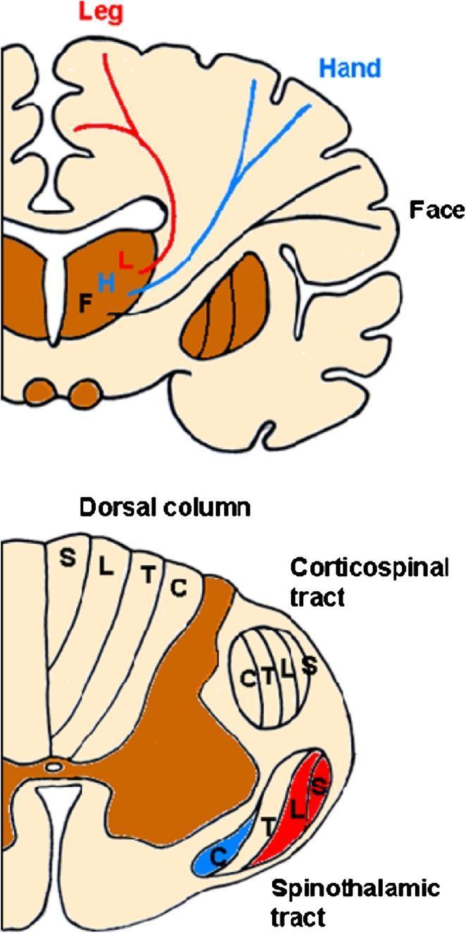
This medical illustration depicts the somatotopic organization (functional mapping) of the human spinothalamocortical system through two anatomical sections. The upper diagram shows a coronal section of the brain, highlighting the thalamocortical fibers. It demonstrates the sensory homunculus where fibers for the face (F) diverge laterally from a medial thalamic position, while fibers for the leg (L) ascend vertically near the lateral ventricle. The lower diagram presents an axial section of the cervical spinal cord, comparing the lamination of major white matter tracts. The dorsal column exhibits a medial-to-lateral arrangement from sacral (S) to cervical (C) fibers. Conversely, both the corticospinal and spinothalamic tracts show an inverse somatotopy where cervical fibers (C) are positioned medially/internally and sacral fibers (S) are positioned laterally/superficially near the spinal cord surface. This educational graphic illustrates the varying spatial relationships of fibers representing different body regions as they ascend or descend the central nervous system.
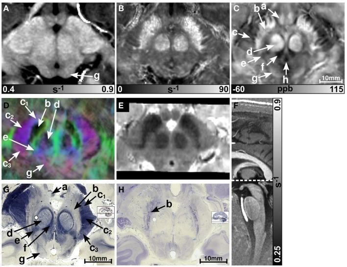
A multi-modal neuroimaging and histological comparison of the human midbrain. Panels A-E display axial sections at the level of the superior colliculus using various MRI contrasts at 7T: (A) R1 relaxation rate map, (B) R2* relaxation rate map, (C) quantitative susceptibility map (QSM), (D) direction-encoded track-density imaging (TDI), and (E) T2-weighted imaging. Panel F provides a sagittal R1 map for anatomical orientation. Panels G and H show corresponding axial histological sections stained for myelin (Luxol Fast Blue) and cellularity (Nissl), respectively. Key anatomical structures labeled include the substantia nigra (b), red nucleus (d), crus cerebri (c) containing corticobulbar (c1), corticospinal (c2), and corticopontine (c3) fibers, and the medial lemniscus (e). The QSM and R2* maps provide superior visualization of the iron-rich substantia nigra and red nucleus compared to R1. The TDI image uses color-coding to represent fiber anisotropy: red for medial-lateral, blue for superior-inferior, and green for anterior-posterior orientations, successfully delineating white matter bundles in the crus cerebri.
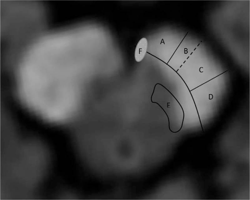
This diagnostic axial MRI scan of the midbrain features a schematic overlay illustrating the topographical functional anatomy of the cerebral peduncle (crus cerebri). The labeled regions (A–F) represent specific descending and ascending tracts. Areas A, B, C, and D are arranged segmentally along the ventral-lateral arc of the peduncle: Area A (medial) contains the corticoponto cerebellar tract (CPCT); Area B corresponds to the corticonuclear tract; Area C represents the corticospinal tract (CST); and Area D (lateral) includes the parieto-, temporo-, and occipitopontine tracts. Region E, located dorsomedial to the main peduncular segments, corresponds to the spinothalamic tract and medial lemniscus. Small oval Area F, situated at the most medial border of the peduncle, indicates the location of the oculomotor nerve (CN III) fibers. This diagram is educationally significant for correlating specific neurological deficits, such as ataxia (A), hemiparesis (C), and sensory disturbances (E), with localized infarcts or lesions within the midbrain peduncular architecture.
- A (medial 1/6) = Corticopontocerebellar (frontopontine) fibres
- B = Corticonuclear (corticobulbar) fibres
- C = Corticospinal fibres
- D (lateral 1/6) = Parieto-/temporo-/occipitopontine fibres
- E = Spinothalamic tract and medial lemniscus (ascending, just dorsomedial)
- F = CN III fibres passing through
The substantia nigra has two divisions. Name them, state the neurotransmitter each uses, and give one key function for each.
The substantia nigra is divided into two functionally and anatomically distinct parts: Division Main neurotransmitter Key function Pars compacta (SNc) Dopamine Supplies dopaminergic input to the striatum (nigrostriatal pathway), helping initiate and modulate voluntary movement. Degeneration of these neurons causes Parkinson's disease. Pars reticulata (SNr) GABA (γ-aminobutyric acid) Acts as a major output nucleus of the basal ganglia, providing inhibitory signals to the thalamus and superior colliculus to regulate movement and control eye movements. Summary Pars compacta (SNc) → Dopamine → Facilitates and modulates voluntary movement via the nigrostriatal pathway. Pars reticulata (SNr) → GABA → Inhibitory output of the basal ganglia, regulating motor activity and saccadic eye movements.
A lesion at the inferior colliculus level of the midbrain is found on imaging. Name five structures you would expect to find at this level, and for each one state its function in one sentence.
At the level of the inferior colliculus, the following important structures are present: Structure Function Inferior colliculus Major relay center of the auditory pathway, integrating sound information and mediating auditory reflexes before projecting to the medial geniculate body. Trochlear nucleus (CN IV) Contains motor neurons that supply the superior oblique muscle, producing depression and intorsion of the eye. Decussation of the trochlear nerve Crossing point of trochlear nerve fibers, so each trochlear nerve innervates the contralateral superior oblique muscle after emerging dorsally. Medial longitudinal fasciculus (MLF) Coordinates conjugate eye movements by interconnecting the nuclei of cranial nerves III, IV, and VI with vestibular nuclei. Cerebral aqueduct Connects the third and fourth ventricles, allowing the flow of cerebrospinal fluid (CSF).
- Correction: The trochlear nerve fibres decussate in the superior medullary velum (the thin roof of the upper fourth ventricle), not at the level of the nucleus itself. The nucleus is in the central grey; the crossing happens just caudally as fibres arch dorsally.
- Bonus structures the lecture specifically lists at this level that you didn't mention:
- Superior cerebellar peduncle (SCP) decussation - dentatorubrothalamic fibres cross here on their way to the contralateral red nucleus and thalamus. Clinically important: an SCP lesion here causes contralateral cerebellar signs.
- Mesencephalic nucleus of CN V - proprioception from jaw muscles (unique: a sensory nucleus housed inside the CNS rather than in a peripheral ganglion).
- Lateral lemniscus - auditory fibres ascending to the inferior colliculus.
Describe the pupillary light reflex pathway step by step — from light entering the eye to bilateral pupil constriction. Then explain: why does shining a light in one eye constrict both pupils?
never mind add this slide and prepare summary followed by a collective MCQs with answers
python3 -c " import pdfplumber pdf_path = '/tmp/workspace/attachments/1bb5e42c-d0b9-4d0c-b099-98580f48c2c9/14@. Hindbrain & Cerebellum - A-Prof. Royhaan.pdf' with pdfplumber.open(pdf_path) as pdf: print(f'Total pages: {len(pdf.pages)}') for i, page in enumerate(pdf.pages): text = page.extract_text() print(f'--- PAGE {i+1} ---') print(text) print() "
python3 << 'PYEOF' from docx import Document from docx.shared import Pt, Inches, RGBColor from docx.enum.text import WD_ALIGN_PARAGRAPH from docx.oxml.ns import qn from docx.oxml import OxmlElement doc = Document() # ── Page margins ────────────────────────────────────────────────────────────── for section in doc.sections: section.top_margin = Inches(1) section.bottom_margin = Inches(1) section.left_margin = Inches(1.1) section.right_margin = Inches(1.1) # ── Helpers ─────────────────────────────────────────────────────────────────── def col(hex_str): h = hex_str.lstrip('#') return RGBColor(int(h[0:2],16), int(h[2:4],16), int(h[4:6],16)) def add_heading(doc, text, level=1, colour="1F4E79"): p = doc.add_heading(text, level=level) for run in p.runs: run.font.color.rgb = col(colour) return p def shade_para(doc, text, fill="E8F4FD", bold_label=None): p = doc.add_paragraph() if bold_label: r = p.add_run(bold_label + " ") r.bold = True; r.font.size = Pt(11) p.add_run(text).font.size = Pt(11) pPr = p._p.get_or_add_pPr() shd = OxmlElement('w:shd') shd.set(qn('w:val'),'clear'); shd.set(qn('w:color'),'auto'); shd.set(qn('w:fill'), fill) pPr.append(shd) p.paragraph_format.left_indent = Inches(0.2) p.paragraph_format.right_indent = Inches(0.2) p.paragraph_format.space_before = Pt(3) p.paragraph_format.space_after = Pt(5) return p def add_table(doc, headers, rows, hdr_col="1F4E79"): t = doc.add_table(rows=1+len(rows), cols=len(headers)) t.style = 'Table Grid' hr = t.rows[0] for i,h in enumerate(headers): c = hr.cells[i]; c.text = h run = c.paragraphs[0].runs[0] run.bold = True; run.font.color.rgb = RGBColor(255,255,255); run.font.size = Pt(10) tcpr = c._tc.get_or_add_tcPr() shd = OxmlElement('w:shd') shd.set(qn('w:val'),'clear'); shd.set(qn('w:color'),'auto'); shd.set(qn('w:fill'), hdr_col) tcpr.append(shd) for ri, row in enumerate(rows): for ci, txt in enumerate(row): cell = t.rows[ri+1].cells[ci] cell.text = txt cell.paragraphs[0].runs[0].font.size = Pt(10) doc.add_paragraph() return t def bullet(doc, text, size=11): p = doc.add_paragraph(text, style="List Bullet") p.runs[0].font.size = Pt(size) return p # ══════════════════════════════════════════════════════════════════════════════ # COVER # ══════════════════════════════════════════════════════════════════════════════ tp = doc.add_paragraph() tp.alignment = WD_ALIGN_PARAGRAPH.CENTER r = tp.add_run("BRAINSTEM & CEREBELLUM") r.bold = True; r.font.size = Pt(26); r.font.color.rgb = col("1F4E79") sp = doc.add_paragraph() sp.alignment = WD_ALIGN_PARAGRAPH.CENTER sp.add_run("Midbrain · Hindbrain · Cerebellum\nCombined Study Guide & MCQ Bank").font.size = Pt(13) doc.add_paragraph() bp = doc.add_paragraph() bp.alignment = WD_ALIGN_PARAGRAPH.CENTER bp.add_run("Based on lectures by A-Prof. Royhaan Folarin").italic = True doc.add_page_break() # ══════════════════════════════════════════════════════════════════════════════ # PART A — MIDBRAIN SUMMARY # ══════════════════════════════════════════════════════════════════════════════ add_heading(doc, "PART A: THE MIDBRAIN — SUMMARY", level=1, colour="1F4E79") add_heading(doc, "A1. Overview & Subdivisions", level=2, colour="1F4E79") doc.add_paragraph("The midbrain is the uppermost brainstem segment, between pons and diencephalon, surrounding the cerebral aqueduct.") add_table(doc, ["Structure","Location","Key Contents"], [ ["Crus cerebri","Ventral","Corticospinal (head med→leg lat), corticonuclear, corticopontine fibres"], ["Substantia nigra","Between crus & tegmen","Pars compacta (DA→striatum); pars reticulata (GABA output ≈ GPi)"], ["Tegmentum","Central","CN III/IV nuclei, red nucleus, lemnisci, MLF, reticular formation"], ["Periaqueductal grey","Around aqueduct","Autonomic integration, pain modulation"], ["Tectum","Dorsal","Superior colliculi (visual); inferior colliculi (auditory)"], ] ) add_heading(doc, "A2. Two Key Levels", level=2, colour="1F4E79") add_table(doc, ["Level","Unique Structures"], [ ["Inferior colliculus (lower midbrain)","Inferior colliculus, CN IV nucleus, SCP decussation, mesencephalic V nucleus, lateral lemniscus"], ["Superior colliculus (upper midbrain)","CN III nuclear complex, Edinger-Westphal nucleus, red nucleus, pretectal nuclei, superior colliculus"], ] ) add_heading(doc, "A3. Classic Midbrain Syndromes", level=2, colour="1F4E79") add_table(doc, ["Syndrome","Lesion Site","Ipsilateral Finding","Contralateral Finding"], [ ["Weber","CN III + crus cerebri","CN III palsy (ptosis, mydriasis, down-and-out)","Hemiplegia (UMN)"], ["Benedikt","CN III + red nucleus","CN III palsy","Rubral tremor / incoordination"], ["Parinaud","Dorsal midbrain / pretectal","Upgaze palsy, light-near dissociation","Bilateral (upgaze palsy)"], ] ) add_heading(doc, "A4. Pupillary Light Reflex", level=2, colour="1F4E79") for i,s in enumerate([ "Light → retina → optic nerve → optic tract", "Pretectal nuclei (dorsal midbrain)", "Bilateral projection → both Edinger-Westphal nuclei", "Preganglionic parasympathetics ride CN III → ciliary ganglion", "Postganglionic → sphincter pupillae → bilateral constriction", ],1): p = doc.add_paragraph(f"{i}. {s}"); p.paragraph_format.left_indent = Inches(0.3); p.runs[0].font.size = Pt(11) shade_para(doc,"Bilateral EW projections explain direct AND consensual constriction from a single eye stimulus.","E8F8F5","Key:") doc.add_paragraph() # ══════════════════════════════════════════════════════════════════════════════ # PART B — HINDBRAIN & CEREBELLUM SUMMARY # ══════════════════════════════════════════════════════════════════════════════ doc.add_page_break() add_heading(doc, "PART B: HINDBRAIN & CEREBELLUM — SUMMARY", level=1, colour="1F4E79") add_heading(doc, "B1. Hindbrain Overview", level=2, colour="1F4E79") doc.add_paragraph("The hindbrain comprises the medulla oblongata, pons, and cerebellum. The medulla and pons form part of the brainstem; the cerebellum lies posterior to them around the fourth ventricle.") add_table(doc, ["Region","Ventral Part","Dorsal Tegmentum"], [ ["Medulla","Pyramids (corticospinal), inferior olivary nucleus","CN nuclei (IX-XII), vestibular nuclei, spinothalamic tract, medial lemniscus"], ["Pons","Pontine nuclei, corticospinal/corticonuclear fibres, transverse pontocerebellar fibres","CN nuclei (V-VIII), ascending tracts, MLF, reticular formation"], ] ) add_heading(doc, "B2. Three Functional Levels of the Medulla", level=2, colour="1F4E79") add_table(doc, ["Level","Key Event","Clinical Significance"], [ ["Pyramidal decussation","Most corticospinal fibres cross → lateral corticospinal tract","Explains contralateral limb control from each hemisphere"], ["Sensory decussation","Internal arcuate fibres cross → form medial lemniscus","Discriminative touch/proprioception fibres cross here"], ["Olive level","Inferior olivary nucleus, lower CN nuclei, open 4th ventricle","Site of lateral/medial medullary syndromes"], ] ) add_heading(doc, "B3. Medullary Syndromes", level=2, colour="1F4E79") add_table(doc, ["Syndrome","Artery","Ipsilateral","Contralateral"], [ ["Medial medullary (Déjerine)","Anterior spinal / vertebral", "CN XII palsy — tongue deviates toward lesion", "Hemiplegia + loss of vibration & proprioception (medial lemniscus)"], ["Lateral medullary (Wallenberg)","PICA / vertebral", "Facial pain-temp loss (CN V), ataxia (ICP), Horner syndrome, dysphagia/hoarseness (NA)", "Body pain-temp loss (spinothalamic), vertigo & nystagmus (vestibular)"], ] ) add_heading(doc, "B4. Pons Organization", level=2, colour="1F4E79") doc.add_paragraph("The pons has a basilar part (motor) and a tegmental part (sensory/CN nuclei). The corticopontocerebellar pathway crosses here: cortex → corticopontine fibres → pontine nuclei → transverse fibres → middle cerebellar peduncle → opposite cerebellar hemisphere.") add_heading(doc, "Pontine Syndromes", level=3, colour="2E74B5") add_table(doc, ["Syndrome","Ipsilateral CN","Contralateral Limb Finding"], [ ["Raymond","Abducens palsy (CN VI)","Hemiplegia"], ["Millard-Gubler","Facial weakness (CN VII)","Hemiplegia"], ["Foville","Facial weakness + horizontal gaze palsy","Variable long-tract deficits"], ] ) add_heading(doc, "B5. Cerebellum — Gross & Functional Organization", level=2, colour="1F4E79") add_table(doc, ["Functional Division","Anatomical Region","Functions"], [ ["Vestibulocerebellum","Flocculonodular lobe","Equilibrium, eye movements, postural stability"], ["Spinocerebellum","Vermis + paravermal zone","Muscle tone, posture, ongoing movement correction"], ["Cerebrocerebellum","Lateral hemispheres","Motor planning, fine voluntary movement, motor learning"], ] ) add_heading(doc, "B6. Cerebellar Cortex Layers", level=2, colour="1F4E79") add_table(doc, ["Layer","Cells","Key Role"], [ ["Molecular (outer)","Stellate, basket cells; Purkinje dendrites; parallel fibres","Modulate Purkinje cell output"], ["Purkinje cell","Purkinje cell bodies","SOLE OUTPUT of cerebellar cortex (inhibitory → deep nuclei)"], ["Granular (inner)","Granule cells, Golgi cells","Relay mossy fibre input via parallel fibres to Purkinje cells"], ] ) add_heading(doc, "B7. Deep Cerebellar Nuclei (medial → lateral)", level=2, colour="1F4E79") add_table(doc, ["Nucleus","Function"], [ ["Fastigial","Balance, axial/truncal control (vestibulo/spinocerebellum output)"], ["Globose + Emboliform (interposed)","Limb movement correction"], ["Dentate","Planning and coordination of skilled movement (cerebrocerebellum output)"], ] ) add_heading(doc, "B8. Cerebellar Peduncles", level=2, colour="1F4E79") add_table(doc, ["Peduncle","Direction","Main Fibres"], [ ["Superior cerebellar peduncle","Mainly OUTPUT","Dentatorubrothalamic tract → red nucleus & thalamus; decussates at inferior colliculus level"], ["Middle cerebellar peduncle","INPUT only","Pontocerebellar fibres (largest peduncle)"], ["Inferior cerebellar peduncle","Mostly INPUT","Vestibulocerebellar, spinocerebellar, olivocerebellar (climbing fibres); small output component"], ] ) add_heading(doc, "B9. Cerebellar Signs (DANISH)", level=2, colour="1F4E79") shade_para(doc, "Cerebellar signs are IPSILATERAL to the lesion (unlike cerebral hemisphere lesions).","FEF9E7","Key rule:") doc.add_paragraph() add_table(doc, ["Sign","Description"], [ ["Dysdiadochokinesia","Impaired rapid alternating movements"], ["Ataxia","Uncoordinated gait / limb movements"], ["Nystagmus","Jerky eye movements (fast phase toward lesion)"], ["Intention tremor","Tremor that worsens on approaching a target"], ["Scanning (staccato) speech","Irregular, explosive speech rhythm"], ["Hypotonia","Reduced muscle tone"], ["(+ Dysmetria)","Past-pointing; unable to judge distance of movement"], ["(+ Rebound phenomenon)","Inability to check a movement"], ] ) # ══════════════════════════════════════════════════════════════════════════════ # PART C — COMBINED CLINICAL VIGNETTES RECAP # ══════════════════════════════════════════════════════════════════════════════ doc.add_page_break() add_heading(doc, "PART C: CLINICAL VIGNETTE QUICK REFERENCE", level=1, colour="1F4E79") add_table(doc, ["Presentation","Syndrome","Lesion Location","Key Artery"], [ ["Ipsilateral CN III palsy + contralateral hemiplegia","Weber","Ventral midbrain — CN III fascicles + crus cerebri","PCA / basilar paramedian branch"], ["Ipsilateral CN III palsy + contralateral rubral tremor","Benedikt","Tegmental midbrain — CN III fascicles + red nucleus","PCA paramedian branch"], ["Upgaze palsy + light-near dissociation + convergence-retraction nystagmus","Parinaud","Dorsal midbrain / pretectal region","Compression (pineal mass) or PCA"], ["Ipsilateral tongue deviation + contralateral hemiplegia + contralateral vibration loss","Medial medullary (Déjerine)","Medial medulla — pyramid + medial lemniscus + CN XII","Anterior spinal / vertebral"], ["Ipsilateral facial pain-temp loss + contralateral body pain-temp loss + ataxia + Horner","Lateral medullary (Wallenberg)","Lateral medulla — PICA territory","PICA or vertebral"], ["Ipsilateral CN VI palsy + contralateral hemiplegia","Raymond","Ventral pons","Paramedian pontine branch"], ["Ipsilateral facial weakness + contralateral hemiplegia","Millard-Gubler","Ventral pons","Paramedian pontine branch"], ["Adduction failure on lateral gaze; convergence preserved","Internuclear ophthalmoplegia","MLF (ipsilateral to adduction failure)","(MS / pontine infarct)"], ], hdr_col="1F4E79" ) # ══════════════════════════════════════════════════════════════════════════════ # PART D — MCQ BANK (30 questions) # ══════════════════════════════════════════════════════════════════════════════ doc.add_page_break() add_heading(doc, "PART D: MCQ BANK — 30 Questions", level=1, colour="1F4E79") doc.add_paragraph("Attempt all questions before checking answers at the end of this section.") doc.add_paragraph() mcqs = [ # ── MIDBRAIN ────────────────────────────────────────────────────────────── { "n":1, "topic":"Midbrain", "q":"Which structure lies immediately between the crus cerebri and the tegmentum of the midbrain?", "opts":["A. Red nucleus","B. Substantia nigra","C. Inferior colliculus","D. Medial lemniscus"], "ans":"B", "exp":"The substantia nigra is wedged between the crus cerebri anteriorly and the tegmentum posteriorly. Its neuromelanin content makes it appear dark in unstained sections." }, { "n":2, "topic":"Midbrain", "q":"Fibres for the lower limb within the corticospinal bundle of the crus cerebri are located:", "opts":["A. Medially within the middle two-thirds","B. Laterally within the middle two-thirds","C. In the medial one-sixth","D. In the lateral one-sixth"], "ans":"B", "exp":"Somatotopic arrangement: head/face fibres lie medially and lower limb fibres lie laterally within the corticospinal region (middle two-thirds of crus)." }, { "n":3, "topic":"Midbrain", "q":"Degeneration of which part of the substantia nigra is responsible for Parkinson disease?", "opts":["A. Pars reticulata","B. Pars compacta","C. Pars diffusa","D. Pars lateralis"], "ans":"B", "exp":"Pars compacta contains dopaminergic neurons projecting to the striatum via the nigrostriatal pathway. Their loss reduces striatal dopamine, producing the cardinal motor features of Parkinson disease." }, { "n":4, "topic":"Midbrain", "q":"At the inferior colliculus level of the midbrain, the superior cerebellar peduncle fibres undergo:", "opts":["A. No crossing","B. Partial decussation","C. Complete decussation","D. Ipsilateral termination in the red nucleus"], "ans":"C", "exp":"The dentatorubrothalamic fibres within the superior cerebellar peduncle fully decussate at the level of the inferior colliculus before terminating in the contralateral red nucleus and thalamus." }, { "n":5, "topic":"Midbrain", "q":"Which cranial nerve is unique in exiting the brainstem from its POSTERIOR surface and fully decussating before exit?", "opts":["A. CN III","B. CN IV","C. CN VI","D. CN XII"], "ans":"B", "exp":"CN IV (trochlear) is the only cranial nerve to exit the brainstem posteriorly (from the dorsal midbrain below the inferior colliculi) and to fully decussate in the superior medullary velum." }, { "n":6, "topic":"Midbrain", "q":"A patient has an ipsilateral CN III palsy and contralateral hemiplegia. Which syndrome is this, and which two structures are damaged?", "opts":["A. Benedikt — red nucleus + CN III","B. Weber — crus cerebri + CN III","C. Parinaud — pretectal + CN III","D. Foville — crus cerebri + CN IV"], "ans":"B", "exp":"Weber syndrome results from a ventral midbrain lesion affecting the CN III fascicles (ipsilateral oculomotor palsy) and the crus cerebri (contralateral UMN hemiplegia)." }, { "n":7, "topic":"Midbrain", "q":"A patient with Benedikt syndrome has contralateral coarse tremor. Which nucleus is damaged?", "opts":["A. Substantia nigra pars compacta","B. Subthalamic nucleus","C. Red nucleus","D. Dentate nucleus"], "ans":"C", "exp":"Benedikt syndrome involves the red nucleus in the midbrain tegmentum. Damage here disrupts cerebellar and extrapyramidal motor signals, producing contralateral Holmes (rubral) tremor." }, { "n":8, "topic":"Midbrain", "q":"Which pretectal finding best distinguishes Parinaud syndrome from other gaze palsies?", "opts":["A. Horizontal gaze palsy","B. Light-near dissociation with convergence-retraction nystagmus","C. Internuclear ophthalmoplegia","D. Skew deviation"], "ans":"B", "exp":"The triad of upgaze palsy, light-near dissociation (pupils react to accommodation not light), and convergence-retraction nystagmus on attempted upgaze defines Parinaud (dorsal midbrain) syndrome." }, { "n":9, "topic":"Midbrain", "q":"The Edinger-Westphal nucleus provides parasympathetic input to which two structures?", "opts":["A. Lateral rectus and levator palpebrae","B. Superior oblique and inferior rectus","C. Sphincter pupillae and ciliary muscle","D. Dilator pupillae and orbicularis oculi"], "ans":"C", "exp":"The Edinger-Westphal nucleus sends preganglionic fibres via CN III to the ciliary ganglion, whose postganglionic fibres supply the sphincter pupillae (pupil constriction) and ciliary muscle (accommodation)." }, { "n":10, "topic":"Midbrain", "q":"The medial longitudinal fasciculus (MLF) connects which nuclei to coordinate horizontal conjugate gaze?", "opts":["A. CN IV nucleus and CN V nucleus","B. CN VI nucleus interneurons and contralateral CN III nucleus","C. Vestibular nuclei and ipsilateral CN IV nucleus","D. Red nucleus and CN III nucleus"], "ans":"B", "exp":"For conjugate horizontal gaze, interneurons from the CN VI (abducens) nucleus project via the MLF to the contralateral CN III (oculomotor) nucleus to coordinate simultaneous abduction and adduction." }, # ── MEDULLA ─────────────────────────────────────────────────────────────── { "n":11, "topic":"Medulla", "q":"At which level of the medulla do the corticospinal fibres cross to form the lateral corticospinal tract?", "opts":["A. Level of the olive","B. Pontomedullary junction","C. Pyramidal decussation","D. Sensory decussation"], "ans":"C", "exp":"The pyramidal decussation occurs in the lower medulla where ~85% of corticospinal fibres cross to descend as the lateral corticospinal tract, explaining contralateral hemisphere control." }, { "n":12, "topic":"Medulla", "q":"The medial lemniscus is formed at which level of the medulla?", "opts":["A. Pyramidal decussation","B. Sensory decussation — crossing of internal arcuate fibres","C. Level of the olive","D. Pontomedullary junction"], "ans":"B", "exp":"At the sensory decussation, second-order neurons from the gracile and cuneate nuclei send internal arcuate fibres that cross the midline to form the contralateral medial lemniscus." }, { "n":13, "topic":"Medulla", "q":"In medial medullary syndrome, a patient's tongue deviates to the LEFT on protrusion. The lesion is most likely:", "opts":["A. Right medial medulla","B. Left medial medulla","C. Right lateral medulla","D. Left lateral medulla"], "ans":"B", "exp":"CN XII palsy causes the tongue to deviate toward the side of the lower motor neuron lesion. Tongue deviation to the left = left CN XII/nucleus lesion = left medial medullary syndrome." }, { "n":14, "topic":"Medulla", "q":"Which artery is most commonly responsible for the lateral medullary (Wallenberg) syndrome?", "opts":["A. Anterior spinal artery","B. Posterior inferior cerebellar artery (PICA) or vertebral artery","C. Anterior inferior cerebellar artery (AICA)","D. Superior cerebellar artery (SCA)"], "ans":"B", "exp":"Lateral medullary syndrome is most frequently caused by occlusion of PICA or the vertebral artery, infarcting the lateral medulla including the nucleus ambiguus, vestibular nuclei, spinal trigeminal nucleus, spinothalamic tract, ICP, and descending sympathetics." }, { "n":15, "topic":"Medulla", "q":"A patient has dysphagia, hoarseness, ipsilateral facial pain-temperature loss, contralateral body pain-temperature loss, and ipsilateral Horner syndrome. Which structure's damage explains the Horner syndrome?", "opts":["A. Nucleus ambiguus","B. Vestibular nuclei","C. Descending hypothalamospinal (sympathetic) fibres","D. Inferior cerebellar peduncle"], "ans":"C", "exp":"In lateral medullary syndrome, descending sympathetic fibres (hypothalamospinal tract) travelling through the lateral tegmentum are interrupted, producing ipsilateral Horner syndrome (ptosis, miosis, anhidrosis)." }, { "n":16, "topic":"Medulla", "q":"The inferior olivary nucleus sends climbing fibres to the cerebellar cortex primarily through which peduncle?", "opts":["A. Superior cerebellar peduncle","B. Middle cerebellar peduncle","C. Inferior cerebellar peduncle","D. Restiform body only"], "ans":"C", "exp":"Olivocerebellar climbing fibres travel via the inferior cerebellar peduncle (specifically the restiform body) to make powerful synapses directly onto Purkinje cell dendrites in the contralateral cerebellar cortex." }, # ── PONS ───────────────────────────────────────────────────────────────── { "n":17, "topic":"Pons", "q":"The largest cerebellar peduncle is the middle cerebellar peduncle (MCP). What is its primary function?", "opts":["A. Cerebellar output to thalamus","B. Vestibular and spinal input to cerebellum","C. Pontocerebellar (corticopontocerebellar) input to cerebellum","D. Olivocerebellar input"], "ans":"C", "exp":"The MCP is the largest peduncle and carries exclusively afferent pontocerebellar fibres arising from the contralateral pontine nuclei — the final leg of the corticopontocerebellar pathway conveying intended movement information." }, { "n":18, "topic":"Pons", "q":"The facial colliculus on the floor of the fourth ventricle is produced by the:", "opts":["A. Facial motor nucleus","B. Abducens nucleus overlaid by facial nerve fibres looping around it","C. Vestibular nuclei","D. Superior olivary complex"], "ans":"B", "exp":"The internal genu of the facial nerve (CN VII) loops around the abducens nucleus (CN VI), creating a surface bulge — the facial colliculus — visible on the floor of the fourth ventricle at the lower pontine level." }, { "n":19, "topic":"Pons", "q":"A patient has right lateral gaze palsy (eyes cannot move right) and right facial weakness, plus left hemiplegia. Which syndrome is this?", "opts":["A. Raymond","B. Millard-Gubler","C. Foville","D. Benedikt"], "ans":"C", "exp":"Foville syndrome involves ipsilateral horizontal gaze palsy (abducens nucleus or PPRF) + ipsilateral facial weakness (CN VII nucleus) + contralateral hemiplegia. This is a lower pontine tegmental lesion." }, { "n":20, "topic":"Pons", "q":"The corticopontocerebellar pathway crosses the midline at which level?", "opts":["A. Midbrain tegmentum","B. Medullary pyramidal decussation","C. Basilar pons (transverse pontine fibres)","D. Inferior cerebellar peduncle"], "ans":"C", "exp":"Axons from pontine nuclei (receiving corticopontine input) cross as transverse pontine fibres in the basilar pons, then enter the middle cerebellar peduncle to reach the contralateral cerebellar hemisphere." }, # ── CEREBELLUM ──────────────────────────────────────────────────────────── { "n":21, "topic":"Cerebellum", "q":"Which functional division of the cerebellum is primarily responsible for equilibrium and eye movement control?", "opts":["A. Spinocerebellum","B. Cerebrocerebellum","C. Vestibulocerebellum","D. Vermis only"], "ans":"C", "exp":"The vestibulocerebellum (flocculonodular lobe) receives strong vestibular input and controls equilibrium, postural stability, and eye movements (vestibulo-ocular reflex)." }, { "n":22, "topic":"Cerebellum", "q":"Which cell is the sole output neuron of the cerebellar cortex?", "opts":["A. Granule cell","B. Basket cell","C. Purkinje cell","D. Golgi cell"], "ans":"C", "exp":"Purkinje cells are the sole efferent neurons of the cerebellar cortex. They are GABAergic (inhibitory) and project to the deep cerebellar nuclei and vestibular nuclei." }, { "n":23, "topic":"Cerebellum", "q":"Climbing fibres, which form powerful excitatory synapses on Purkinje cells, originate from:", "opts":["A. Pontine nuclei","B. Inferior olivary nucleus","C. Vestibular nuclei","D. Dentate nucleus"], "ans":"B", "exp":"Climbing fibres arise exclusively from the inferior olivary nucleus (contralateral side, via the inferior cerebellar peduncle). They are critical for motor learning and error correction." }, { "n":24, "topic":"Cerebellum", "q":"The dentate nucleus is the largest deep cerebellar nucleus. It primarily receives input from and contributes to:", "opts":["A. Flocculonodular lobe → balance control","B. Lateral cerebellar hemispheres → motor planning and skilled movement","C. Vermis → axial muscle control","D. Interposed nuclei → limb correction"], "ans":"B", "exp":"The dentate nucleus receives Purkinje cell inhibition from the lateral cerebellar hemispheres (cerebrocerebellum) and projects via the superior cerebellar peduncle to the contralateral red nucleus and thalamus, supporting planning and coordination of skilled movement." }, { "n":25, "topic":"Cerebellum", "q":"A patient with a right cerebellar hemisphere lesion will most likely show signs:", "opts":["A. Ipsilateral (right-sided) — intention tremor, dysmetria, dysdiadochokinesia","B. Contralateral (left-sided) — intention tremor, dysmetria","C. Bilateral — scanning speech only","D. Contralateral — hemiplegia"], "ans":"A", "exp":"Cerebellar lesions produce IPSILATERAL signs because the cerebellum controls the same side of the body (double decussation: corticopontocerebellar and dentatorubrothalamic both cross, cancelling out)." }, # ── INTEGRATED / CLINICAL ───────────────────────────────────────────────── { "n":26, "topic":"Clinical Integration", "q":"A 55-year-old has internuclear ophthalmoplegia (INO) — failure of left eye adduction on rightward gaze, with right abducting nystagmus; convergence is normal. The lesion is in the:", "opts":["A. Right MLF","B. Left MLF","C. Right CN III nucleus","D. Left CN VI nucleus"], "ans":"B", "exp":"INO on rightward gaze with LEFT adduction failure = left MLF lesion. The left MLF carries the signal from right abducens interneurons to the left CN III nucleus for left eye adduction. Convergence is spared as it uses a different pathway." }, { "n":27, "topic":"Clinical Integration", "q":"Which of the following is the most common cause of bilateral INO in a young adult?", "opts":["A. Vertebrobasilar stroke","B. Multiple sclerosis","C. Wernicke encephalopathy","D. Pineal germinoma"], "ans":"B", "exp":"Bilateral INO in a young adult is the classic presentation of multiple sclerosis, which causes demyelination of the MLF. Unilateral INO in older patients is more commonly due to brainstem infarction." }, { "n":28, "topic":"Clinical Integration", "q":"The key principle unifying Weber, Benedikt, medial medullary, Raymond, and Millard-Gubler syndromes is:", "opts":["A. All involve the cerebellum","B. All produce ipsilateral cranial nerve deficit + contralateral long-tract deficit (crossed syndrome)","C. All involve the thalamus","D. All are caused by vertebral artery occlusion"], "ans":"B", "exp":"A unilateral brainstem lesion above the pyramidal decussation interrupts the CN at the same level (ipsilateral LMN deficit) while the corticospinal/lemniscal fibres have not yet crossed (contralateral UMN/sensory deficit) — the hallmark crossed brainstem syndrome." }, { "n":29, "topic":"Clinical Integration", "q":"A patient presents with vertigo, nystagmus, and ipsilateral ataxia after a posterior fossa stroke. Which cerebellar peduncle is most likely involved by the infarcted lateral medulla?", "opts":["A. Superior cerebellar peduncle","B. Middle cerebellar peduncle","C. Inferior cerebellar peduncle","D. All three equally"], "ans":"C", "exp":"The inferior cerebellar peduncle runs through the lateral medulla (PICA territory). Its involvement in lateral medullary syndrome contributes the ipsilateral limb and truncal ataxia that accompanies the other features of Wallenberg syndrome." }, { "n":30, "topic":"Clinical Integration", "q":"A 22-year-old presents with upgaze palsy and pupils that react to accommodation but not to light. MRI shows a pineal region mass. Which nucleus receives impaired input from the compressed pretectal nuclei?", "opts":["A. Trochlear nucleus","B. Edinger-Westphal nucleus","C. Abducens nucleus","D. Red nucleus"], "ans":"B", "exp":"In Parinaud syndrome, pretectal nucleus compression blocks the light-reflex arc (retina → pretectal → Edinger-Westphal → pupil constriction). The accommodation arc bypasses the pretectal nuclei and remains intact, producing light-near dissociation." }, ] for mcq in mcqs: p = doc.add_paragraph() r = p.add_run(f"Q{mcq['n']}. [{mcq['topic']}] {mcq['q']}") r.bold = True; r.font.size = Pt(11) p.paragraph_format.space_before = Pt(8) for opt in mcq['opts']: op = doc.add_paragraph(opt) op.paragraph_format.left_indent = Inches(0.3) op.runs[0].font.size = Pt(10) doc.add_paragraph() # ── Answer Key ──────────────────────────────────────────────────────────────── doc.add_page_break() add_heading(doc, "ANSWER KEY & EXPLANATIONS", level=1, colour="1F4E79") # Quick answer grid cols = 5 rows_needed = (len(mcqs) + cols - 1) // cols t = doc.add_table(rows=rows_needed+1, cols=cols) t.style = 'Table Grid' hr = t.rows[0] for i in range(cols): hr.cells[i].text = f"Q{i*rows_needed+1}–Q{min((i+1)*rows_needed, 30)}" r = hr.cells[i].paragraphs[0].runs[0] r.bold = True; r.font.size = Pt(10); r.font.color.rgb = col("1F4E79") for idx, mcq in enumerate(mcqs): ri = idx % rows_needed + 1 ci = idx // rows_needed cell = t.rows[ri].cells[ci] cell.text = f"Q{mcq['n']}: {mcq['ans']}" cell.paragraphs[0].runs[0].font.size = Pt(10) doc.add_paragraph() add_heading(doc, "Detailed Explanations", level=2, colour="1F4E79") for mcq in mcqs: p = doc.add_paragraph() r = p.add_run(f"Q{mcq['n']} — Answer: {mcq['ans']}") r.bold = True; r.font.color.rgb = col("C00000"); r.font.size = Pt(11) exp = doc.add_paragraph(mcq['exp']) exp.paragraph_format.left_indent = Inches(0.3) exp.runs[0].font.size = Pt(10) exp.paragraph_format.space_after = Pt(6) # ══════════════════════════════════════════════════════════════════════════════ # SAVE # ══════════════════════════════════════════════════════════════════════════════ out = "/tmp/workspace/midbrain-study-guide/Brainstem_Cerebellum_StudyGuide_MCQ.docx" doc.save(out) print(f"Saved: {out}") PYEOF
Brainstem Cerebellum StudyGuide MCQ
Word Document · DOCX
What's inside (4 parts):
- Subdivisions table, crus cerebri fibre map, substantia nigra, two key levels, classic syndromes (Weber/Benedikt/Parinaud), pupillary light reflex pathway
- Medulla (3 levels + decussations), medullary syndromes (Déjerine, Wallenberg), pons organization + crossed pontine syndromes, cerebellum (gross + functional divisions, cortex layers, deep nuclei, peduncles, DANISH signs)
- 8 syndromes side by side: presentation → syndrome → lesion site → artery
| Topic | Questions |
|---|---|
| Midbrain | Q1-Q10 |
| Medulla | Q11-Q16 |
| Pons | Q17-Q20 |
| Cerebellum | Q21-Q25 |
| Clinical Integration | Q26-Q30 |